Preparing method of micro-fluidic chip with embedded hyaluronic acid functionalized nanofiber membrane

A nanofiber membrane and microfluidic chip technology, which is applied in the field of microfluidic chips and cell sorting, can solve the problems of difficult storage, high cost, and easy inactivation price, and achieve the effects of low cost, easy modification, and simple device

Active Publication Date: 2018-02-02
DONGHUA UNIV
View PDF6 Cites 9 Cited by
  • Summary
  • Abstract
  • Description
  • Claims
  • Application Information

AI Technical Summary

Problems solved by technology

[0005] Most current methods for capturing CTCs are based on antibody-antigen specific binding,...

Method used


Image

  • Preparing method of micro-fluidic chip with embedded hyaluronic acid functionalized nanofiber membrane
  • Preparing method of micro-fluidic chip with embedded hyaluronic acid functionalized nanofiber membrane
  • Preparing method of micro-fluidic chip with embedded hyaluronic acid functionalized nanofiber membrane

Examples


Embodiment 1

[0059] (1) Dissolve 180mg of chitosan and 20mg of polyethylene oxide PEO in 85% acetic acid solution, and react with magnetic stirring for 8h at room temperature until chitosan becomes a uniform and stable solution, then cool to room temperature to obtain a concentration of 3.0wt% chitosan / PEO spinning solution was stored in a refrigerator at 4°C for use. Slowly absorb the spinning solution obtained above into a syringe, and use a high-voltage electrospinning machine to perform electrospinning. The spinning conditions are: spinning voltage 30kV, flow rate 0.1mL / h, spinning distance 12cm, ambient temperature 20-30 ℃, humidity 20-40%, take the plate device covered with aluminum foil as the receiving device, and then place it in a vacuum drying oven for 24 hours to obtain a chitosan nanofiber membrane, which is cross-linked with glutaraldehyde in a desiccator for 6 hours to obtain Water-insoluble cross-linked chitosan nanofibrous membrane CNFs.

[0060] (2) 200.94 mg of hyaluron...

Embodiment 2

[0067] The present invention uses scanning electron microscopy (SEM), attenuated total reflection-Fourier transform infrared spectroscopy (ATR-FTIR), hydrogen nuclear magnetic resonance spectroscopy ( 1 H NMR), ultraviolet-visible absorption spectroscopy (UV-vis), anti-protein adsorption test, water contact angle test, blood compatibility test, anticoagulation test, static / dynamic capture and release test of cancer cells, and immunostaining test To characterize various properties of the hyaluronic acid functionalized nanofibrous membrane prepared in the present invention and its application potential combined with microfluidic chip in sorting and non-destructive release of circulating tumor cells.

[0068] Scanning Electron Microscopy Test:

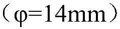
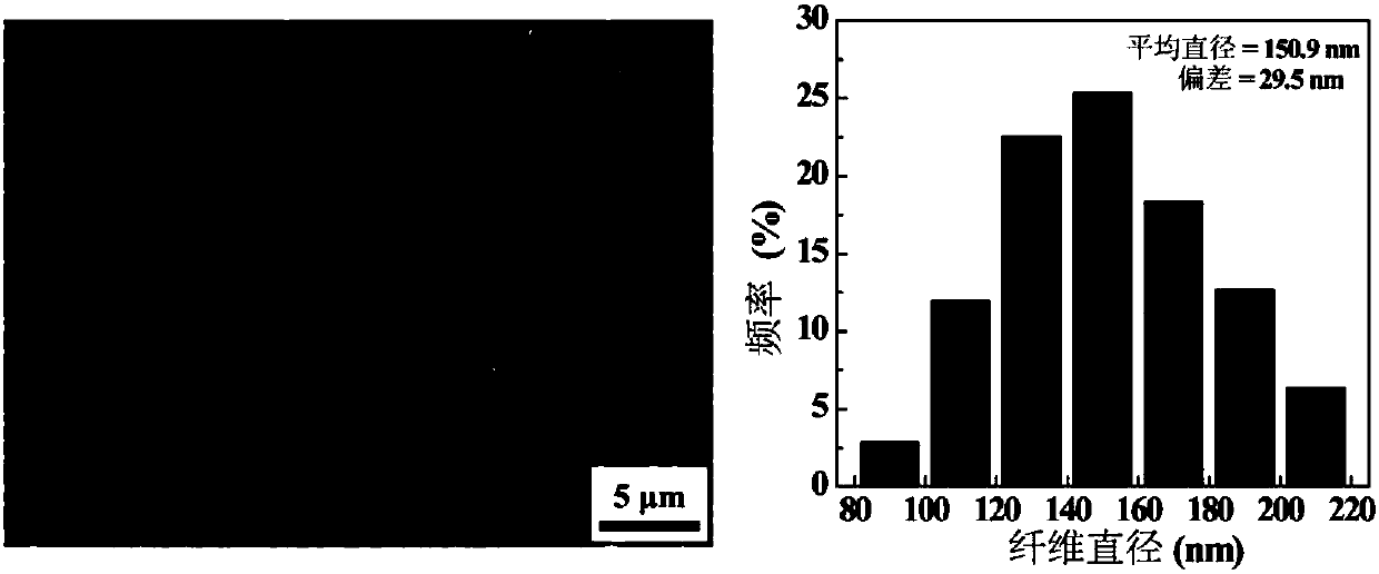
[0069] Adopt the morphology and the diameter distribution of the chitosan nanofiber that embodiment 1 step (1) obtains of SEM characterization, SEM result is as follows figure 1 As shown, the chitosan nanofibers prepared by electrospinn...

Embodiment 3

[0079] Anti-protein adsorption test:

[0080] CNFs, CBAA-CNFs and HA-CBAA-CNFs nanofibers in Example 1 were evaluated against protein (BSA and fibronectin) adsorption for characterizing the properties of CNFs, CBAA-CNFs and HA-CBAA-CNFs nanofiber membranes Anti-BSA adsorption ability and anti-fibronectin adsorption ability, firstly, the standard curve equations of different concentrations of BSA and fibronectin were measured by ultraviolet light. On this basis, choose the concentration of BSA as 2mg / mL and the concentration of BSA as 1mg / mL as the test concentration. Take 10 mg of CNFs, CBAA-CNFs and HA-CBAA-CNFs nanofibers respectively, put each sample into a 24-well plate in quadruplicate, and then add 1 mL of BSA and fibronectin solution to each well plate, at room temperature After co-incubating for 1 h, take out the nanofibers, take the supernatant and test the absorbance of the supernatant at 278nm with a Lamada 25 UV spectrophotometer, and calculate the adsorption rate...


PUM

PropertyMeasurementUnit
Concentrationaaaaaaaaaa

Abstract

The invention relates to a preparing method of a micro-fluidic chip with embedded hyaluronic acid functionalized nanofiber membrane. The preparing method comprises the steps that chitosan/PEO spinningsolution is subjected to static spinning, vacuum drying and crosslinking, and chitosan nanofiber membrane CNFs is obtained; zwitter-ions CBAA and targeting ligands HA-Cys-MPTMS are modified, and HA functionalized nanofiber membrane HA-CBAA-CNFs is obtained; the nanofiber membrane HA-CBAA-CNFs and a PDMS micro-fluidic channel cover plate are subjected to plasma bonding, and the micro-fluidic chipwith embedded hyaluronic acid(HA) functional nanofiber membrane is obtained, wherein the micro-fluidic chip can be used for sorting and non-destructive releasing of circulating tumor cells. The preparing method has the advantages that the method is simple in technology, easy to operate and high in specificity, marker-free and high-throughput sorting and non-destructive releasing of the circulatingtumor cells can be completed in a short time, accordingly efficient separation for the circulating tumor cells is achieved, and the preparing method has good application prospects in early detectionof tumor patients.

Description

technical field [0001] The invention belongs to the technical field of microfluidic chips and cell sorting, and in particular relates to a method for preparing a microfluidic chip embedded with a hyaluronic acid functionalized nanofiber membrane. Background technique [0002] Cancer is the primary cause of human death. According to World Health Organization (WHO) experts' forecast, the number of global cancer incidence will reach 20 million in 2020, and the death toll will reach 12 million. Cancer will become a major cause of human survival and health in the 21st century. main threat. Studies have shown that 90% of the deaths of cancer patients are closely related to tumor metastasis. As the key link between the primary tumor and metastases, circulating tumor cells (Circulating tumor cells, CTCs) have attracted widespread attention at home and abroad in recent years. In the early stage of tumor metastasis, tumor cells detach from the primary solid tumor and enter the blood...

Claims


Application Information

Patent Timeline
IPC IPC(8): D04H1/728D04H1/4258C12N5/09
CPCC12N5/0693C12N2533/72C12N2533/80D04H1/4258D04H1/728
Inventor 史向阳王梦媛肖云超朱晓玥
Owner DONGHUA UNIV
Why Eureka
Try Eureka