
A cell culture dish used for staining observation, and a cell culture staining and observing method

A technology of cell culture and petri dish, which is applied in the field of observation, cell culture dish, and cell culture staining. It can solve the problems of cells deviating from normal cell shape, poor control of strength, and floating slides, etc., so as to facilitate cell staining and follow-up observation. , Conducive to observation and photography, the effect of smooth surface

Pending Publication Date: 2018-02-16
INST OF ANIMAL SCI & VETERINARY MEDICINE SHANDONG ACADEMY OF AGRI SCI
View PDF0 Cites 1 Cited by
  • Summary
  • Abstract
  • Description
  • Claims
  • Application Information

AI Technical Summary

Problems solved by technology

However, in the experiment, it was found that no matter whether the cover glass or the special glass slide was used, the following three problems existed: (1) the slide was placed in the culture well, and after adding the cell suspension, it was found that the slide would float, causing the cells to Adhesive growth on the upper and lower sides of the glass slide, which is not conducive to observing cell morphology under a microscope
Even if a small amount of culture solution is first dropped on the place where the slides are to be placed in the culture wells, and then the slides are placed gently, so that the slides and the culture wells seem to stick together, but some slides float after adding the cell suspension
(2) There are slight differences in shape and function between the cells grown on the glass slide and the cells cultured in the culture well. The cells grown on the glass slide deviate from the normal cell shape and their secretory function is reduced. For example, the formation of lipid droplets in adipocytes slows down.
(3) Take out the glass slide with tweezers after the staining process is completed. Since the glass slide is thin, it is difficult to control the strength when clamping.

Method used


Image

  • A cell culture dish used for staining observation, and a cell culture staining and observing method
  • A cell culture dish used for staining observation, and a cell culture staining and observing method
  • A cell culture dish used for staining observation, and a cell culture staining and observing method

Examples


Embodiment 1

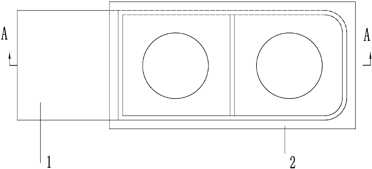
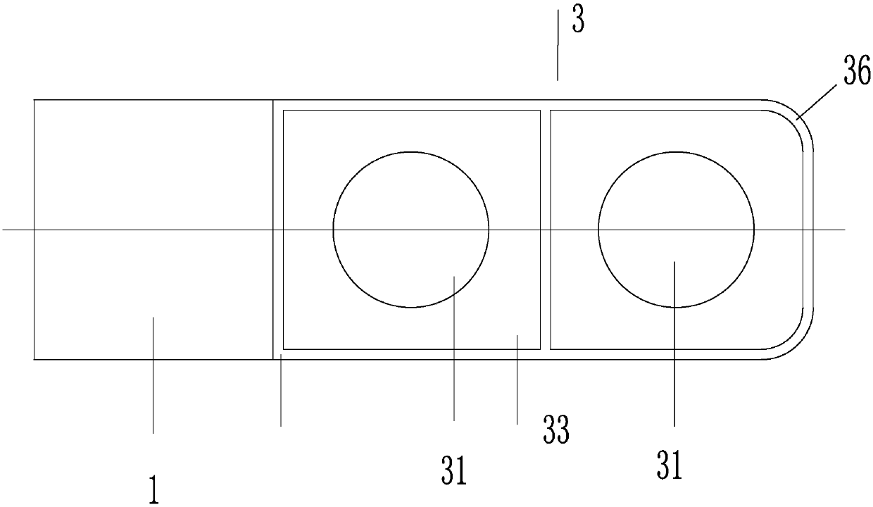
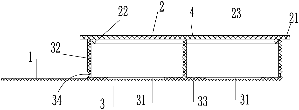
[0041] combine Figure 1 to Figure 5, a cell culture dish, comprising a culture dish body 3, a culture dish cover 2, a handle 1 is provided on one side of the culture dish body 3, the culture dish body 3 includes a base plate 33, and several base plate grooves 31 are provided on the base plate 33 The upper periphery of the bottom plate 33 is provided with a closed vertical sealing plate 32, the handle 1 is connected to the bottom plate 33, and the lower end surface of the culture dish cover 2 is matched with the upper end surface of the vertical sealing plate 32.

[0042] The vertical sealing plate 32 is provided with a sealing plate groove outside the upper part where it connects with the bottom plate 33 , and the sealing plate groove is an arc-shaped groove 34 or an inclined surface 35 from top to bottom. By providing a sealing plate groove on the outside of the vertical sealing plate 32, the thickness of the sealing plate groove portion becomes smaller, and the vertical sea...

Embodiment 2

[0050] A cell culture staining and observation method, specifically comprising the following steps:

[0051] 1) Culture medium preparation

[0052] Add 50-60 ml of fetal bovine serum (FBS), 3-5 ml of penicillin-streptomycin mixture, 0.01-0.02 g of type I collagen to every 450-500 ml of cell culture medium, and add appropriate types and concentration of growth factors or energy substances, and then filtered through a 0.22 micron filter membrane and then packed into sterilized centrifuge tubes, 40-45 ml per tube, stored at 4°C and used up within two weeks; before cell culture 0.5-1 Place the culture medium in a 37°C water bath to preheat;

[0053] 2) Cell culture

[0054] After 80-90% of the cells cultured in vitro adhere to the wall, add PBS solution to the culture bottle to wash once, then add 0.25% trypsin containing 0.05% EDTA and digest in a 37°C incubator for 3-5 minutes, then add Gently blow the culture medium into a single free cell, adjust the cell density to 2-5×10 ...

Embodiment 3

[0060] A cell culture staining and observation method, specifically comprising the following steps:

[0061] (1) Preparation of culture medium

[0062] Add 50 ml of FBS, 3 ml of penicillin-streptomycin mixed solution, 0.01 g of type I collagen, and 0.1 g of taurine to each 450 ml of DMEM / F12 solution, then filter through a 0.22-micron filter membrane and pack into a sterilized centrifuge. tubes, 40ml per tube, store at 4°C and use up within two weeks. Preheat the culture medium in a 37°C water bath 0.5 hours before cell culture.

[0063] (2) Bovine fat cell culture

[0064] After 80%-90% of bovine adipocytes cultured in vitro adhere to the wall, add 2 ml of PBS solution to the T25 culture flask to wash once, then add 1 ml of 0.25% trypsin (containing 0.05% EDTA) and place in a 37°C incubator Digest within 4 minutes, then add the culture medium and blow gently to form a single free cell, adjust the cell density to 3×10⁴ cells / ml, add 4 ml of cell suspension in a special cell...


PUM


Abstract

A cell culture dish used for staining observation, and a cell culture staining and observing method are disclosed. The cell culture dish includes a culture dish body and a culture dish cover. One sideof the cover dish body is provided with a handle. The culture dish body includes a bottom plate. The bottom plate is provided with a plurality of bottom grooves. The upper periphery of the bottom plate is provided with an enclosed vertical sealing plate. The handle is connected to the bottom plate. The lower end surface of the culture dish cover is matched with the upper end surface of the vertical sealing plate. The method includes 1) preparing a culture liquid, 2) culturing cells, 3) staining, and 4) performing microscopic examination. A step of adding a glass sheet into a culture hole is not needed, and a cell suspension is directly added into the cell culture dish specially manufactured, and therefore glass sheet floating and glass sheet clamping problems are avoided. After staining is finished, the bottom wall of the culture dish is put below and observed by an inverted microscope directly, or the bottom wall of the culture dish is turned over and observed by an upright microscope, and therefore cell layers and a microscope lens are very close. The cell culture dish and the method are particularly suitable for observation by high-power oil immersion objectives.

Description

technical field [0001] The invention relates to the technical field of cell culture, in particular to a cell culture dish for staining and observation and a cell culture staining and observation method. Background technique [0002] As an important research method, cell culture is widely used in nutrition, pathology, environmental toxicology, cell biology, oncology, genomics, genetics and resource protection, and has become an indispensable technical means in scientific research . In experiments, staining (such as immunochemistry, immunofluorescence, hematoxylin-eosin, etc.) is often required to observe cell morphology and structure, so it is necessary to culture cells on a suitable carrier to facilitate observation under a microscope . [0003] At present, the method of cell "climbing" is usually used for staining and observation, that is, the coverslip is first rinsed, sterilized and placed in the well of the culture plate or culture dish, and the cell suspension is adde...

Claims


Application Information

Patent Timeline
IPC IPC(8): C12M3/00C12M1/22G01N1/30
CPCG01N1/30C12M23/10C12M23/20
Inventor 谭秀文刘桂芬游伟靳青刘倚帆赵红波刘晓牧万发春张相伦
Owner INST OF ANIMAL SCI & VETERINARY MEDICINE SHANDONG ACADEMY OF AGRI SCI
Learn More