A Histological Method to Distinguish Three Kinds of Muscle Cells in Carp
A technique of muscle cells and histology, which is applied in the field of microscopic observation of samples, can solve the problems of long preparation time and achieve the effect of easy observation and maintaining integrity
- Summary
- Abstract
- Description
- Claims
- Application Information
AI Technical Summary
Problems solved by technology
Method used
Image
Examples
Embodiment 1
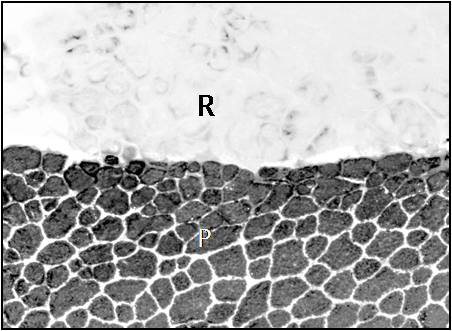
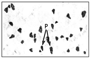
[0020] A histological method for distinguishing three kinds of carp muscle cells, including freezing, freezing, pretreatment, ATPase reaction, color development, and optical microscope observation, the specific steps are:
[0021]1) Freezing: Wash the sample block taken from the back of the carp with a cryoprotectant with a concentration of 2.1% for 12 minutes, put it on the sample stage, add the frozen section embedding agent dropwise, and then put the sample and the sample stage into a container of isoamyl In the container of alkane, put the container into liquid nitrogen and freeze it to the freezing point quickly, the above-mentioned cryoprotectant is trehalose and glycoside, and its weight ratio is 1:0.62, and the ratio of levoside and dextran in glycoside is 1:90, during the freeze-drying process, a reasonable ratio of levoside and dextran can increase the viscosity of the solution, making the glucoside produce steric hindrance between protein molecules in muscle cells, a...
Embodiment 2
[0030] A histological method for distinguishing three kinds of carp muscle cells, the specific steps are:
[0031] 1) Freezing: Wash the sample block taken from the back of the carp with a cryoprotectant with a concentration of 3% for 9 minutes, put it on the sample stage, add the frozen section embedding agent dropwise, and then put the sample and the sample stage into a container of isoprene. In the container of alkane, put the container into liquid nitrogen and quickly freeze to the freezing point. The above-mentioned cryoprotectant is trehalose and glycoside, and its weight ratio is 1:0.8. The ratio of levoside and dextran in glycoside is 1:84;
[0032] 2) Slicing: Put the sample that reached the freezing point into a cryostat at -28°C for 55 minutes, then fix the sample stage to a cryostat and cut out a slice with a thickness of 11 µm, collect the cut sample on a glass slide, Dry at room temperature to obtain sample slices;
[0033] 3) Pretreatment: Fill the pretreatmen...
Embodiment 3
[0038] A histological method for distinguishing three kinds of carp muscle cells, the specific steps are:
[0039] 1) Cut a 1×1×1cm sample block from the back pectoral fin of carp, wash it with a cryoprotectant with a concentration of 2.5% for 10 minutes, put it on the sample stage, add the frozen section embedding agent dropwise, and place the sample and the sample stage. Put into the container that isopentane is housed, and put the container into liquid nitrogen and quickly freeze it to the freezing point. The ratio of glycosides is 1:88;
[0040] 2) Put the sample that reached the freezing point into a cryostat at -25°C for 60 minutes, then fix the sample stage to a cryostat and cut out a slice with a thickness of 10 µm, collect the cut sample on a glass slide, and store it at room temperature Dried to obtain sample slices;
[0041] 3) Fill the pretreatment container with 0.1M tryprotinin buffer and 18mM CaCl 2 The pH of the pretreatment solution is 10.60, and the pretre...
PUM
 Abstract
Description
Claims
Application Information
 - R&D
- Intellectual Property
- Life Sciences
- Materials
- Tech Scout
- Unparalleled Data Quality
- Higher Quality Content
- 60% Fewer Hallucinations
Browse by: Latest US Patents, China's latest patents, Technical Efficacy Thesaurus, Application Domain, Technology Topic, Popular Technical Reports.
© 2025 PatSnap. All rights reserved.Legal|Privacy policy|Modern Slavery Act Transparency Statement|Sitemap|About US| Contact US: help@patsnap.com


