
Method for evaluation and quality control of vesicle separating efficiency based on liposome

A liposome and lipid technology, applied in the field of analysis and testing, can solve the problems of large sample size, inability to effectively evaluate and quality control the vesicle separation efficiency of clinical samples, and cumbersome operations.

Active Publication Date: 2018-05-29
北京恩泽康泰生物科技有限公司
View PDF2 Cites 4 Cited by
  • Summary
  • Abstract
  • Description
  • Claims
  • Application Information

AI Technical Summary

Problems solved by technology

These methods are either time-consuming or require special equipment, requiring a large sample size, cumbersome operations, and cannot be accurately quantified, and cannot be effectively used for the evaluation and quality control of vesicle isolation efficiency in clinical samples

Method used


Image

  • Method for evaluation and quality control of vesicle separating efficiency based on liposome
  • Method for evaluation and quality control of vesicle separating efficiency based on liposome
  • Method for evaluation and quality control of vesicle separating efficiency based on liposome

Examples


Embodiment 1

[0038] The preparation of embodiment 1 fluorescent liposome

[0039] 1. Preparation of liposomes by constant pressure controlled extrusion

[0040] Transfer 100 μL of chloroform to a glass vial (capacity 20 mL) using a 250 μL sealed glass syringe; add 30 μL of methanol to the same glass vial using a 100 μL sealed glass syringe; transfer 216 μL of 10 mg / mL phosphatidylcholine, 42 μL of 10 mg / mL phospholipid Acetyl ethanolamine and 20 μL 11 mg / mL cholesteryl ester and 0.2 mmol Fluorescein DHPE were added to the above glass vial; the organic solvent was evaporated using a slow flow of nitrogen gas until the formation of a film lipid was observed at the bottom of the vial; the vial was placed in a vacuum desiccator for at least 30 Minutes to remove residual solvent; add 2ml of PBS buffer, pass through a filter with a pore size of 100nm to obtain liposomes with a particle size similar to vesicles (exosomes).

[0041] 2. Removal of fluorescent dyes free from liposomes

[0042] Tra...

Embodiment 2

[0043] The identification of embodiment 2 liposomes

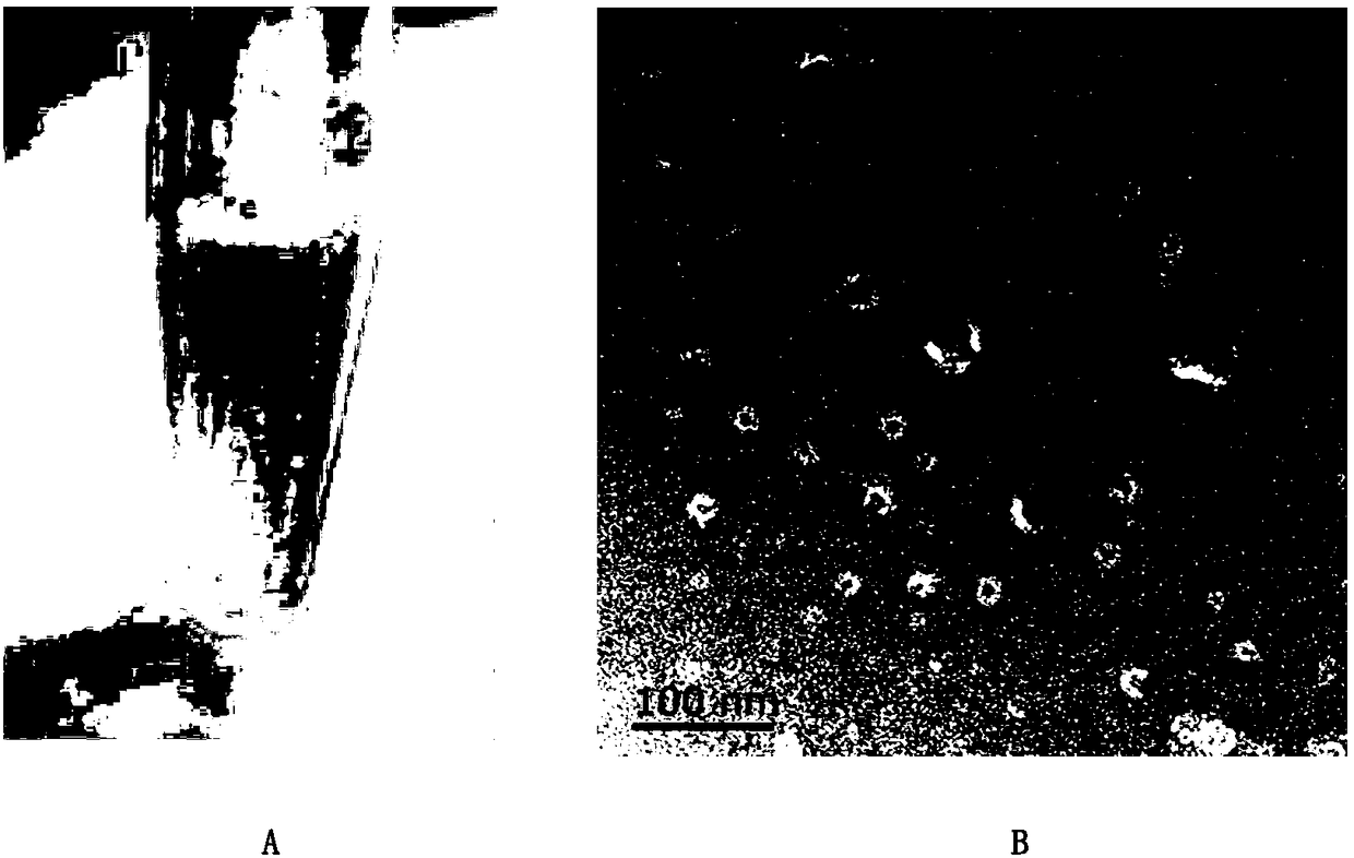
[0044] Take 40 μL of the liposome prepared in Example 1, dilute it to 200 μL with PBS buffer, measure the particle size of the liposome with a Malvern laser particle size analyzer Zetasizer Nano ZS, and record the results. Take 10 μL liposomes with ddH 2 O was diluted at a volume ratio of 1:10; the diluted liquid was transferred to the carbon film for adsorption for 10 minutes, and then washed with DI water, and uranyl acetate UO 2 (CH 3 COO) 2 2H 2 O, after staining for 1 minute, use a transmission electron microscope (TEM), model: JEOL-JEM1400 to carry out visual detection of liposome morphology, and take pictures for recording.

[0045] The results showed that the obtained liposome had a particle size of 30-150nm and an average density of 1.06-1.23g / mL.

Embodiment 3

[0046] Example 3 Liposomes are used to evaluate the extraction efficiency of different vesicle isolation methods

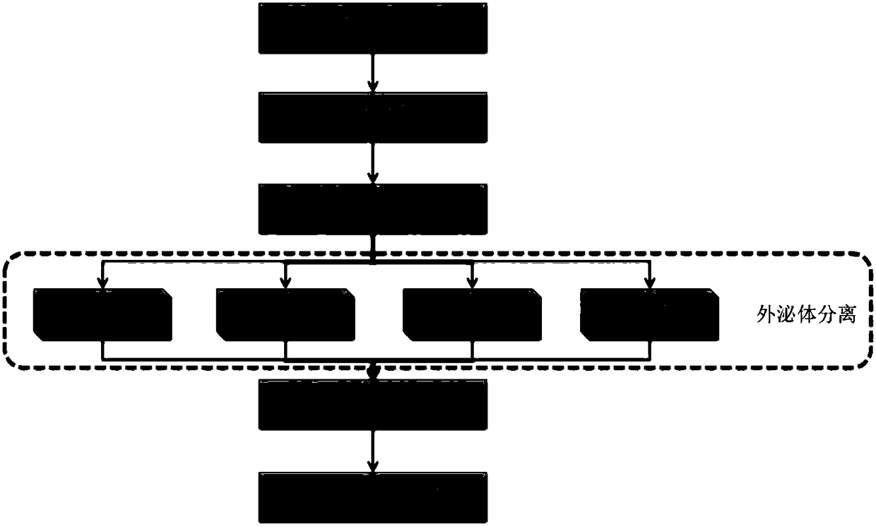
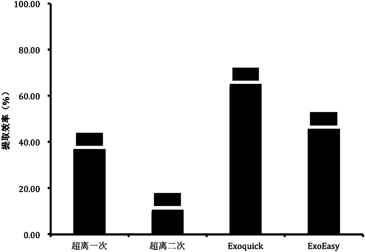
[0047] Take 40 μL of 1 mg / ml liposome prepared in Example 1, dilute to 250 μL with PBS, add 750 μL normal human serum, and ultracentrifuge respectively [see Théry C, Amigorena S, Raposo G, et al.Isolation and Characterization of Exosomes from Cell Culture Supernatants and Biological Fluids[M] / / Current Protocols in Cell Biology.John Wiley&Sons, Inc.2006:Unit3.22.], membrane affinity (Qiagen exoEasy) [see Enderle D, Spiel A, Coticchia C M, etal.Characterization of RNA from Exosomes and Other Extracellular VesiclesIsolated by a Novel Spin Column-Based Method[J].Plos One,2015,10(8):e0136133.], polymer precipitation (SBI Exoquick)[see Tang Y T, Huang Y Y, Zheng L, etal.Comparison of isolation methods of exosomes and exosomal RNA from cellculture medium and serum:[J].International Journal of Molecular Medicine,2017,40(3):834.], the vesicles were isolated according to th...


PUM

PropertyMeasurementUnit
particle sizeaaaaaaaaaa
particle diameteraaaaaaaaaa

Abstract

The invention provides a method for evaluation and quality control of the vesicle separating efficiency based on liposome. The method comprises the following steps of: firstly utilizing lipids such asphosphatidylcholine, phosphatidyl ethanolamine and cholesteryl ester and the like and fluorescent dyes to prepare fluorescent liposome; after respectively mixing the fluorescent liposome with serum,plasma and urine samples, using different separation methods including ultracentrifugation, a membrane affinity method, polymer precipitation and chromatography and the like to separate vesicles; carrying out fluorimetric determination on the separated vesicles. The method provided by the invention has the beneficial effects that the extraction efficiency of the vesicles in different methods and the extraction efficiency in the treatment process of different clinical samples can be calculated by the fluorescence intensity of the original liposome and the fluorescence intensity of the separatedvesicles. The method utilizes the fluorescent liposome to evaluate the extraction efficiency of the vesicles, provides powerful technical support for applying the vesicles in a stable quality-controlsystem with clinical needs, and simultaneously provides an effective method for observing and tracking the separation process of the vesicles by naked eyes.

Description

technical field [0001] The invention relates to the technical field of analysis and testing, in particular to a method for evaluating and quality-controlling vesicle separation efficiency based on liposomes. Background technique [0002] Extracellular vesicles (Extracellular Vesicles, EVs; the following vesicles represent extracellular vesicles) refer to vesicle-like bodies with a double-membrane structure that are shed from the cell membrane or secreted by cells, with diameters ranging from 30-1000nm , Extracellular vesicles are mainly composed of microvesicles (MicroVesicles, MVs) and exosomes (exosomes). Microvesicles are small vesicles that are shed from the cell membrane after cell activation or injury. Due to the unique biological characteristics of extracellular vesicles, they are of great significance in disease diagnosis, especially the exosomes. [0003] Exosomes are a kind of membranous vesicles with a particle size of 30-150 nm secreted into the extracellular en...

Claims


Application Information

Patent Timeline
Patent Type & Authority Applications(China)
IPC IPC(8): G01N21/64G01N1/28G01N1/34
CPCG01N1/28G01N1/34G01N21/6428G01N21/6486
Inventor 赵立波孔关义李志
Owner 北京恩泽康泰生物科技有限公司
Learn More