Novel composite biological tissue repairing material as well as preparation method and application thereof
A technology for repairing materials and biological tissues, applied in medical science, prosthesis, etc., can solve the problems of insufficient mechanical properties of single-layer amniotic membrane, fast degradation time, peripheral nerve defects, etc., achieve broad clinical application and market prospects, and improve tensile strength. and tensile strength, reducing the effect of inflammatory response
- Summary
- Abstract
- Description
- Claims
- Application Information
AI Technical Summary
Problems solved by technology
Method used
Image

Examples
Embodiment 1
[0040] Example 1 Preparation of the composite biological tissue repair material of the present invention
[0041] 1.1 Preparation of experimental group A:
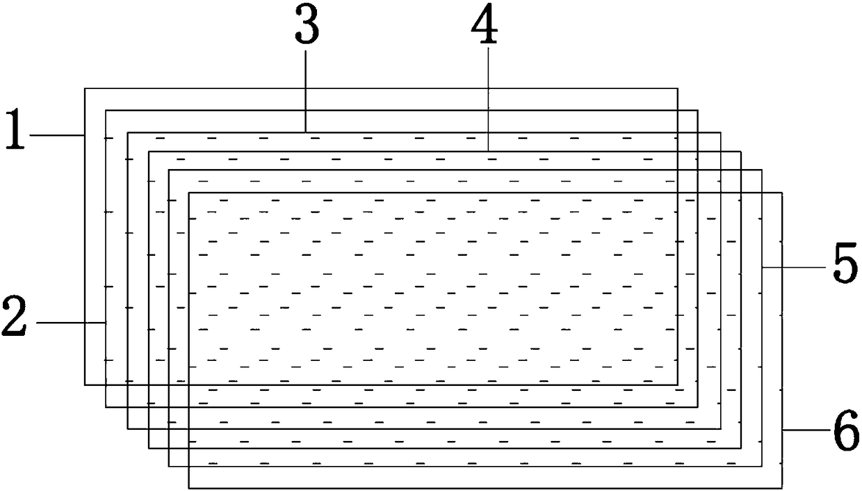
[0042] (1) Take the placental amniotic membrane of healthy puerperae, undergo defatting, virus inactivation, descaling, decellularization, and freeze-drying (prepared according to Example 1 of Publication No. CN1618954A), and then cut the acellular amniotic membrane after freeze-drying It is a rectangular unit of size 2cm x 4cm.
[0043] (2) Take a stainless steel flat plate and lay it flat on the operating table, first lay the first rectangular unit on the flat plate, and evenly apply 5% collagen solution on the upper surface of the material (0.2-0.3ml per square centimeter), The second rectangular unit is then overlapped with the first rectangular unit, and the air bubbles between the layers of each unit are removed to make them fit tightly.
[0044] (3) Repeat the above step (2) to overlap to 5 layers.
[0045] (4) a...
Embodiment 2
[0065] Example 2 Preparation of the composite biological tissue repair material of the present invention
[0066] Preparation of experimental group B:
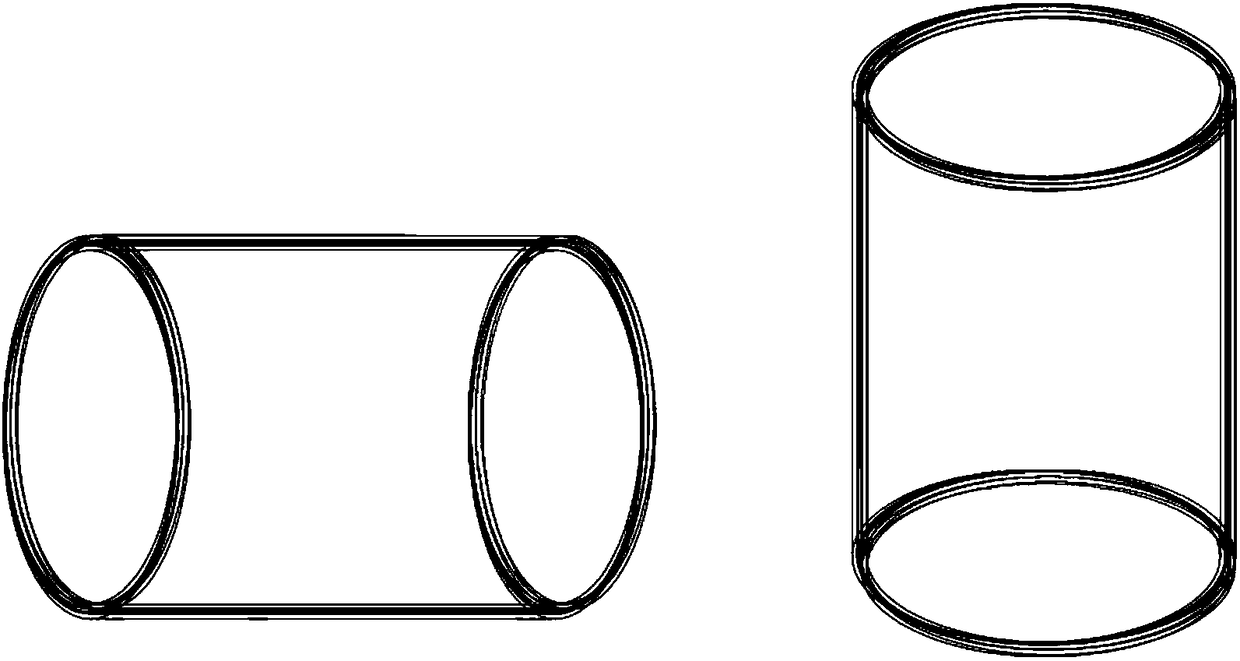
[0067] (1) Take the placental amniotic membrane of healthy puerperae, undergo defatting, virus inactivation, descaling, decellularization, freeze-drying treatment (can be prepared according to Example 1 of publication number CN1618954A), and then cut the acellular amniotic membrane after freeze-drying It is a rectangular unit with a size of 4cm×8cm.
[0068] (2) Before wrapping, evenly spray α-cyanoacrylate medical adhesive on one side of each unit (smear 0.1-0.15ml per square centimeter), denoted as the Y side, and the one side that is not sprayed with adhesive The side is marked as N side.
[0069] (3) Take a cylindrical mold with a diameter of 1 mm, place the N side of the first rectangular unit towards the surface of the mold, and wrap the mold in a ring direction along the short side to remove the air bubbles between th...
Embodiment 3
[0072] Example 3 Preparation of the composite biological tissue repair material of the present invention
[0073] Preparation of experimental group C:
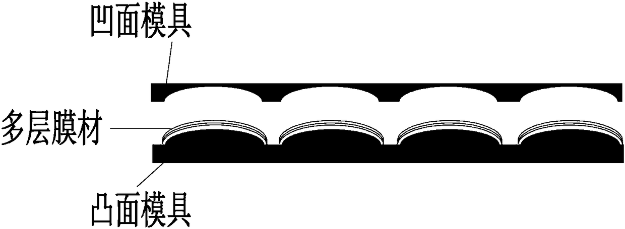
[0074] (1) Take the placental amniotic membrane of healthy puerperium, defatting, virus inactivating, decontaminating, and decellularizing to obtain an intermediate pint of cellular amniotic membrane (which can be prepared according to Example 1 of Publication No. CN1618954A).
[0075] (2) Take a concave mold, spread the first layer of intermediate product on the mold, and evenly apply 2% sodium hyaluronate solution (0.3-0.4ml per square centimeter) on the upper surface of the intermediate product; apply the second layer The intermediate product is laid flat on the first layer of material and stacked, and the air bubbles between the layers are eliminated to make it closely fit.
[0076] (4) Repeat step (2) to overlap and wrap to 4 units.
[0077] (5) another convex mold is taken and covered on the above-mentioned material, p...
PUM
 Abstract
Description
Claims
Application Information
 - R&D
- Intellectual Property
- Life Sciences
- Materials
- Tech Scout
- Unparalleled Data Quality
- Higher Quality Content
- 60% Fewer Hallucinations
Browse by: Latest US Patents, China's latest patents, Technical Efficacy Thesaurus, Application Domain, Technology Topic, Popular Technical Reports.
© 2025 PatSnap. All rights reserved.Legal|Privacy policy|Modern Slavery Act Transparency Statement|Sitemap|About US| Contact US: help@patsnap.com