Protein misfolding-based establishing method of cell model for antioxidant drug screening

A cell model and misfolding technology, applied in the field of cell biology, can solve problems such as misfolding and quantitative evaluation

Active Publication Date: 2018-11-16
SHANXI UNIV
View PDF1 Cites 1 Cited by
  • Summary
  • Abstract
  • Description
  • Claims
  • Application Information

AI Technical Summary

Problems solved by technology

[0006] Aiming at the defect that protein misfolding cannot be quantitatively evaluated in real time in living cells, the...

Method used


Image

  • Protein misfolding-based establishing method of cell model for antioxidant drug screening
  • Protein misfolding-based establishing method of cell model for antioxidant drug screening
  • Protein misfolding-based establishing method of cell model for antioxidant drug screening

Examples


Embodiment 1

[0020] Example 1: Packaging lentiviral particles

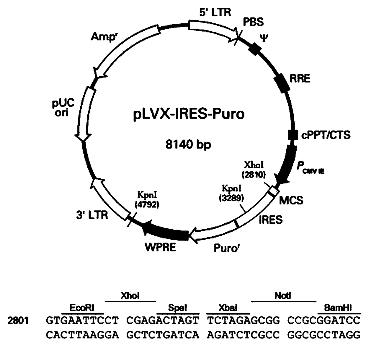
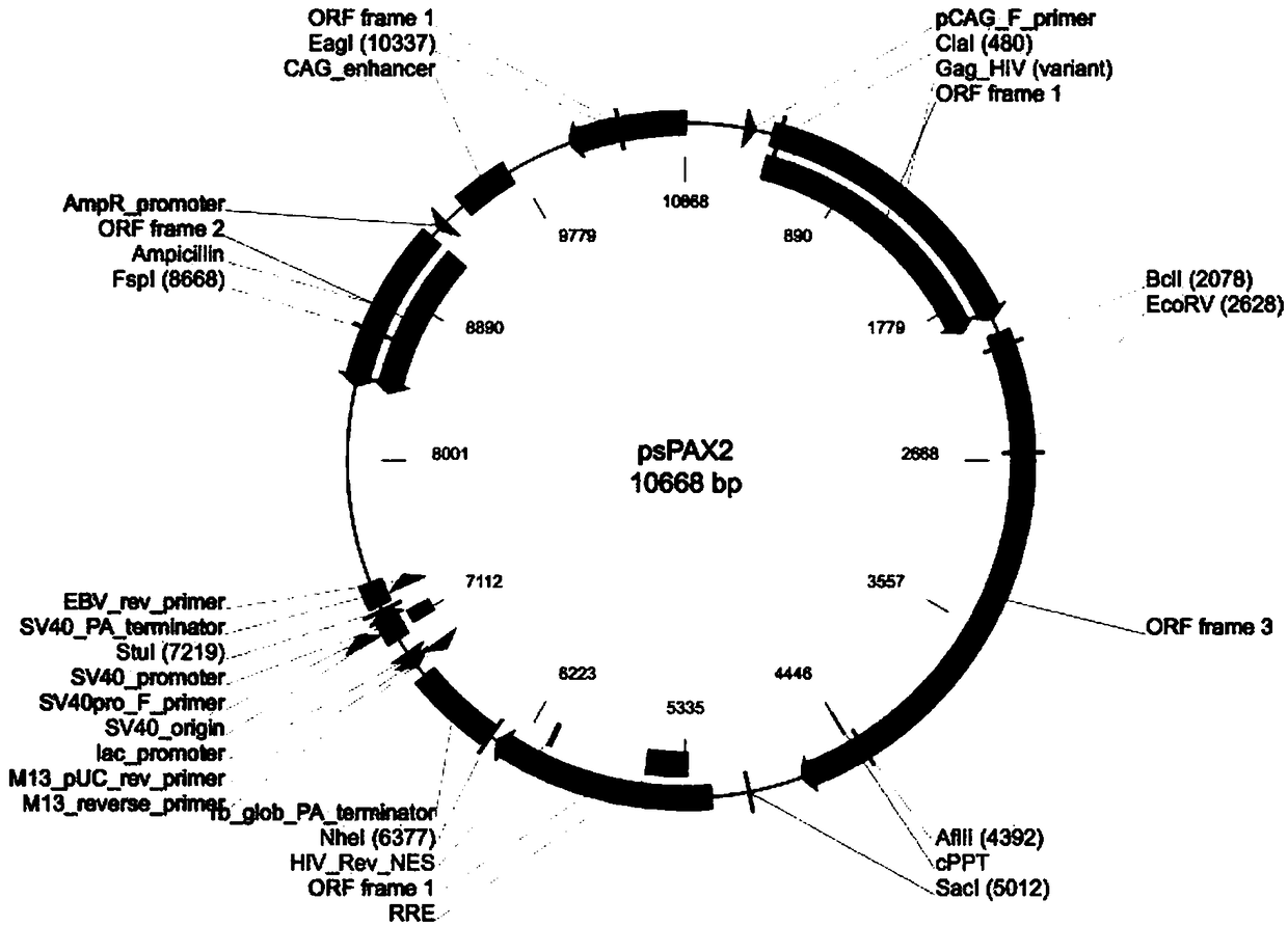
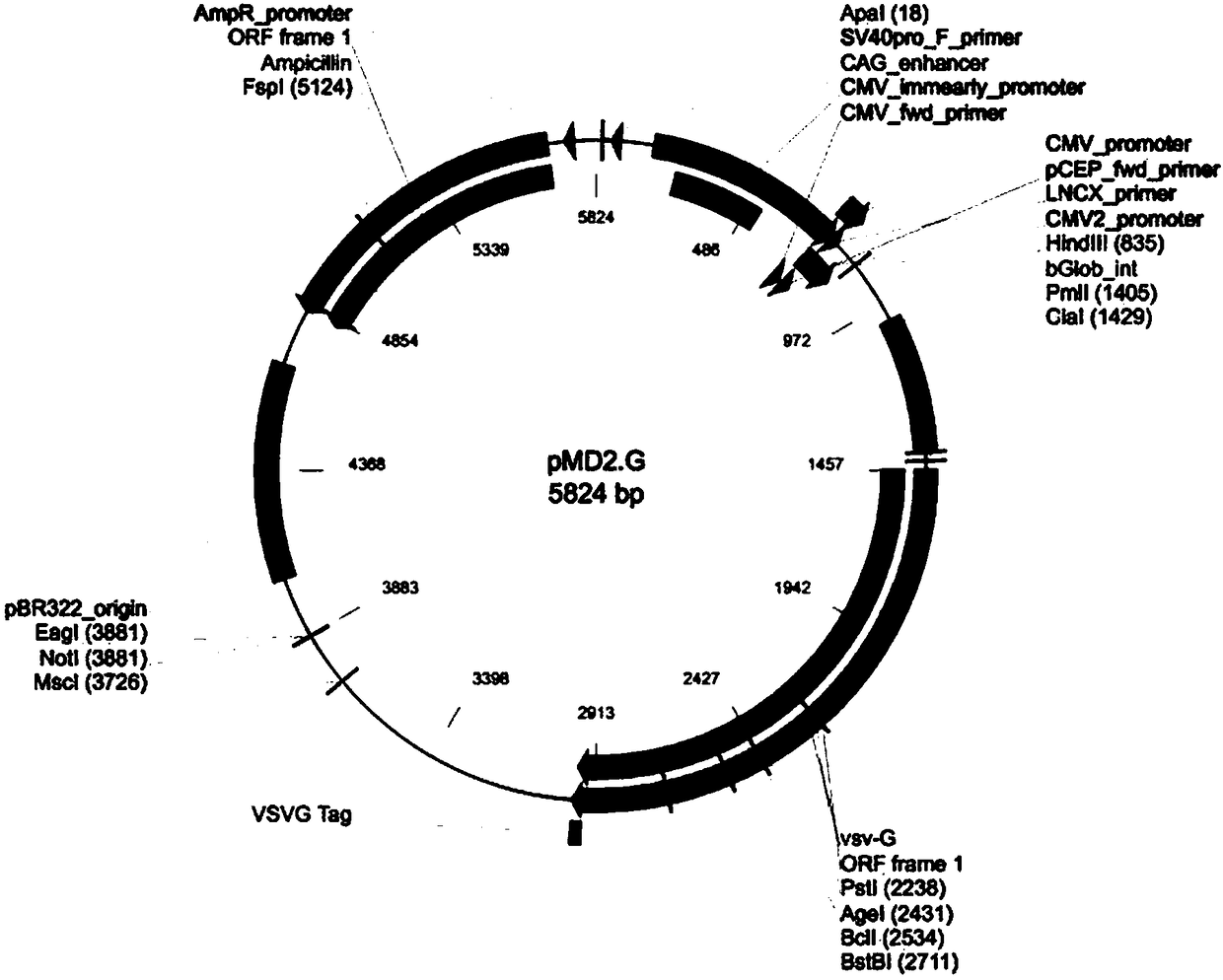
[0021] The lentiviral system includes three plasmids: Plvx-IRES-Puro plasmid expressing reporter gene ( figure 1 ), packaging plasmid psPAX2 ( figure 2 ), envelope protein particle pMD2.G ( image 3 ). Among them, the Plvx-IRES-Puro plasmid contains the puromycin resistance gene and the ampicillin resistance gene, the packaging plasmid psPAX2 contains the gag gene of the HIV virus, which encodes the main structural protein of the virus, and the envelope protein particle pMD2.G contains the herpes simplex The derived VSV-G gene provides the required envelope protein for viral packaging.

[0022] Reporter gene construction: use the known eGFP-COMP fusion gene plasmid as a template, perform polymerase chain reaction to amplify the target fragment (TAKARA company), recover the target fragment from the kit (Shanghai Sangon Bioengineering Co., Ltd.), enzyme The excised fragment and the vector were ligated with T4 DNA Ligase, o...

Embodiment 2

[0024] Embodiment 2: virus transduction HeLa cell

[0025] The collected supernatant contains the recombinant COMP lentivirus, and the packaged virus is used to further transduce HeLa cells, and the stably transfected cell lines are screened. The specific operations are as follows:

[0026] HeLa cells were plated in 6-well plates 24 h before transduction, and when the cells adhered to the wall and grew to about 80%, the old medium was sucked off, and fresh MEM medium (10% fetal bovine serum, 1% Penicillin- Streptomycin) 1.8 mL, recombinant COMP lentivirus 200 μL, and Hexadimethrine bromide (transfection enhancer) 1.6 μL, mixed gently, and cultured in an incubator at 37 °C with 5% CO2;

[0027] After 24 h, the medium containing the virus was sucked off, and 2 mL of fresh medium was replaced, and the culture was continued;

[0028] After 48 h, replace the medium, add 2 mL of fresh medium and add Puromycin (2 μg / mL);

[0029] Afterwards, the medium (containing puro) was changed e...

Embodiment 3

[0031] Example 3: Evaluation of the intracellular COMP content of a cell model using a fluorescence spectrophotometer

[0032] In the stably transfected HeLa cell line, COMP protein and eGFP are fused and expressed, so the content of COMP protein can be indirectly reflected by detecting the content of GFP.

[0033] We used the Cary Eclipse Fluorescence Spectrophotometer (Agilent Technologies) to detect the content of GFP. Initially, we found that GFP can best reflect the relative fluorescence intensity of GFP when the excitation wavelength is 485 nm and the emission wavelength is 530 nm.

[0034] We collected wild-type and mutant cells and cell culture fluid, diluted them with Tris-EDTA solution, and measured the fluorescence intensity of GFP using Cary Eclipse Fluorescence Spectrophotometer (Agilent Technologies). The results were analyzed using the ratio of GFP in cells and GFP in cell culture fluid Ratio to represent changes in intracellular COMP.

[0035] The results show...


PUM


Abstract

The invention belongs to the field of cytobiology, and provides a protein misfolding-based establishing method of a cell model for antioxidant drug screening aiming at the defect that real-time quantitative establishment on protein misfolding cannot be carried out in living cells at present. The protein misfolding-based establishing method comprises the following steps: establishing fluorescent protein labeled wild and mutant reporter gene COMP (Cartilage Iligomeric Matrix Protein) gene lentiviral vector, packaging virus in human embryo kidney 293T cells, transducing HeLa cells, and screeningpuromycin, thus obtaining the cell model for stably expressing COMP cells. A drug screening cell model which is in international level and independent innovation is established, not only key parameters can be provided for a protein misfolding drug screening industrialization study in the further, but also correlated scientific basis is provided for a sustainable study on serious illness; the cellmodel has the characteristics of high chromosome integration degree, high fluorescence strength, stable inheritable character and antioxidant drug sensitiveness, and is an ideal model for studying protein misfolding drug screening.

Description

technical field [0001] The invention belongs to the field of cell biology, and in particular relates to a method for constructing a cell model for screening antioxidant drugs based on protein misfolding. Background technique [0002] Proteins are the executors of all functions in living organisms. Any function in the human body, from catalyzing chemical reactions to defending against foreign aggressors, is the result of proteins. Proteins must fold precisely into the correct three-dimensional structure to function in human cells. In recent years, it has been found that protein misfolding can lead to some diseases. The relationship between protein misfolding and disease has become a new research frontier in molecular biology. Constructing a simple, safe and efficient drug screening cell model is the solution to the prevention and treatment of protein misfolding diseases such as Alzheimer's disease, Parkinson's disease, genetic bone disease and phenylketonuria, which are po...

Claims


Application Information

Patent Timeline
IPC IPC(8): C12N5/10C12N15/867C12N15/65C12N15/12C12Q1/02G01N21/64
CPCC07K14/47C12N15/65C12N15/86C12N2740/15043G01N21/6428G01N33/5044
Inventor 肖涵王晶吴长新
Owner SHANXI UNIV
Why Eureka
Try Eureka