
Fusion protein, preparation method and application thereof, expression system and vaccine

A fusion protein and protein technology, applied in the biological field, can solve the problems of decreased ability of lymphocyte antibodies, decreased cellular immunity, decreased body resistance to disease, etc., to achieve high expression, enhanced immunogenicity, and stable titer Effect

Inactive Publication Date: 2019-01-04
TECON BIOLOGY CO LTD
View PDF4 Cites 5 Cited by
  • Summary
  • Abstract
  • Description
  • Claims
  • Application Information

AI Technical Summary

Problems solved by technology

[0005] After PCV infection, the ability of lymphocytes to produce antibodies decreases, the cellular immunity decreases, and the ability of alveolar macrophages to phagocytize and clear pathogens also decreases, and the activity of suppressor T cells increases, resulting in a decrease in the body's disease resistance, thereby making other pathogens more vulnerable. Easy to invade, can be secondary infected with porcine reproductive and respiratory syndrome, mycoplasma, etc., resulting in the failure of various vaccinations

Method used


Image

  • Fusion protein, preparation method and application thereof, expression system and vaccine
  • Fusion protein, preparation method and application thereof, expression system and vaccine
  • Fusion protein, preparation method and application thereof, expression system and vaccine

Examples


preparation example Construction

[0064] A method for preparing the above fusion protein, comprising expressing the gene encoding the fusion protein in a host.

[0065] The method has the advantages of simple preparation method, low cost, powerful vaccine function and the like.

[0066] The host in the present invention can be, but not limited to, an Escherichia coli expression system, a yeast expression system, an insect expression system, a plant expression system or a mammalian expression system. Since the protein expressed by mammalian cells is translated and processed, its structure and biological properties are closer to natural proteins, so the present invention preferably uses a mammalian expression system to express the gene encoding the fusion protein.

[0067] In a preferred embodiment of the present invention, a mammalian expression system is used to express the gene encoding the fusion protein. Since the protein expressed by mammalian cells is translated and processed, its structure and biologica...

Embodiment 1

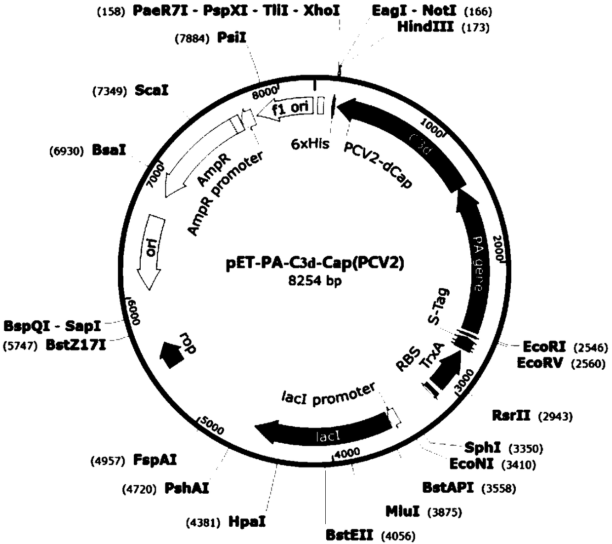
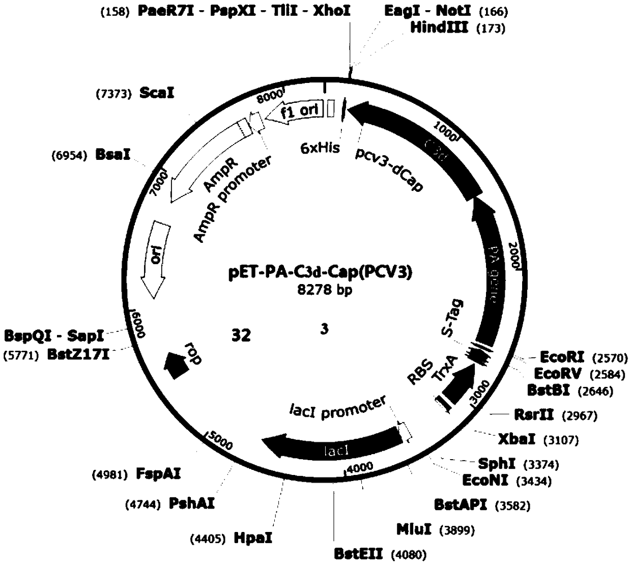
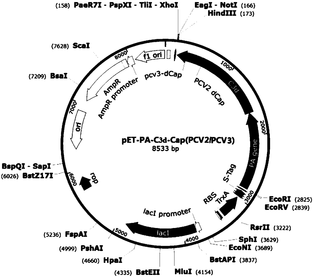
[0101] Example 1 Expression, identification and purification of fusion protein PA-C3d-Cap-K

[0102] According to NCBI reports, Pseudomonas aeruginosa PA (Pseudomonas aeruginosa PA, accession number: CP007224.1, see SEQ ID NO.1) gene sequence, porcine complement C3d (see SEQ ID NO.8) gene sequence, PCV2-ADDLPP The Cap protein gene sequence (see SEQ ID NO.2) of the 10069 strain (accession number: EU594437.1) and the Cap protein gene sequence (see SEQ ID NO.2) of the PCV3-US / MN strain (NCBI accession number: KX898030.1) .3) Analyze and finally design Pseudomonas exotoxin A (PEA) domain I and II protein, porcine complement C3d, porcine circovirus Cap protein dominant epitope and carboxyl terminal part containing SEQ ID NO.4 The polypeptide selects flexible amino acids (SEQ ID NO.7) and connects them in series in a certain combination to express a fusion protein with good immunogenicity.

[0103] 1.1 Construction and identification of PEA domains I and II and Cap gene cloning vec...

Embodiment 2

[0127] The preparation of embodiment 2 porcine circovirus vaccine

[0128] Dilute the fusion protein prepared in Example 1 with PBS solution, mix the diluted fusion protein solution with SEPPICISA201R VG adjuvant at a mass fraction of 50%, stir at a speed of 8000r / min for 10min, and add 0.01% (volume ratio) thimerosal solution, so that the final concentration does not exceed 1 / 10,000. After fully oscillating and mixing, according to the requirements of the appendix of the current version of the Chinese Veterinary Pharmacopoeia, after passing the sterility test, viscosity measurement, and stability measurement, place it in 4 ℃ for later use.


PUM


Abstract

The invention provides a fusion protein and a preparation method and application thereof, an expression system and a vaccine, and relates to the technical field of biology. The fusion protein providedherein includes Pseudomonas exotoxin A domain section, porcine circovirus type 2 Cap protein dominant epitope section, porcine circovirus type 3 Cap protein dominant epitope section and carboxy terminus. The epitopes of PCV2 (porcine circovirus type 2) and PCV3 Cap proteins are screened out; the epitopes have good antigenicity and are easy to highly express; the epitopes are fused to partial functional fragments of PEA (Pseudomonas exotoxin A); carboxy terminal sequence and enhanced immunogenicity are imparted to C end of the fusion protein; therefore, the fusion protein is more soluble. Thefusion protein herein has the advantages of targeted positioning, good antigenicity, and high expression quantity. The fusion protein is advantageous in good safety, stable titer, zero toxic and sideeffects and cellular immune response activation when applied as an antigen to prepare vaccines.

Description

technical field [0001] The invention relates to the field of biotechnology, in particular to a fusion protein and its preparation method, application, expression system and vaccine. Background technique [0002] Porcine circovirus (Porcine circovirus, PCV) belongs to Circoviridae, single-stranded, circular DNA virus. It is mainly divided into non-pathogenic porcine circovirus type (PCV1) and pathogenic porcine circovirus type (PCV2). It is currently known that the Cap protein is a structural protein of PCV and has good immunogenicity. In October 2016, R. Palinski of Kansas State University in the United States and T.G.Phan of the University of California, San Francisco reported a new PCV genotype, called PCV3. The virus was isolated from diseased sows or piglets, and the PCV2 test was negative. Genome sequence analysis found that the PCV3 genome contains 2000 bases, has a similar genome structure to PCV1 and PCV2, and mainly encodes two genes, cap and rep. Porcine circov...

Claims


Application Information

Patent Timeline
IPC IPC(8): C07K19/00C07K16/08C12N15/62C12N15/85G01N33/569A61K39/12A61K39/295A61P31/20
CPCA61K39/12A61K2039/552A61K2039/70A61P31/20C07K14/005C07K14/21C07K16/081C07K2319/55C12N15/85C12N2750/10022C12N2750/10034G01N33/56983
Inventor 贺笋张伟李俊辉李延涛程兰玲高窦潘晓梅徐龙飞师小潇
Owner TECON BIOLOGY CO LTD
Learn More