Feline panleukopenia virus (FPV) VP2 protein and prepared virus-like particles
A feline parvovirus, virus-like technology, applied in the field of genetic engineering, to achieve the effect of improving antibody titer and preventing infection
- Summary
- Abstract
- Description
- Claims
- Application Information
AI Technical Summary
Problems solved by technology
Method used
Image

Examples
Embodiment 1
[0019] Example 1: Amplification and sequence analysis of VP2 gene
[0020] In 2018, the suspected feline parvovirus materials were collected for processing, and the disease materials were routinely separated and identified. It was determined that the feline parvovirus was tested for HA with porcine red blood cells, and the hemagglutination value was 7log2. The HI test with known feline parvovirus sera showed poor cross-reactivity; it proved that the pathogenic feline parvovirus strain had mutated.
[0021] 1. Amplification of feline parvo VP2 gene
[0022] According to the feline small VP2 gene sequence published in NCBI, primers were designed and synthesized. The sequence information of the primers is as follows:
[0023] primer1: 5′-ATGAGTGATGGAGCAGTTCAACC-3′;
[0024] primer2: 5'-TTAATATAATTTTCTAGGTGCTAGTTG-3'.
[0025] Extract the isolated feline small nucleic acid as a template, and use primers primer1 and primer2 to amplify the target fragment by PCR. After sequence d...
Embodiment 2
[0029] Embodiment 2, the construction of the bacmid expressing VP2 gene
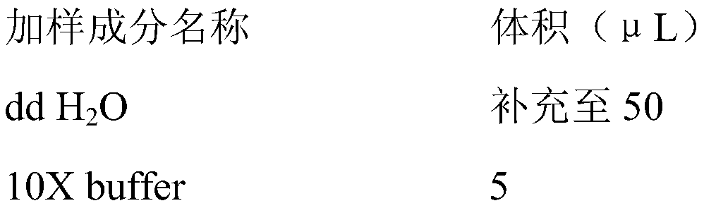
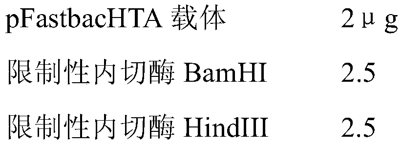
[0030] 2.1 Enzyme digestion reaction
[0031] 2.1.1 Mark the 1.5mL EP tube to be used, and add and mix the sample according to the following table in the 1.5mL EP tube: the reaction system is 50 μL, and the sample addition is shown in the table below:
[0032]
[0033]
[0034] 2.1.2 Place the 1.5mL EP tube in step 2.2.1 in a constant temperature water bath at 37°C for 2-3 hours.
[0035] 2.1.3 Gel recovery of double enzyme digestion products
[0036] Take out the above-mentioned double digestion system and perform agarose gel electrophoresis to recover the DNA fragments therein.
[0037] (1) Mark the sample collection EP tube, adsorption column and collection tube.
[0038] (2) Weigh the marked empty EP tube and record the value.
[0039] (3) Carefully cut out a single target DNA band from the agarose gel on a gel cutter with a scalpel and put it into a clean 1.5mL centrifuge tube.
[0040] (...
Embodiment 3
[0100] Embodiment 3 SF9 cell transfection
[0101] (1) Preparation: UV sterilization in a biosafety cabinet for 30 minutes; TNM-FH culture solution was placed in a 27°C water bath and preheated to 27°C.
[0102] (2) Add 2 μg of recombinant DNA to 100 μl of TNM-FH medium without serum and double antibody, and mix well. Add 9 μl Cellfectin Reagent to 100 μl TNM-FH medium without serum and double antibody, and mix well. The liposomes were mixed with the recombinant DNA and allowed to stand at room temperature for 40 min.
[0103] (3) Take out the 6-well plate cells from the incubator at 27°C, discard the supernatant medium, wash the cells three times with pre-warmed TNM-FH culture medium, and discard the TNM-FH culture medium.
[0104] (4) Add 2 ml of 10% fetal bovine serum TNM-FH culture solution to each cell well.
[0105] (5) Gently add the mixture of recombinant DNA and liposomes into each well of cells, mix gently, and culture statically at 27°C for 5-6 hours.
[0106] (...
PUM
 Abstract
Description
Claims
Application Information
 - R&D
- Intellectual Property
- Life Sciences
- Materials
- Tech Scout
- Unparalleled Data Quality
- Higher Quality Content
- 60% Fewer Hallucinations
Browse by: Latest US Patents, China's latest patents, Technical Efficacy Thesaurus, Application Domain, Technology Topic, Popular Technical Reports.
© 2025 PatSnap. All rights reserved.Legal|Privacy policy|Modern Slavery Act Transparency Statement|Sitemap|About US| Contact US: help@patsnap.com