Cytomembrane nano-vesicles and production method thereof

A nanovesicle, cell membrane technology, applied in the field of biomedicine, can solve the problems of inability to cross the blood-brain barrier, time-consuming, large particle size, etc., and achieve the effects of good product uniformity, high production yield, and stable vesicle properties.

Pending Publication Date: 2020-06-05
FIRST AFFILIATED HOSPITAL OF DALIAN MEDICAL UNIV
View PDF3 Cites 4 Cited by
  • Summary
  • Abstract
  • Description
  • Claims
  • Application Information

AI Technical Summary

Problems solved by technology

[0005] 1×10 7 Each cell was cultured for 24 hours, and the output was about 2.1×10 11 vesicles, the yield is low and time-consuming, which limits its clinical application
Adding chemical reagents or nanomaterials to induce cells can increase the yield appropriately, but the vesicles formed are micron-sized and large in size, which cannot cross the blood-brain barrier

Method used


Image

  • Cytomembrane nano-vesicles and production method thereof
  • Cytomembrane nano-vesicles and production method thereof
  • Cytomembrane nano-vesicles and production method thereof

Examples


Embodiment 1

[0034] Preparation and characterization of cell membrane nanovesicles derived from RAW264.7 cells

[0035] 1. The best preparation method of cell membrane nanovesicles derived from RAW264.7 cells

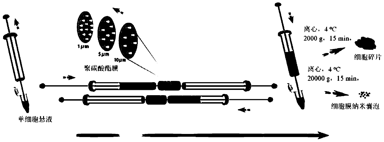
[0036] The mouse mononuclear macrophage leukemia cell RAW264 used in the present invention was purchased from the Institute of Basic Medical Sciences, Chinese Academy of Medical Sciences. RAW246.7 cells were cultured in a 25T culture flask to 80% density, digested, centrifuged at 800 g for 5 min, discarded the supernatant, and blown the pellet in 1 mL of PBS solution to make a single cell suspension.

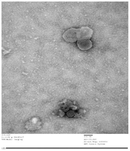
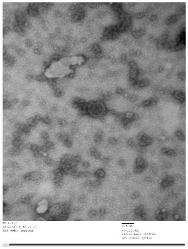
[0037] Method I: The cell suspension was continuously extruded 11 times through a 100 μm polycarbonate membrane filter using the Mini Extruder provided by Avanti Polar Lipids, USA. Separation and purification: After centrifugation at 4°C, 20,000g, 15min, take the supernatant at 4°C, 100,000g, ultracentrifuge for 1h, discard the supernatant, and pipette the pellet repeatedly with ster...

Embodiment 2

[0045] 1. Preparation method of NVs derived from umbilical cord mesenchymal stem cells

[0046] Cultivation of umbilical cord Mesenchymal Stem Cells (UMSCs): Rinse the umbilical cord tissue with PBS buffer, cut the tissue into 2cm-3cm sections with sterile scissors, wash the blood clot with PBS buffer, repeat 2 -3 times until the washing liquid is clear. Cut the umbilical cord, remove two arteries and one vein in turn. Trim the tissue to a surface area of ​​approximately 10 mm 2 ~25mm2 tissue block, the colloid side is spread on the bottom of the culture dish, let it stand for 15min~20min, then slowly add an appropriate amount of medium along the side wall, and place it at 37°C, 5% CO 2 cultured in an incubator. When the cell confluence reaches 80%-90%, discard the medium, wash with PBS buffer, add trypsin to digest the cells until most of the cells become round and begin to fall off. Add appropriate amount of culture medium to stop digestion. After the cells were blown a...

Embodiment 3

[0053] 1. Preparation method of cell membrane nanovesicles derived from neural stem cells

[0054] Neural Stem Cells (Neural Stem Cells, NSCs) culture: The tissue used is aborted fetuses (medical waste) at 7-14 weeks of gestation. The cerebral cortex is separated, placed in the complete culture medium of NSCs, and divided into small tissue pieces. Blow and suck 20-30 times, let it stand for 10 minutes, absorb the supernatant, centrifuge at 820rpm for 5 minutes, and discard the supernatant. Add special digestive solution to the precipitation, place the culture dish in an incubator for digestion for 1-2 minutes, centrifuge at 820 rpm for 5 minutes, and discard the supernatant. Add NSCs complete culture solution to the pellet and add it to the centrifuge tube, blow gently for 10-15 times to form a single cell suspension and then inoculate. placed in a carbon dioxide incubator at a temperature of 35°C and CO 2 The concentration is 5%. After the NSCs grow into neurospheres of su...


PUM

PropertyMeasurementUnit
particle diameteraaaaaaaaaa
particle sizeaaaaaaaaaa

Abstract

The invention discloses cytomembrane nano-vesicles and a production method thereof. The cytomembrane nano-vesicles are composed of bi-molecule-layer lipid membranes and contents wrapped by the bi-molecule-layer membranes, wherein the membranes and the contents of the cytomembrane nano-vesicles all come from metrocytes used for production. The production method of the cytomembrane nano-vesicles comprises the following step: sequentially and repeatedly extruding a single-cell suspension to pass through polycarbonate membranes, with different pore diameters, of an extruder to obtain a clear solution, and conducting gradient centrifugation, separation and purification on the clear solution to obtain the cytomembrane nano-vesicles. The production method of the cytomembrane nano-vesicles is highin yield, the nano-vesicles are stable in property, the product uniformity is good, operation is simple and convenient, the quality is controllable, and the repeatability is high, so that large-scaleproduction is facilitated, and the cytomembrane nano-vesicles can be applied to treatment of Parkinson's disease.

Description

technical field [0001] The invention belongs to the technical field of biomedicine, and in particular relates to a cell membrane nanovesicle and a preparation method thereof. Background technique [0002] Parkinson's Disease (PD) is a chronic neurodegenerative disease that endangers the health of middle-aged and elderly people, and the incidence rate of people over 60 years old is about 1%. The main pathological feature of PD is the progressive loss of dopaminergic neurons (Dopamine neuron, DN) in the substantia nigra of the brain, resulting in a lack of dopamine content in the striatum, thereby causing movement disorders. The motor symptoms of PD patients mainly include resting tremor, muscle rigidity, bradykinesia and postural balance disturbance. At present, the main methods for the clinical treatment of PD are levodopa preparations, anticholinergics, receptor agonists, drugs that promote DA release, and deep brain stimulation (Deep Brain Stimulation, DBS) surgery. Earl...

Claims


Application Information

Patent Timeline
Patent Type & Authority Applications(China)
IPC IPC(8): C12N5/0786C12N5/0775C12N5/0797A61K35/15A61K35/28A61K35/30A61P25/16
CPCC12N5/0645C12N5/0668C12N5/0623A61K35/30A61K35/28A61K35/15A61P25/16
Inventor 刘晶沈丽明
Owner FIRST AFFILIATED HOSPITAL OF DALIAN MEDICAL UNIV
Why Eureka
Try Eureka