
High-brightness lipid droplet cell nucleus multicolor imaging fluorescent probe

A fluorescent probe, high-brightness technology, applied in the field of fluorescence imaging, can solve the problems of signal overlap, increase the experimental cost, unknown cytotoxicity, etc., achieve good rigidity and flatness, simple spatiotemporal resolution, excellent photostability Effect

Active Publication Date: 2020-06-26
DALIAN INST OF CHEM PHYSICS CHINESE ACAD OF SCI
View PDF3 Cites 2 Cited by
  • Summary
  • Abstract
  • Description
  • Claims
  • Application Information

AI Technical Summary

Problems solved by technology

This puts forward high requirements on the fluorescent properties of the dyes. The dyes not only need to have high brightness and photostability, but also need to have a narrow emission spectrum and a large Stokes shift to avoid signal overlap during imaging. Most of the fluorescent dyes are difficult to meet such stringent requirements; and a variety of dyes need to be added in the process of incubating cells, which not only increases the cost of the experiment, but also has unknown cytotoxicity
If only one dye is used for multicolor fluorescence imaging, in order to distinguish the fluorescent signals produced by different organelles, it is necessary to quench all the dyes after marking and imaging one organelle, and then add a new dye to mark and image the second organelle. The experimental process is very complicated, and the quenching process needs to add some chemical reagents, which will cause cell death, and it is impossible to perform multicolor imaging on living cells

Method used


Image

  • High-brightness lipid droplet cell nucleus multicolor imaging fluorescent probe
  • High-brightness lipid droplet cell nucleus multicolor imaging fluorescent probe
  • High-brightness lipid droplet cell nucleus multicolor imaging fluorescent probe

Examples


Embodiment 1

[0034] Synthesis of the probe OMor-Aze.

[0035] The intermediate Naph-Aze synthesis route and product structure are as follows:

[0036]

[0037] Weigh 4-bromo-1,8-naphthalene anhydride (3.0g, 10.9mmol), anhydrous copper sulfate (5.2g, 32.7mmol) in 10mL dry DMF, add azetidine (1.9mmol) under nitrogen protection g, 32.7 mmol), the temperature was raised to 140° C. to react for 20 h, and after cooling the reaction solution to room temperature, the reaction solution was slowly poured into ice water, and a large amount of orange solids were precipitated. The filtrate was removed by suction filtration, and the filter residue was dissolved in dichloromethane and purified by silica gel column chromatography (petroleum ether / dichloromethane=1 / 10, V / V) to obtain 1.1 g of orange solid powder with a yield of 40%.

[0038] Its high-resolution mass spectrometry data are as follows:

[0039] HRMS(ESI): m / z: [M+H] + : Calculated: 254.0817, Experimented: 254.0883.

[0040] After detec...

Embodiment 2

[0055] The synthesis route and product structure of the probe Mor-Aze are as follows:

[0056]

[0057] Weigh Naph-Aze (0.1g, 0.4mmol) in 5mL of absolute ethanol, add 4-(2-aminoethyl)morpholine (0.15g, 1.2mmol), heat the reaction solution to 80°C for 12h, The reaction solution was cooled to room temperature, and the solvent was distilled off under reduced pressure. The residue was separated through a silica gel column (dichloromethane / methanol=50 / 1, V / V) to obtain the product, which was 0.12 g of an orange solid, with a yield of 87%.

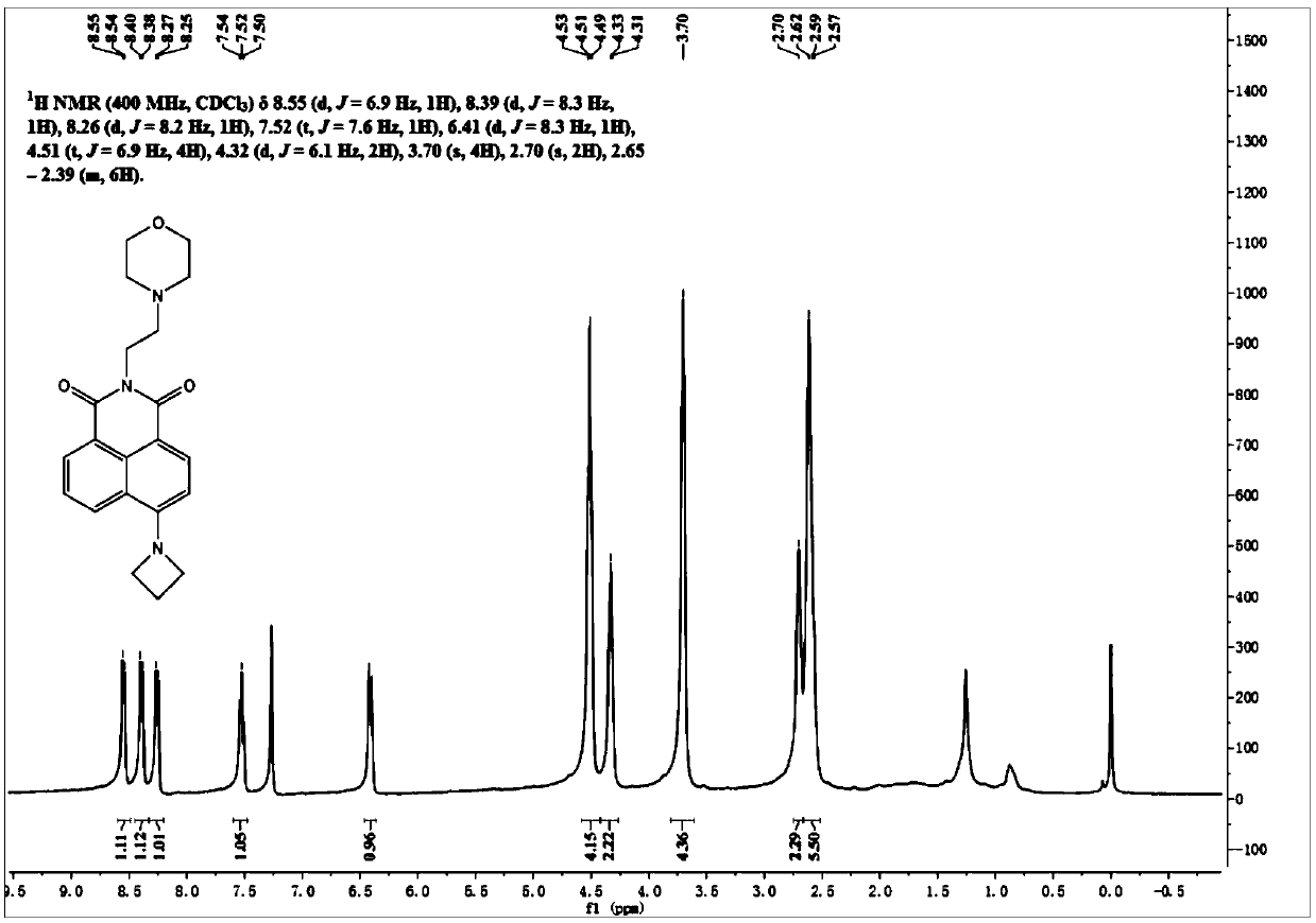
[0058] Its H NMR spectrum is as follows figure 1 As shown, the specific data are as follows:

[0059] 1 H NMR (400MHz, CDCl 3 , ppm): δ=8.55(d, J=6.9Hz, 1H), 8.39(d, J=8.3 Hz, 1H), 8.26(d, J=8.2Hz, 1H), 7.52(t, J=7.6Hz ,1H),6.41(d,J=8.3Hz,1H), 4.51(t,J=6.9Hz,4H),4.32(d,J=6.1Hz,2H),3.70(s,4H),2.70(s ,2H),2.65-2.39 (m,6H).

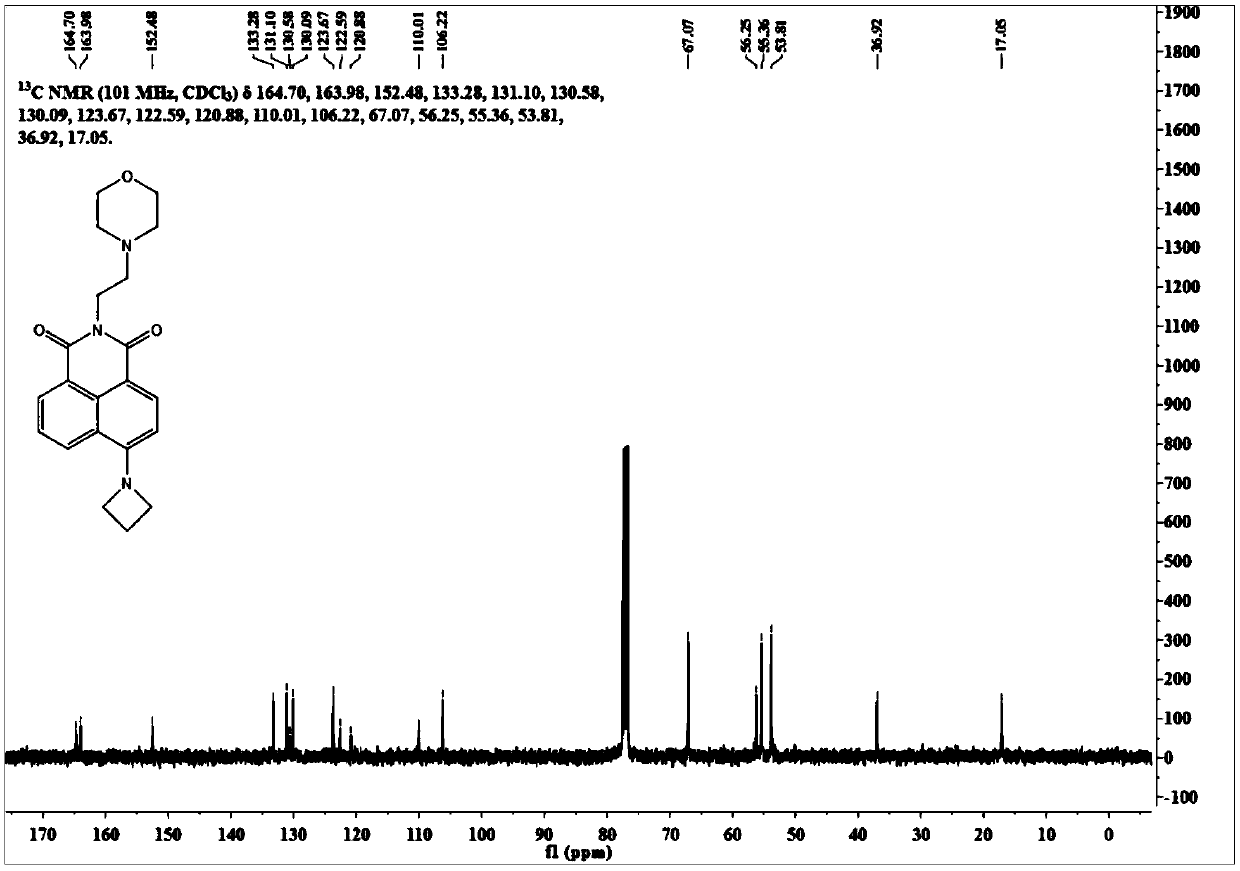
[0060] Its carbon NMR spectrum is as follows figure 2 As shown, the specific data are as follows:

[0061] 13 C...

Embodiment 3

[0069] The synthesis route and product structure of the probe DEma-Aze are as follows:

[0070]

[0071] Weigh Naph-Aze (0.1g, 0.4mmol) in 5mL of absolute ethanol, add N,N-dimethylethylamine (0.1g, 1.2mmol), heat the reaction solution to 80°C for 10h, the reaction solution After cooling to room temperature, the solvent was distilled off under reduced pressure, and the residue was separated through a silica gel column (dichloromethane / methanol=50 / 1, V / V) to obtain 0.11 g of an orange solid with a yield of 86%.

[0072] Its nuclear magnetic spectrum hydrogen spectrum data are as follows:

[0073] 1 H NMR (400MHz, CDCl 3 )δ8.51(dt, J=29.2, 14.7Hz, 1H), 8.40-8.29(m, 1H), 8.24-8.14(m, 1H), 7.49(td, J=8.5, 2.3Hz, 1H), 6.44 -6.29(m,1H),4.61-4.41(m,4H),4.32(t,J=7.2Hz,2H),2.66(t,J=7.2Hz,2H),2.61-2.52(m,2H), 2.37(s, 6H).

[0074] Its nuclear magnetic spectrum carbon spectrum data are as follows:

[0075] 13 C NMR (101MHz, CDCl 3 )δ164.77, 164.06, 152.51, 133.34, 131.16, 130.6...


PUM

PropertyMeasurementUnit
Molar extinction coefficientaaaaaaaaaa

Abstract

The invention provides a high-brightness lipid droplet cell nucleus multicolor imaging fluorescent probe. The probe is 4-substituted naphthalimide dye which can be used for multicolor fluorescence imaging of lipid droplets and cell nucleuses in living cells, and the dye has the advantages of low synthesis raw material cost, simple method, easiness in derivation and the like. Researches show that an azetidine structure is introduced to the 4-site of a naphthalimide matrix in the dye, so that the rigidity and planarity of the dye are improved, the TICT process is effectively inhibited, the molarextinction coefficient of the dye in ethanol is about 15,000 M<-1>cm<-1>, the fluorescence quantum yield is about 0.6, and the dye has very high brightness and light stability. According to the present invention, a morpholine ring, N,N-dimethyl and other basic groups are introduced to the dye, such that the dye can target the cell nucleus, and the molecule has the appropriate lipid solubility soas to achieve the precise positioning of the cell nucleus and the lipid droplet; the dye has very high brightness and light stability, can rapidly mark cell nucleuses and lipid droplets, and can be used in the fields of multicolor fluorescence imaging and the like.

Description

technical field [0001] The invention belongs to the technical field of fluorescence imaging, in particular to a class of high-brightness fluorescent probes for multicolor imaging of lipid droplet nuclei. Background technique [0002] The nucleus is the most important organelle in the cell, which regulates physiological processes such as cell inheritance and metabolism; lipid droplets are the main storage place for neutral lipids in the cell, and are closely related to fat metabolism and steatosis. Recent studies have shown that in some physiological activities, there is a close relationship between the nucleus and lipid droplets. For example, in the process of cell apoptosis, the chromatin in the nucleus will be condensed and then cleaved, which will be accompanied by lipid droplet fusion and other processes; and the metabolites of fatty acids in lipid droplets will activate certain proteins, thereby inducing increased apoptosis. The advent of multicolor fluorescence imagin...

Claims


Application Information

Patent Timeline
IPC IPC(8): C09B57/08C09K11/06G01N21/64C07D401/04
CPCC07D401/04C09B57/08C09K11/06C09K2211/1029G01N21/6428G01N21/643
Inventor 徐兆超陈婕乔庆龙
Owner DALIAN INST OF CHEM PHYSICS CHINESE ACAD OF SCI
Learn More