
Application of functionalized graphene film in three-dimensional reconstruction of cyro-electron microscope

A graphene film and three-dimensional reconstruction technology, which is applied in the field of high-resolution structure analysis of biomolecules, can solve the problems of easy breakage, inconsistent thickness of micro-sieve edges, poor mechanical rigidity and ductility, etc., and achieve the effect of reducing background noise

Active Publication Date: 2020-07-31
TSINGHUA UNIV +1
View PDF7 Cites 2 Cited by
  • Summary
  • Abstract
  • Description
  • Claims
  • Application Information

AI Technical Summary

Problems solved by technology

[0004] At present, the commonly used supporting membrane material for cryo-electron microscopy is micro-sieve carbon film, which has a series of disadvantages in the field of cryo-electron microscopy, including uneven surface properties, inconsistent thickness at the edge of micro-sieve holes, and amorphous carbon skeleton at liquid nitrogen temperature. Poor electrical conductivity, poor mechanical rigidity and ductility, easy to break, etc., the gas-liquid interface of the solution film suspended in the micro-sieve has adverse effects on the structural stability, particle orientation, and distribution uniformity of biomacromolecules
Ultra-thin carbon film (thickness 3-5nm) supported by micro-sieve is also a commonly used support material for frozen sample preparation, which reduces the influence of solution gas-liquid interface on biomolecules to a certain extent, but amorphous carbon Membrane background noise is high, limiting high-resolution structural studies of smaller molecular structures
The above problems have greatly reduced the repeatability, general applicability, success rate, and uniformity of sample quality of frozen samples.
In the past few years, several research groups have been exploring frozen sample grids made of new materials, including silicon nitride micro-sieves, gold micro-sieves, graphene oxide films, etc., but they have not been widely used so far. The main reason is that These materials themselves are not ideal support membrane materials for the grid, and have problems such as poor conductivity, poor rigidity at liquid nitrogen temperature, and easy rupture, and the repeatable preparation process is not mature in practice.

Method used


Image

  • Application of functionalized graphene film in three-dimensional reconstruction of cyro-electron microscope
  • Application of functionalized graphene film in three-dimensional reconstruction of cyro-electron microscope
  • Application of functionalized graphene film in three-dimensional reconstruction of cyro-electron microscope

Examples


Embodiment 1

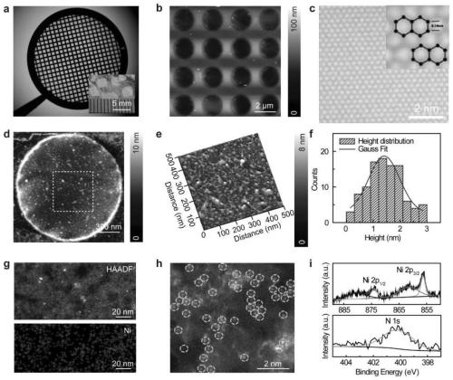
[0065] Embodiment 1, the preparation of bioactive ligand functionalized graphene film

[0066] 1. Preparation

[0067] Add 7 μL of a mixed solution of 0.40M potassium permanganate and 0.20M sodium hydroxide dropwise to the surface of the transferred graphene film, let it stand for a period of time (about 50 minutes), suck off the mixed solution with filter paper, and rinse with 1M Sodium bisulfite is fully cleaned until there is no residual treatment solution on the surface of the graphene film. Next, pass through 5.0 mM 1-ethyl-3-(3-dimethyl-aminopropyl) carbodiimide hydrochloride (EDC), 5.0 mM N-hydroxy-sulfosuccinimide (sulfo- NHS) and 0.10M 2-(N-morpholino)ethanesulfonic acid (MES) mixed solution (pH 5.0) to modify the graphene surface and keep it for about 50 minutes. Afterwards, the graphene grid was activated for a certain period of time (about 2 hours) in 50mM TBE buffer (pH 8.5) containing 11.3mM nitrilotriacetic acid, and then 11.3mM nickel sulfate (NiSO 4 ) solut...

Embodiment 2

[0073] Example 2, Research on biological application performance of bioactive ligand functionalized graphene film


PUM

PropertyMeasurementUnit
thicknessaaaaaaaaaa

Abstract

The invention discloses an application of a functionalized graphene film in three-dimensional reconstruction of a cyro-electron microscope. The bioactive ligand functionalized graphene thin film can be used for three-dimensional reconstruction of a cyro-electron microscope; the bioactive ligand functionalized graphene thin film is formed by connecting a bioactive ligand to a graphene thin film, and the bioactive ligand is a Ni-nitrilotriacetic acid ligand, wherein the bioactive ligand functionalized graphene film is used as or used for preparing a support film of a cyro-electron microscope; the bioactive ligand functionalized graphene film specifically captures proteins or biomacromolecules with His tags. The bioactive ligand functionalized graphene film can be expected to combine biomolecule purification and cyro-electron microscope sample preparation into one step by directly positioning or capturing a target biomolecule in a cell lysate in future optimization.

Description

technical field [0001] The invention relates to a method for realizing high-resolution structure analysis of biomolecules by using functionalized graphene, and belongs to the technical field of biological cryo-electron microscopy. Background technique [0002] Since its appearance in the 1970s, cryo-electron microscopy has become the most important means of structural biology research after a long period of development, especially in recent years with scientific and technological breakthroughs. Cryo-electron microscopy quickly freezes aqueous biological samples under physiological conditions into amorphous glassy ice, and then uses transmission electron microscopy to observe the samples in the frozen state, and combines image processing technology to analyze the structure of biological macromolecules. Unique advantages not available. The revolutionary breakthroughs in instrument hardware and structural analysis software of cryo-electron microscopy technology in recent years...

Claims


Application Information

Patent Timeline
Patent Type & Authority Applications(China)
IPC IPC(8): G16B5/00
Inventor 王宏伟彭海琳陈亚楠刘楠张金灿
Owner TSINGHUA UNIV
Learn More