Bionic periosteum, periosteum-bone substitute and preparation method

A bone substitute and bionic bone technology, applied in biochemical equipment and methods, microorganisms, tissue regeneration, etc., can solve problems such as nonunion and delayed healing, and achieve bone regeneration promotion, good application prospects, and good osteogenic induction effect Effect

Pending Publication Date: 2020-08-11
SUZHOU UNIV
View PDF2 Cites 6 Cited by
  • Summary
  • Abstract
  • Description
  • Claims
  • Application Information

AI Technical Summary

Problems solved by technology

[0002] The repair of large bone defects caused by severe trauma, tumor or infection is still a big challenge in orthopedics, and there are often delayed healing or even non-union in the repair process

Method used


Image

  • Bionic periosteum, periosteum-bone substitute and preparation method
  • Bionic periosteum, periosteum-bone substitute and preparation method
  • Bionic periosteum, periosteum-bone substitute and preparation method

Examples


Embodiment 1

[0040] (1) Mix the prepolymer and curing agent in the Dow Corning 184 kit at a mass ratio of 10:1, and place it at 60°C for 2 hours to cure to prepare PDMS. Place PDMS in a plasma cleaner to clean it under an oxygen atmosphere. Before PDMS cell culture, UV sterilize the PDMS substrate, and then incubate with 20μg / mL fibronectin for 6-8h;

[0041] (2) Inoculate MC3T3-E1 cells on the incubated PDMS at a density of 0.5×10 4 / cm 2 , when the cells grow to 80% of the surface, add 20 μg / mL ascorbic acid to the medium, and continue to cultivate;

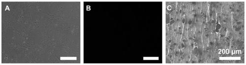
[0042] (3) After 14 days, wash the cultured cell sheet 3 times with PBS, first use 0.25% Triton-X100 and 25mM NH 4 OH solution was used to decellularize the cells for 10min, and then treated with 25U / mL DNase I and 1.5μL / mL RNase A at 37°C for 2h to remove residual cellular DNA and RNA, and the prepared ECM sheets were stored at 4°C for later use. The shape of the sheet is shown in the appendix of the instruction manual. figure 1 ;

...

Embodiment 2

[0046] (1) The prepolymer and curing agent in the Dow Corning 184 kit were mixed at a mass ratio of 20:1, and then cured to obtain PDMS, which was treated with plasma. Before cell culture, the prepared PDMS was sterilized by ultraviolet light, and then incubated with gelatin solution for 8h;

[0047] (2) Inoculate bone marrow mesenchymal stem cells on the hatched PDMS at a density of 2×10 4 / cm 2 , when the cells grow to 90% of the surface, add 50 μg / mL ascorbic acid to the medium to promote the secretion of extracellular matrix, and continue to cultivate until the cell sheet matures;

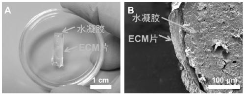
[0048] (3) Wash the cultured cell sheet 3 times with PBS, first use 0.25% Triton-X100 and 50mM NH 4 OH solution was used to decellularize the cells for 5 minutes, and then treated with 50U / mL DNaseI and 2.5μL / mL RNase A at 37°C for 2h to remove residual cellular DNA and RNA. The ECM sheet has good biocompatibility;

[0049] (4) Add PBS to the GelMA solid sponge, then place it in a 37°C water...

Embodiment 3

[0052] (1) The prepolymer and curing agent in the Dow Corning 184 kit were mixed at a mass ratio of 5:1, and cured to prepare PDMS, which was treated by plasma. Before cell culture, UV sterilize the PDMS substrate, and then incubate with 10μg / mL fibronectin for 6-8h;

[0053] (2) Adipose-derived mesenchymal stem cells were inoculated on the hatched PDMS at a seeding density of 1×10 4 / cm 2 , when the cells grow to 90% of the surface, add 40 μg / mL ascorbic acid to the medium to promote the secretion of extracellular matrix, and continue to cultivate until the cell sheet matures;

[0054] (3) Wash the cultured cell sheet 3 times with PBS, first use 0.1% Triton-X100 and 40mM NH 4 OH solution decellularized the cells for 20 minutes, then treated them with 40 U / mL DNase I and 2.0 μL / mL RNase A at 37 °C for 2 h to remove residual cellular DNA and RNA, and stored the prepared ECM sheets at 4 °C for later use;

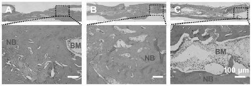
[0055] (4) Prepare 2% sodium alginate solution and 10mM calcium chlori...


PUM

PropertyMeasurementUnit
thicknessaaaaaaaaaa

Abstract

The invention discloses a bionic periosteum for repairing a segmental bone defect, a periosteum-bone substitute and a preparation method. The bionic periosteum is prepared by culturing osteogenic precursor cells on the surface of polydimethylsiloxane, and removing the cells after forming a cell sheet to obtain an extracellular matrix (ECM) sheet. The bionic periosteum winds the surface of a degradable bone scaffold material to form the periosteum-bone substitute. The prepared osteogenic precursor cell-derived ECM sheet not only has biological activity, but also shows a bone induction effect invitro and in vivo. In addition, the ECM sheet has a chemotactic effect on mesenchymal stem cells. Through combination of the cell-derived ECM sheet and the degradable bone scaffold material, an existing membrane induction technique for repairing the large-area segmental bone defect in clinic can be improved; the preparation method is simple; and the periosteum-bone substitute can be used as an excellent periosteum-bone substitute material.

Description

technical field [0001] The invention relates to the technical field of biomedical materials, in particular to a bionic periosteum, a periosteum-bone substitute and a preparation method for repairing large segmental bone defects. Background technique [0002] The repair of large bone defects caused by severe trauma, tumor or infection is still a big challenge in orthopedics, and there are often delayed or even non-union in the repair process. It is well known that when a defect exceeds a critical size, it cannot be fully repaired by its own healing process. [0003] Periosteal integrity is critical for fracture healing and bone regeneration. The periosteum is a thin tissue membrane covering the outer surface of the bone, which transports 70-80% of the blood supply to the bone cortex, provides osteoblasts, precursor cells, and periosteum stem cells, and plays an important role in bone formation and regeneration. The periosteum can promote healing through a variety of biologi...

Claims


Application Information

Patent Timeline
Patent Type & Authority Applications(China)
IPC IPC(8): C12N5/077A61L27/20A61L27/22A61L27/28A61L27/36A61L27/50A61L27/52A61L27/54
CPCC12N5/0654A61L27/3633A61L27/365A61L27/3687A61L27/50A61L27/28A61L27/52A61L27/54A61L27/222A61L27/20C12N2533/30A61L2430/02A61L2430/40A61L2300/412C08L5/04
Inventor 李斌韩凤选余颖康
Owner SUZHOU UNIV
Why Eureka
Try Eureka