In-situ brain glioma microenvironment responsive nano material and preparation method and application thereof

A technology of glioma and nanomaterials, which is applied to the magnetic resonance responsive imaging and therapeutic effects of albumin-based nano-therapeutic agents and its preparation, in situ glioma microenvironment-responsive nanomaterials and its preparation , can solve the problems of no magnetic resonance enhancement ability, inability to enhance tumor tissue-specific imaging, insoluble in water, etc., and achieve good biocompatibility, easy operation, good dispersion and stability

Pending Publication Date: 2020-11-03
AFFILIATED HUSN HOSPITAL OF FUDAN UNIV
View PDF2 Cites 1 Cited by
  • Summary
  • Abstract
  • Description
  • Claims
  • Application Information

AI Technical Summary

Problems solved by technology

Although it has been reported in recent years that nanocarriers can be loaded with magnetic resonance contrast agents for enhanced magnetic resonance imaging of glioma, most studies focus on Gd-based contrast agents, which are characterized by enhancement in both tumor and normal tissues, and cannot be used for tumor tissue enhancement. Specific Enhanced Imaging
and MnO 2 As a new tumor microenvironment-responsive MRI contrast agent, but MnO 2 Insoluble in water, and MnO 2 Not inherently MRI-enhanced

Method used


Image

  • In-situ brain glioma microenvironment responsive nano material and preparation method and application thereof
  • In-situ brain glioma microenvironment responsive nano material and preparation method and application thereof
  • In-situ brain glioma microenvironment responsive nano material and preparation method and application thereof

Examples


preparation example Construction

[0048] The preparation method of the albumin-based nanometer material is simple, the reaction of the preparation method is simple, the operation is easy, the target product is easy to be obtained, and it is suitable for mass production. The preparation method of albumin-based nanomaterials is exemplarily described as follows:

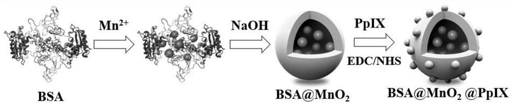
[0049] A. BSA@MnO 2 Preparation of nanoparticles: Slowly add manganese chloride tetrahydrate solution dropwise into bovine serum albumin (bovine serum albumin, hereinafter referred to as BSA) aqueous solution while stirring. Among them, the concentration of albumin solution is preferably 2mg / ml, albumin and MnCl 2 4H 2 The dosage ratio of O is preferably 60 mg:7.9 mg, and the stirring time is 5-10 min. Slowly add NaOH solution dropwise into the mixed solution to adjust the pH of the mixed solution to 8-10, and continue vigorously stirring for 0.5-1 h after the dropwise addition. Then use a 220um microporous filter to filter the mixed solution, and u...

Embodiment 1

[0053] An in situ glioma microenvironment-responsive nanomaterial comprises the following components in parts by weight:

[0054] 60 parts of nanocarriers;

[0055] Manganese dioxide (MnO 2 ) 0.3 copies;

[0056] Protoporphyrin (PpIX) 2.4 parts;

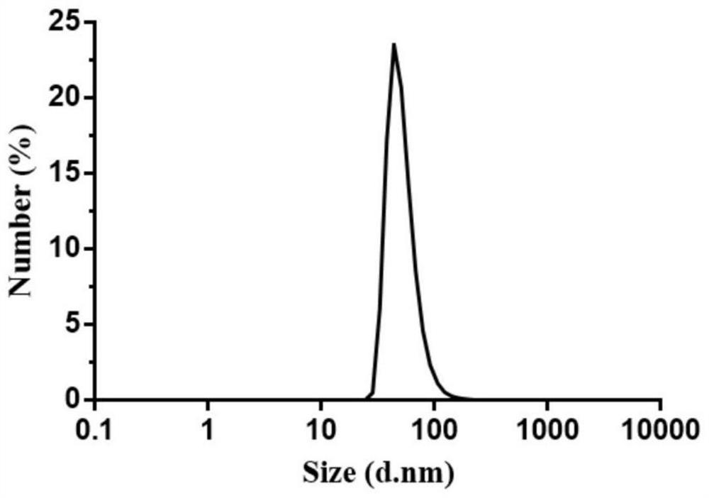
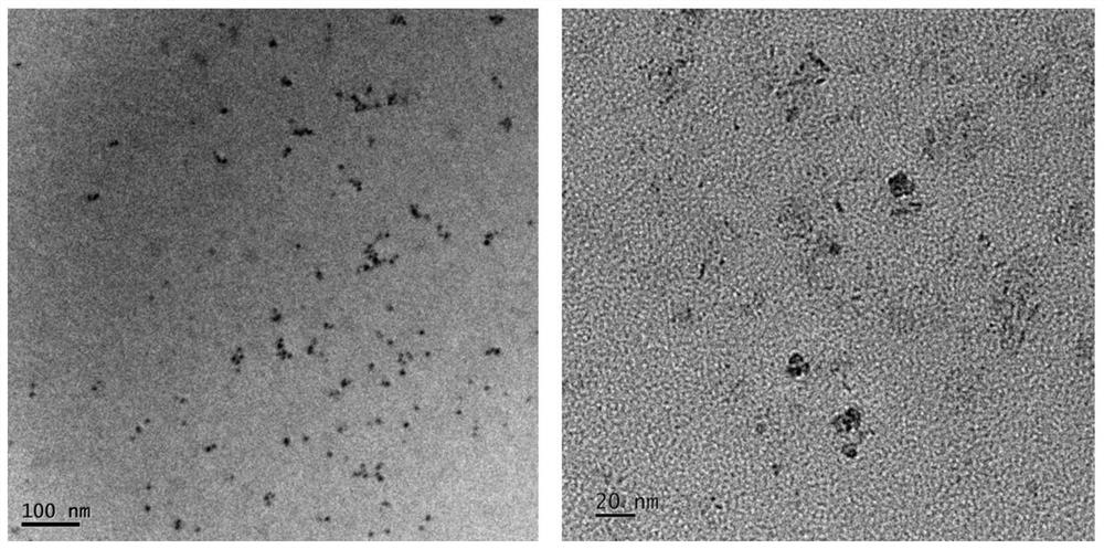
[0057] The nanocarriers are albumin, manganese dioxide (MnO 2 ) nanocrystals and protoporphyrin are entrapped in the bovine serum albumin cavity. Wherein the loading amount of the manganese dioxide nanocrystal is 0.5wt%, and the loading amount of the protoporphyrin is 3.8wt%; the hydrated particle size of the albumin-loaded manganese dioxide nanomaterial is 4.19nm.

[0058] Its preparation method comprises the following steps:

[0059] A. BSA@MnO 2 Preparation of nanoparticles: Accurately weigh 60mg of albumin (Bovine SerumAlbumin, hereinafter referred to as BSA) and dissolve it in 30ml of double distilled water, slowly add 0.1ml of manganese chloride tetrahydrate solution (0.1M) while stirring, stir for 5min, albumin and The ...

Embodiment 2

[0063] An in situ glioma microenvironment-responsive nanomaterial comprises the following components in parts by weight:

[0064] 60 parts of nanocarriers;

[0065] Manganese dioxide (MnO 2 ) 1.2 copies;

[0066] Protoporphyrin (PpIX) 2.4 parts;

[0067] The nanocarriers are albumin, manganese dioxide (MnO 2 ) nanocrystals and protoporphyrin are entrapped in the bovine serum albumin cavity. Wherein the loading amount of manganese element is 1.89wt%, and the loading amount of protoporphyrin is 3.8wt%; the hydrated particle size of albumin-loaded manganese dioxide nanometer material is 24.4nm.

[0068] Its preparation method comprises the following steps:

[0069] A. BSA@MnO 2 Preparation of nanoparticles: Precisely weigh 60mg of albumin (Bovine SerumAlbumin, hereinafter referred to as BSA) and dissolve it in 30ml of double distilled water, slowly add 0.4ml of manganese chloride tetrahydrate solution (0.1M) while stirring, stir for 5min, albumin and The reaction mass rati...


PUM

PropertyMeasurementUnit
particle sizeaaaaaaaaaa
concentrationaaaaaaaaaa

Abstract

The invention discloses an in-situ brain glioma microenvironment responsive nano material, and a preparation method and application thereof, and relates to the technical field of biology. The in-situbrain glioma microenvironment responsive nano material comprises 60 parts of a nano carrier, 0.3-4 parts of manganese dioxide, and 1.2-3.6 parts of protoporphyrin; albumin serves as a bionic carrier,MnO2 nanocrystals serve as an MRI contrast agent and are loaded in an albumin cavity in a biomineralization manner, protoporphyrin serves as a therapeutic agent, and the protoporphyrin is entrapped inthe albumin cavity through an amide reaction. The loading capacity of manganese dioxide nanocrystals in albumin nanoparticles is 0.5-6.7 wt%, and the loading capacity of the protoporphyrin is 2-6wt%.After intravenous injection, the nano material can accurately and specifically display an in-situ glioma focus, and is helpful for assisting in clinical improvement of treatment accuracy and safety;Besides, oxygen is generated to improve a tumor hypoxic microenvironment, and the protoporphyrin generates active oxygen under ultrasonic excitation to specifically kill tumor cells.

Description

technical field [0001] The invention belongs to the field of biotechnology, and specifically relates to an in situ glioma microenvironment-responsive nanomaterial and its preparation method and application, in particular to the magnetic resonance responsive imaging and therapeutic effects of an albumin-based nano-diagnostic and therapeutic agent And its preparation method and application. Background technique [0002] As the most common primary brain tumor, glioma has the characteristics of infiltrative growth, high recurrence rate and high mortality rate, and the median survival time is only 15 months. Surgery combined with radiotherapy and chemotherapy is the standard clinical treatment for glioma, but the effect is not satisfactory. [0003] Sonodynamic therapy is a new type of tumor treatment method, which uses ultrasound to activate the sonosensitizer to catalyze oxygen to generate highly toxic singlet oxygen, so as to achieve the purpose of killing tumors. Compared wi...

Claims


Application Information

Patent Timeline
Patent Type & Authority Applications(China)
IPC IPC(8): A61K49/14A61K49/18A61K41/00A61K9/14A61K47/42A61P35/00
CPCA61K49/143A61K49/1818A61K41/0033A61K9/146A61P35/00
Inventor 尹波佘德君黄慧慧刘莉耿道颖庞志清陆逸平
Owner AFFILIATED HUSN HOSPITAL OF FUDAN UNIV
Why Eureka
Try Eureka