Pig liver tissue organoid model and in-vitro construction method thereof

A construction method and organoid technology, which is applied in the field of in vitro construction of pig liver tissue organoid models, can solve the problems of lack of intercellular matrix in organoids and the inability to truly simulate the physiological functions of the liver, achieving rapid construction, simple methods, and reduced The effect of sampling

Active Publication Date: 2020-11-24
YANGZHOU UNIV
View PDF13 Cites 3 Cited by
  • Summary
  • Abstract
  • Description
  • Claims
  • Application Information

AI Technical Summary

Problems solved by technology

[0003] The purpose of the present invention is to provide a method for constructing a porcine liver tissue organoid model in vitro, so as to solve the current human-derived or mouse-derived liver organoids existing in the

Method used


Image

  • Pig liver tissue organoid model and in-vitro construction method thereof
  • Pig liver tissue organoid model and in-vitro construction method thereof
  • Pig liver tissue organoid model and in-vitro construction method thereof

Examples


Embodiment 1

[0031] Example 1 In vitro construction of porcine liver organoids

[0032] In this example, 3-day-old male piglet liver tissue was used to establish pig liver organoids for experiments.

[0033] Specific steps are as follows:

[0034] 1), three-day-old newborn piglets were anesthetized in a sterile laboratory, and about 500 mg of liver tissue was removed. The collected fresh piglet liver tissue was repeatedly washed with PBS cleaning solution containing 5 times of penicillin, streptomycin, gentamycin and amphotericin B, cut into pieces, and then washed with 5 times of penicillin, streptomycin, gentamicin The PBS washing solution of amphotericin and amphotericin B was centrifuged until the supernatant was clear.

[0035] 2) Transfer the shredded and cleaned tissue to a 50mL sterile centrifuge tube, add 10mL of digestion solution, and seal the centrifuge tube with parafilm. Digestion was incubated at 200 rpm in a constant temperature shaking incubator at 37°C; the digestion s...

Embodiment 2

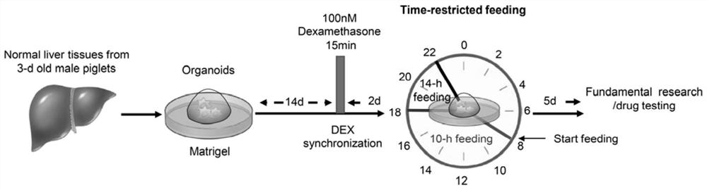
[0045] Example 2 Dexamethasone Treatment and Organoid Biological Cycle Reset

[0046] On day 15 after organoid inoculation, the organoids were treated with 100 nM (final concentration) of dexamethasone (DEX, Sigma-Aldrich) for 15 min to reset the organoid clock, synchronizing it to 0 o'clock. Organoids were then washed three times with PBS wash (37°C) and cultured in expansion medium. After 48 hours of DEX treatment, the organoids in 6 wells were used as the control group, exposed to the expansion medium from 8:00 am to 10:00 pm, and then exposed to the basal medium at 10:00 pm ~ 8:00 am, the total time is 24 hours + 1 hour. Organoids from the other 6 wells served as the limiting nutrient group and were exposed to expansion medium for 10 h from 8:00 am to 6:00 pm and +1 to basal from 6:00 pm to 8:00 am in a 24 hour cycle medium for 14 hours. Exposure was continued for 5 days in a 24-hour cycle, after which organoids were harvested and analyzed.

Embodiment 3

[0047] Example 3 Detection of organoid cell viability

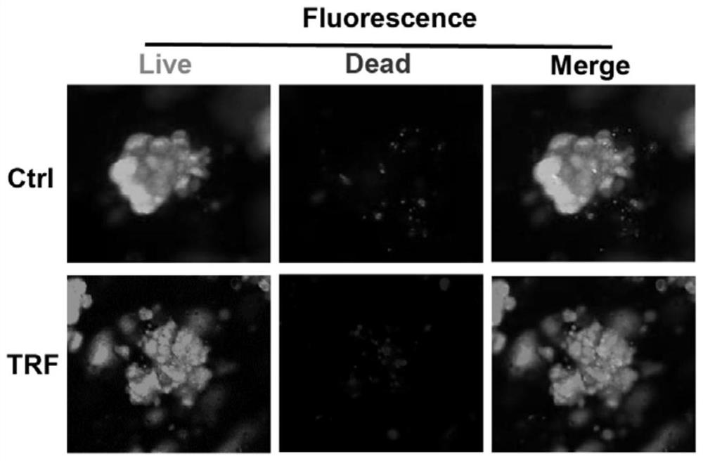
[0048] (1) Detection of organoid viability by staining

[0049] Add 100 μL live / dead cell double staining kit (calcein-AM / ethidium bromide homodimer-1, Thermofisher Scientific) to decibels in control and nutrient-restricted group organoids and incubate at room temperature for 30 min. Fluorescence microscopy was used to capture the signal of calcein-AM to represent live cells and the signal of ethidium bromide homodimer-1 to identify dead cells.

[0050] Fluorescent staining results such as figure 2 As shown, the results showed that nutrient restriction did not affect the viability of liver cells.

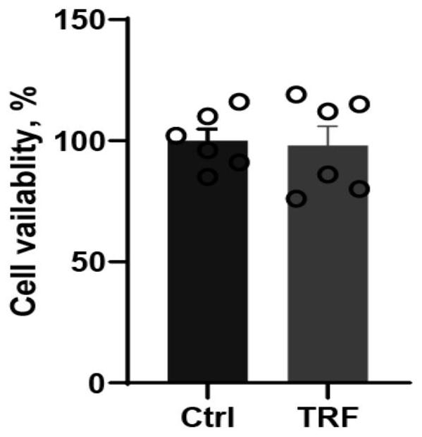
[0051] (2) Cell-Titer GLO detection of cell activity

[0052] Organoid cell viability assays were performed on the control and nutrient-restricted groups separately by adding Cell-Titer GLO reagent (Promega) and measuring luminescence on a GLOMAX microplate luminometer (Promega) according to the manufacturer's instructions....


PUM


Abstract

The invention discloses an in-vitro construction method of a pig liver tissue organoid model. The method comprises the steps of shearing and cleaning the liver tissues of a newborn piglet, and addingdigestive juice for incubation and digestion to obtain organoid fine particles; mixing the organoid fine particles with matrix gel, and performing inoculation; and after the matrix gel is solidified,performing incubation for 4 days by using an induction culture medium, then performing culture by using an expansion culture medium, and replacing the expansion culture medium once every 3-4 days, soas to obtain the pig liver tissue organoid model. The construction method is simple, rapid in construction and high in operability; the organoid model is constructed directly through the organoid fineparticles of a newborn animal; the organoid model not only can truly reflect the internal environment in a living pig model, but also can also be used for studying liver growth and lipid metabolism in the living pig model at the molecular cell level; and sampling of living pigs is reduced.

Description

technical field [0001] The invention belongs to the technical field of animal cell engineering and relates to an in vitro construction method of a porcine liver tissue organoid model. Background technique [0002] Organoids are micro-organs constructed in vitro by 3D culture technology. Because it has the complex three-dimensional structure of real organs and the physiological functions highly similar to the original organs, it has been widely used in the field of biology and gradually replaced cell lines. Liver organoid culture, which uses a unique system of extracellular matrices such as Matrigel to bring organoids closer to the structural and functional properties of in vivo tissue, is becoming a popular alternative to primary cell culture for reproducing in a dish To organize and study liver physiology and disease pathogenesis in humans and mice. However, although this method can eliminate many interfering effects in vitro and provide a simplified model of tissues in v...

Claims


Application Information

Patent Timeline
IPC IPC(8): C12N5/071
CPCC12N5/0671C12N2533/90
Inventor 蔡德敏刘好雨李豪辛子蒙张可欣顾昊天李艳伟
Owner YANGZHOU UNIV
Why Eureka
Try Eureka