Fluorescent imaging probe and preparation method and application thereof

A fluorescence imaging agent and probe technology, applied in the field of fluorescence imaging, can solve the problems of unpredictable cancer cells, easy transmembrane transport into tumor cells, tumor cell retention time, easy aggregation, etc. The effect of strong membrane transport and long residence time

Active Publication Date: 2021-02-09
JIANGSU INST OF NUCLEAR MEDICINE
View PDF2 Cites 4 Cited by
  • Summary
  • Abstract
  • Description
  • Claims
  • Application Information

AI Technical Summary

Problems solved by technology

However, the probes in the above literatures have only done in vitro probe-labeled protein experiments, but have not been carried out in vivo experiments. It is difficult to predict whether they are suitable for the labeling of cancer cells in vivo, and because the environment in vivo is very complicated, cyanine Fluorescent dye-based probes not only face the problem of easy aggregation in vivo, but also face the problems of tumor cell targeting, whether it is easy to tr

Method used


Image

  • Fluorescent imaging probe and preparation method and application thereof
  • Fluorescent imaging probe and preparation method and application thereof
  • Fluorescent imaging probe and preparation method and application thereof

Examples


Embodiment 1

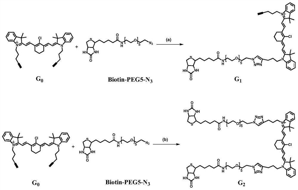
[0063] Example 1 Compound G 1 and G 2 Synthesis

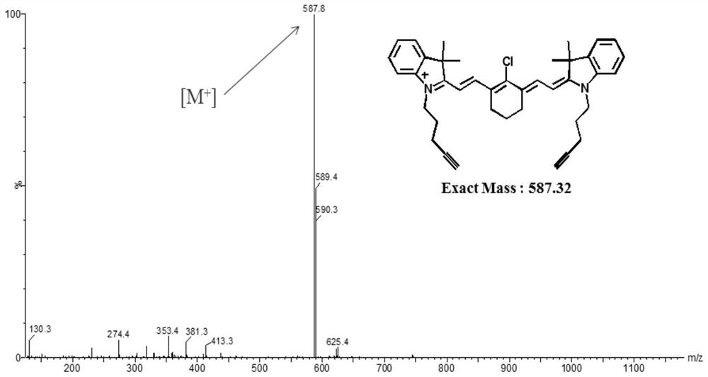
[0064] 1 Compound G 0 Synthesis

[0065] According to the reported method (Chemical communications2017,53,6117-6120), G 0, specific method: KI (5.8g, 34.93mmol, 6eq) was dissolved in acetonitrile (25mL); then 5-chloro-1-pentyne (1841 μL, 17.45mmol, 3eq) was added. The reaction solution was stirred at 50°C for 40 minutes. To this was added 2,3,3-trimethylindoline (933 μL, 5.82 mmol, 1 eq). The reaction mixture was heated at 85°C for 24 hours. The reaction liquid was filtered, and then acetonitrile was removed by distillation under reduced pressure. The resulting solid was purified by column chromatography (mobile phase: MeOH and DCM volume ratio: 2-5%) to obtain compound 1 in a yield of 17% (224mg, 0.991mmol). Compound 1 (221 mg, 0.977 mmol, 1 eq) and 2-chloro-1-formyl-3-(hydroxymethylene)-1-cyclohexene (84.1 mg, 0.489 mmol, 0.5 eq) were dissolved in anhydrous acetonitrile (3 mL). Acetic anhydride (123 μL) and sodium a...

Embodiment 2

[0078] Determination of photochemical and photophysical properties of embodiment 2 compounds G1 and G2

[0079] 1 Determination of absorption spectrum and fluorescence spectrum

[0080] Methanol was chosen as the solvent to prepare G 0 ,G 1 and G 2 The stock solution (20mM) was used to measure the spectrum. Dilute G with methanol and water, respectively 0 , G 1 and G 2 The stock solutions were diluted to different concentrations (4 μM, 3 μM, 2 μM, 1 μM, 0.5 μM) to obtain absorption spectra. Absorbance fit straight lines at λmax and concentration were obtained. Prepare 4 µM G 0 , G 1 and G2 solution to measure the fluorescence spectrum. The present invention uses methanol and water as solvents respectively to compare the fluorescence intensity of probes in different solvents.

[0081] 2 Determination of photon quantum yield

[0082] Prepare G with methanol and water as solvents, respectively 1 , G 2 and indocyanine green (ICG) solution (1 μM); ICG was used as a re...

Embodiment 3

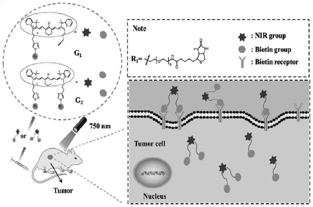
[0095] Example 3 Tumor cell-specific imaging in vitro

[0096] 1 cell culture

[0097] To verify the specific accumulation of the probe at the tumor site, HeLa cells were selected as BR-positive tumor cells, and LO2 cells were selected as BR-negative normal cells. HeLa and LO2 cells were placed in high glucose DMEM medium containing 10% (v / v) fetal bovine serum (FBS) in 5% (v / v) CO 2 Cultivated in a humid and stable environment. The incubation temperature was 37°C.

[0098] 2 Cytotoxicity assay

[0099] Perform MTT assay to evaluate probe G 1 and G 2 cytotoxicity. HeLa and LO2 cells were seeded into 96-well plates, and the density was controlled at 10,000 cells per well. Cells were incubated at 37°C for 24h to allow cell attachment. Will G 1 and G 2 respectively dissolved in DMF to obtain 20mM G 1 and G 2 Stock solution, G 1 and G 2 The stock solution was diluted to the target concentration. After removing the medium, probe solutions at different concentrations ...


PUM

PropertyMeasurementUnit
Molar extinction coefficientaaaaaaaaaa

Abstract

According to the invention, two NIRF probes (G1 and G2) with good water solubility are designed. The PEG structure of the probe improves the hydrophilicity of the probe, and an in vitro spectrum and alipid-water distribution coefficient prove that G1 and G2 have a relatively low aggregation trend. The biotin structure enhances the targeting of the probe to BR positive tumor cells, and G1 and G2 show good fluorescence imaging effects in in-vitro cell experiments and in-vivo fluorescence imaging. Due to the cooperation of a specially selected hydrophilic group-PEG structure and targeting group-biotin, the probe G1 and the probe G2 can be balanced between hydrophilicity and targeting property, so that balance is achieved in multiple aspects such as fluorescence intensity, tumor targeting property, transmembrane transport property, residence time in tumor cells, and the prepared probes G1 and G2 have the advantages of high fluorescence intensity, strong targeting, strong transmembrane transportation and long residence time in tumor cells.

Description

technical field [0001] The invention belongs to the technical field of fluorescence imaging, and in particular relates to a fluorescence imaging probe and its preparation method and application. Background technique [0002] Today, fluorescence imaging has emerged as an effective tumor imaging technique. Its advantages are many, such as: high sensitivity, low cost, non-invasive and non-radioactive. However, the shallow tissue penetration depth and high background signal limit the further development of fluorescence imaging. In the past decade, near-infrared fluorescence (NIRF) imaging has broken through both of the aforementioned barriers. The near infrared (NIR) region (700-900nm) is the ideal imaging region. Biological tissue absorbs very little near-infrared light relative to visible light; therefore, near-infrared light has a deeper tissue penetration depth. Moreover, in the NIR region, the autofluorescence of organisms is almost non-existent, so the background signa...

Claims


Application Information

Patent Timeline
IPC IPC(8): C07D495/04C07D519/00C09K11/06A61K49/00G01N21/64
CPCC07D495/04C07D519/00C09K11/06A61K49/0032A61K49/0052G01N21/6428G01N21/6456G01N21/6458G01N21/6486C09K2211/1029C09K2211/1051C09K2211/1059
Inventor 谢敏浩林建国邱玲李航刘清竹李珂
Owner JIANGSU INST OF NUCLEAR MEDICINE
Why Eureka
Try Eureka