Single cell lysis buffer and application thereof
A lysate, single-cell technology, applied in the biological field, can solve the problems of simultaneous rupture of cell nucleus and mitochondria, and achieve the effect of high commercial application value, short time consumption and moderate lysis intensity
- Summary
- Abstract
- Description
- Claims
- Application Information
AI Technical Summary
Problems solved by technology
Method used
Image
Examples
Embodiment 1
[0020] Example 1: Configuration of single cell lysate
[0021] The composition of the single-cell lysate, in specific applications, can use the ratio of the single-cell lysate components in the following example: Prepare 1mL single-cell lysate, and use a pipette to draw 50 uL of 2 mol / L Tris-Hcl into a centrifuge tube , add 5 mol / L Licl 100 uL, add 20% Triton X100 5uL, add 1 mol / L EDTA 20 uL, add 100mmol / LDTT50uL, blow and aspirate to mix, add sterile water 775 uL to make up to 1mL, mix and set aside.
Embodiment 2
[0022] Example 2: Configuration of single cell lysate
[0023] The composition of the single-cell lysate, in specific applications, can use the ratio of the single-cell lysate components in the following example: Prepare 1mL single-cell lysate, and use a pipette to draw 50 uL of 2 mol / L Tris-Hcl into a centrifuge tube , add 5 mol / L Licl 100 uL, add 20% Triton X100 5uL, add 1 mol / L EDTA 20 uL, add 100mmol / LDTT50uL, add sterile water 775 uL, mix and set aside.
Embodiment 3
[0024] Example 3: Lysis of 293T cells
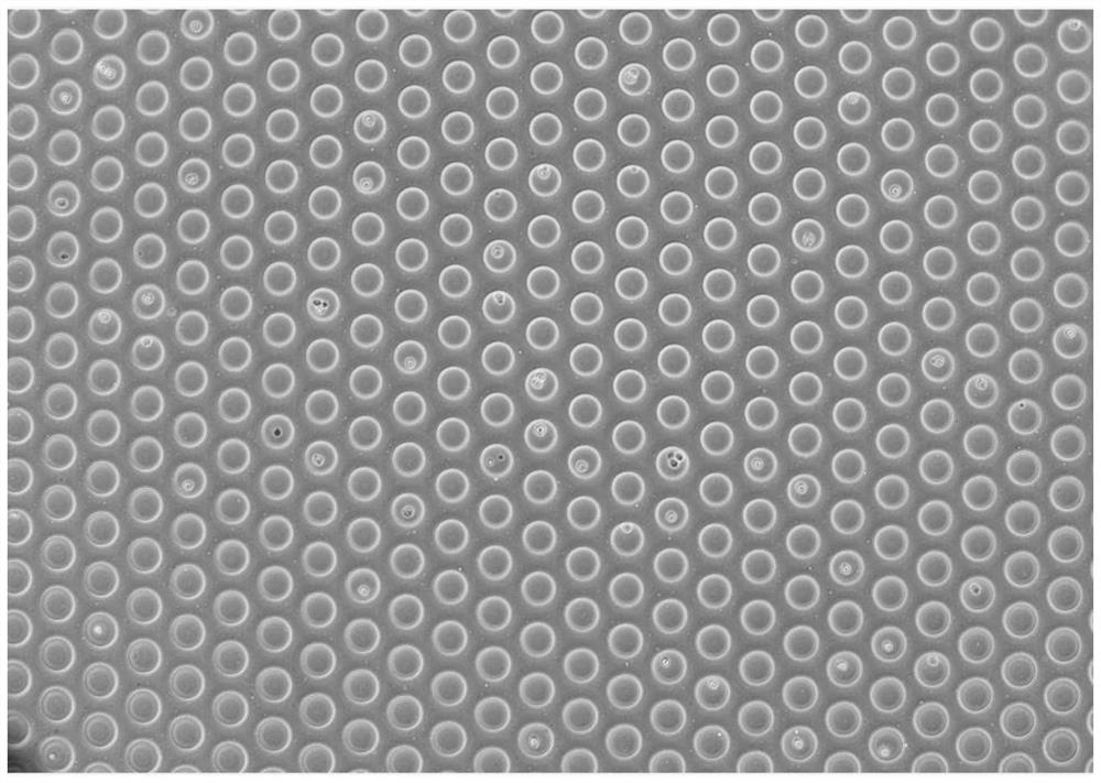
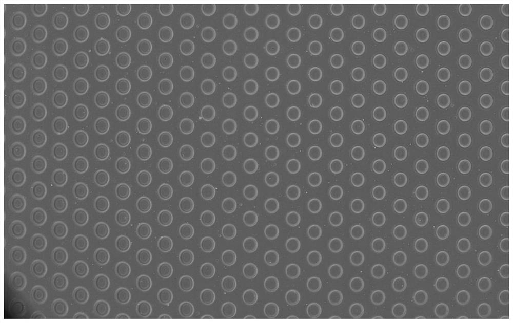
[0025] The growth rate of 293T cells is fast, and the doubling time generally does not exceed 1 day. Subsequent experiments can be carried out after the recovery of the primary cells. Add 100 uL of the single-cell lysate prepared in Example 1 to the microfluidic chip, observe the cell lysis before and after addition under a microscope, record the lysis time, and take pictures to record the lysis process.
[0026] figure 1 is the microscope image of cells in the lysed microfluidic chip before lysis; figure 2 is the microscopic image of cells lysed in the lysed microfluidic chip; figure 1 , figure 2 It can be seen that before adding single-cell lysate, the outline of 293T cells falling into the microfluidic chip is clear, and the nucleus is clearly visible. After adding the single-cell lysate, the cells were completely lysed and there was almost no mitochondrial lysis, and the time for all the cells to be lysed was not more than 2 mi...
PUM
 Abstract
Description
Claims
Application Information
 - R&D
- Intellectual Property
- Life Sciences
- Materials
- Tech Scout
- Unparalleled Data Quality
- Higher Quality Content
- 60% Fewer Hallucinations
Browse by: Latest US Patents, China's latest patents, Technical Efficacy Thesaurus, Application Domain, Technology Topic, Popular Technical Reports.
© 2025 PatSnap. All rights reserved.Legal|Privacy policy|Modern Slavery Act Transparency Statement|Sitemap|About US| Contact US: help@patsnap.com


