
Active tuberculosis marker, kit, detection method and model construction method

A technology of tuberculosis and markers, applied in the field of biomedicine, can solve the problems of low sensitivity and poor specificity, and achieve the effect of high sensitivity, good specificity and good clinical application value

Pending Publication Date: 2021-07-30
SHANGHAI PUBLIC HEALTH CLINICAL CENT
View PDF7 Cites 6 Cited by
  • Summary
  • Abstract
  • Description
  • Claims
  • Application Information

AI Technical Summary

Problems solved by technology

[0004] In order to solve the problems of poor specificity and low sensitivity in the existing tuberculosis diagnostic technology, the present invention provides a rapid detection model of active tuberculosis based on plasma proteomics technology and machine learning algorithm and its construction method, which is effective for tuberculosis Detection has good application value

Method used


Image

  • Active tuberculosis marker, kit, detection method and model construction method
  • Active tuberculosis marker, kit, detection method and model construction method
  • Active tuberculosis marker, kit, detection method and model construction method

Examples


Embodiment 1

[0054] Isolation of plasma samples from whole blood

[0055] The fasting peripheral blood of patients with active pulmonary tuberculosis was collected in the morning with EDTA anticoagulant tubes, centrifuged at 3000 rcf for 15 min, and the plasma was separated into a new 1.5 mL centrifuge tube within 6 hours. Plasma samples were stored in a -80°C freezer.

Embodiment 2

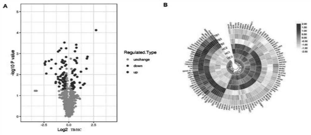
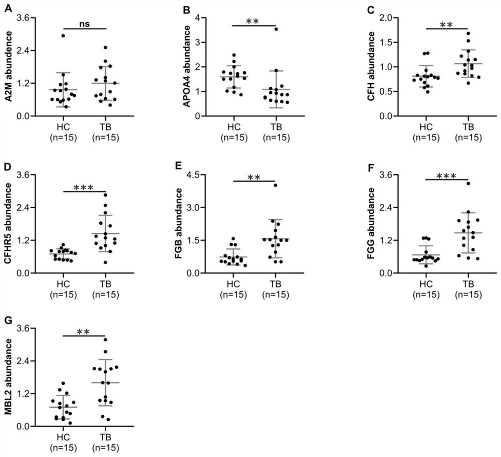
[0057] Quantitative proteomic analysis of plasma samples

[0058] High-abundance plasma proteins were removed using Pierce Top 12 Abundant Protein Depletion SpinColumns Kit (Thermo, USA) according to the kit instructions. The protein solution was reduced with 5mM dithiothreitol, digested at 56°C for 30min, alkylated with 11mM iodoacetamide, and digested at room temperature for 15min in the dark. Then add 100mM TEAB to dilute the protein sample to a urea concentration of less than 2M. Finally, trypsin was added at a trypsin-protein mass ratio of 1:50 for the first digestion, and a trypsin-protein mass ratio of 1:100 was added for the second digestion for 4 h. After trypsinization, the samples were desalted using a Strata X C18 solid-phase extraction column (Phenomenex) and dried in vacuo. Sample peptides were reconstituted in 0.5M TEAB and labeled with TMT kit. Agilent 300Extend C18 column was used for high performance liquid chromatography (HPLC) separation. The processed ...

Embodiment 3

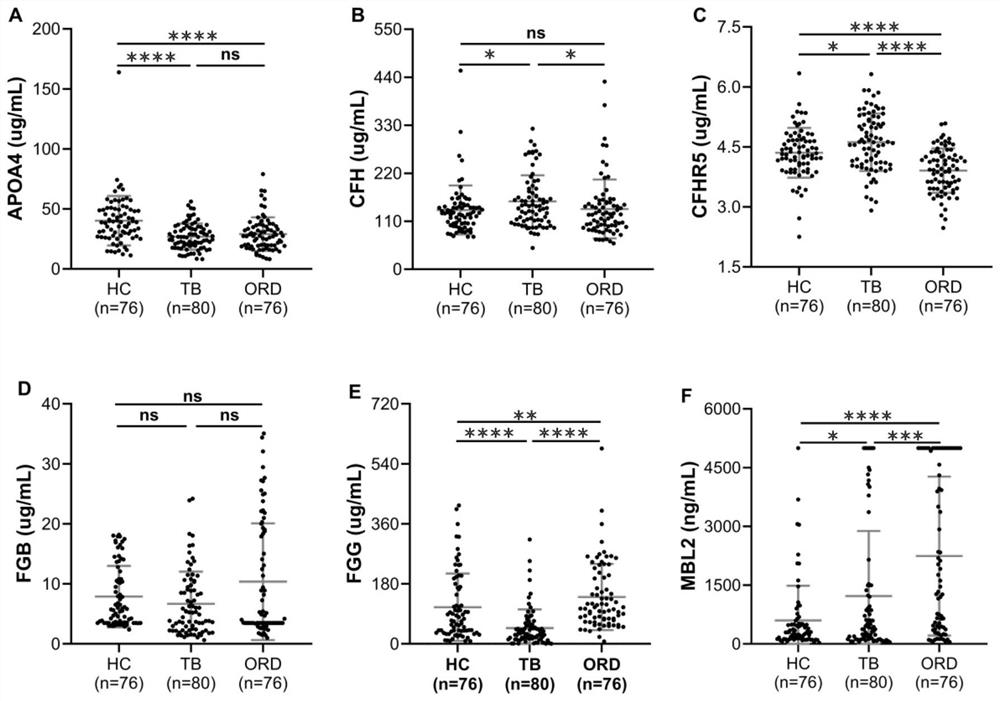
[0060] Determination of Protein Expression Level in Plasma Samples by ELISA

[0061] Sample collection: 80 cases of active tuberculosis patients, 76 cases of healthy controls, and 76 cases of non-tuberculosis pulmonary respiratory diseases.

[0062] Human APOA4 and MBL2 ELISA kits (Elabscience, WuHan, CN) and human CFH, CFHR5, FGB, FGG ELISA kits (Cloud-Clone Corp, WuHan, CN) were used to detect the expression levels of plasma protein markers according to standard operations. The t test or one-way ANOVA in GraphPad Prism 8 software was used for statistical analysis (when P<0.05, there was a significant difference, and when P<0.01, there was an extremely significant difference). Differentially expressed plasma protein data were expressed as mean±SEM, and a scatter plot containing error bars was drawn.


PUM


Abstract

The invention provides an active tuberculosis marker, a kit, a detection method and a model construction method. An active tuberculosis diagnostic marker is a plasma protein biomarker in a blood sample, wherein the plasma protein biomarker is a combined marker comprising one or more of APOA4 protein, CFH protein, CFHR5 protein, FGG protein and MBL2 protein. According to the invention, the combined marker of APOA4, CFH, CFHR5 and FGG derived from plasma protein is used as the biomarker for active tuberculosis detection for the first time, a rapid diagnosis model for active tuberculosis detection is constructed, and a new direction is provided for clinical diagnosis of tuberculosis. Therefore, the invention overcomes the defects of low detection rate, long time consumption and the like of the existing active tuberculosis diagnosis, has the characteristics of good specificity and high sensitivity, and has good clinical application value for the auxiliary diagnosis of the active tuberculosis.

Description

technical field [0001] The invention belongs to the technical field of biomedicine, and more specifically relates to proteomics technology and machine learning algorithms, and constructs an active tuberculosis detection model involving plasma protein biomarkers. Background technique [0002] Tuberculosis (TB) is still a major global public health problem, an infectious disease with the highest mortality rate caused by a single pathogen, and a serious threat to human health. According to World Health Organization (WHO) estimates, in 2019, about 10 million people were newly infected with tuberculosis worldwide, and the death toll was nearly 1.4 million. The current situation of tuberculosis in China is severe, and it is the second largest tuberculosis country after India. The high morbidity and mortality of tuberculosis, as well as airborne transmission, imposes a considerable burden on both patients and society. Therefore, timely and accurate detection of tuberculosis infec...

Claims


Application Information

Patent Timeline
Patent Type & Authority Applications(China)
IPC IPC(8): G16B5/00G16B40/10G16H50/20G16H50/50G06K9/62G06N20/00
CPCG16B5/00G16B40/10G16H50/20G16H50/50G06N20/00G06F18/24G06F18/214
Inventor 张舒林何俊才宋言峥温子禄
Owner SHANGHAI PUBLIC HEALTH CLINICAL CENT
Learn More