Manufacturing method of blood circulating tumor cell pathological chip

A tumor cell and chip technology, which is used in the detection of circulating tumor cells in the blood, can solve the problems of false positives, affecting the observation of morphological diagnosis, and unable to provide cell morphological characteristics.

Pending Publication Date: 2022-03-01
王道祥
View PDF3 Cites 0 Cited by
  • Summary
  • Abstract
  • Description
  • Claims
  • Application Information

AI Technical Summary

Problems solved by technology

[0007] 1) The dyes of conventional cytopathological staining methods will affect the binding of the first antibody or the first probe to the relevant target, so these stains need to be washed to better realize the immunohistochemical staining of biomarkers
2) After cleaning, a higher concentration of protein antibody or nucleic acid probe is often required for staining than conventional staining, which is likely to cause false positives and mislead clinical diagnosis, treatment and prognosis
4) Even if routine staining does not need to be removed in individual cases, the process of immunohistochemical staining or chromogenic in situ hybridization staining can lead to the loss of conventional dyes, resulting in poor observation of cell morphology under conventional staining
5) The usual immunohistochemistry or nucleic acid chromogenic in situ hybridization staining, in order to improve the sensitivity of the staining, the process of chemical staining will be improved as much as possible, which will cause excessive accumulation of chromogenic products and cover the cells, affecting the diagnosis of observation morphology
However, in this method, the cells are first fixed on the glass slide, and the slide is washed several times in an open system before staining. In this way, the cells will be lost during the staining process, which makes the rare CTC cells become fewer
In addition, the patent application uses immunofluorescence staining, which requires a fluorescent microscope and other special scanning equipment, and cannot provide morphological characteristics of cells that can be used for pathological diagnosis

Method used


Image

  • Manufacturing method of blood circulating tumor cell pathological chip
  • Manufacturing method of blood circulating tumor cell pathological chip

Examples


Embodiment 1

[0095] Blood sample preparation

[0096] 1. EDTA anticoagulant tube (purple) 10ml blood collection tube for taking patient's venous blood. A cell suspension containing 100 human urothelial carcinoma cell line T24 was added.

[0097] 2. Add 1 ml BD Lysing Solution 10X concentrate and incubate for 5 minutes to lyse red blood cells.

[0098] 3. Add 1 ml anti-human CD45 monoclonal antibody at a concentration of 0.5 μg / ml and incubate for 30 minutes.

[0099] 4. Add 1 ml of liquid guinea pig serum (G9774, Sigma-Aldrich) and incubate for 1 minute.

[0100] 5. Transfer the reaction liquid into a round bottom centrifuge tube, centrifuge at 1000rpm for 5 minutes, remove the supernatant, and obtain the cell pellet.

Embodiment 2

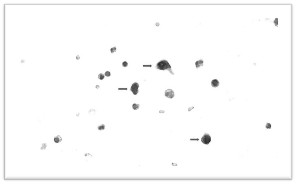
[0102] Liquid-based Immunochemical Staining Method for Circulating Tumor Cells

[0103] 1. The circulating tumor cell pellet obtained in Example 1 was diluted and suspended in 1 ml of PBS (pH7.4) buffer solution, and placed in a 10 ml plastic test tube.

[0104] 2. Add 20 microliters of Tween20 (to increase the permeability of the cell membrane, which is beneficial to the staining of the cell membrane and nucleus).

[0105] 3. Add 100 microliters of Dual Endogenous Enzyme Block (Dako, Carpinteria, Ca) to remove endogenous peroxidase (peroxidase) and alkaline phosphatase (alkline phosphatase) activity in cells, and incubate for 5 minutes.

[0106] 4. After centrifuging at 1000 rpm for 5 minutes, remove the supernatant and remove excess Dual Endogenous EnzymeBlock.

[0107] 5. Add 200 microliters of Serum-Free Block (Dako) and incubate for 10 minutes (to fill in non-specific protein binding sites).

[0108] 6. Add the primary antibody, ie, 200 microliters of mouse anti-human C...

Embodiment 3

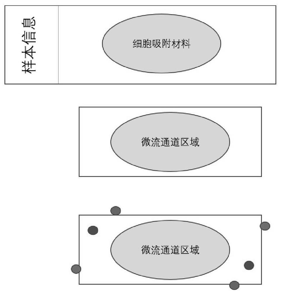
[0116] Fabrication of Pathology Chip

[0117] The design of the chip is as follows:

[0118] 1. Pathological slides of regular size, 75 mm X 25 mm;

[0119] 2. The middle 40X20mm area (rectangle) contains cell adsorption material (100nm rough surface);

[0120] 3. 50X25 mm cover glass, 0.2 mm thick, containing microfluidic channels 1 mm wide and 50-100 microns high;

[0121] 4. The microfluidic channel is a serpentine single channel;

[0122] 5. The glass slide and the cover glass form a closed system, and the cell suspension passes through the microfluidic channel to ensure that the rare tumor cells are fully contacted with the adsorption material and are adsorbed on the glass slide;

[0123] 6. The valve parts of the liquid inlet and the liquid outlet are protruding, with a height of 0.5 cm, which is convenient for the equipment to pump the cell suspension;

[0124] 7. The position of the liquid inlet and the liquid outlet does not block the moving track of the conventio...


PUM

PropertyMeasurementUnit
Thicknessaaaaaaaaaa
Heightaaaaaaaaaa

Abstract

The invention relates to a manufacturing method of a blood circulating tumor cell pathological chip. Specifically, the invention relates to liquid-based staining of circulating tumor cells in a blood sample, and detection of the circulating tumor cells in a pathological section. The invention also relates to a pathological chip for carrying out the above method.

Description

technical field [0001] The present invention relates to the detection of blood circulating tumor cells (Circulating Tumor Cells, CTCs), more specifically to the staining of circulating tumor cells in blood samples, and then making them into a pathology chip for morphological diagnosis and biological identification. The present invention also relates to a pathology chip and a kit for performing the above method. Background technique [0002] Conventional cytopathology (cytology) pathological slides are generally prepared by using patient blood and other body fluids (1) to smear directly on the slide, (2) or to fix the cells in the liquid on the slide by centrifugal force ( cytospin), (3) or through the centrifugation process, the cells in the liquid are formed into a sediment (cell block), and then subjected to routine pathological section processing, and then the pathologist observes the cells under a microscope for cytopathological diagnosis. [0003] Circulating tumor cel...

Claims


Application Information

Patent Timeline
IPC IPC(8): G01N1/30G01N1/34G01N1/28G01N21/84
CPCG01N1/30G01N1/34G01N1/28G01N21/84G01N33/574
Inventor 楚文江王剑
Owner 王道祥
Why Eureka
Try Eureka