


Quick Research
Generate reliable direction feasibility study reports for your R&D in just a few steps.

Technical Q&A
Discover and master advanced knowledge NOW. Basics, ideas, possibilities, all at once.

Find Solutions
As an expert in R&D theories, this can generate solutions to your technical problems instantly.

Evaluate Feasibility
Analyze your overall solution with one click, know your potential R&D risks in advance.

Monitor Landscape
Get weekly tech updates, stay abreast of the latest tech innovations and key insights.
Reagent-free electrochemiluminescence detection method for cell imaging
A detection method and cell imaging technology, applied in chemiluminescence/bioluminescence, analysis by chemical reaction of materials, measurement devices, etc. and other problems, to achieve the effect of uniform particle size, good dispersion and good stability
- Summary
- Abstract
- Description
- Claims
- Application Information
AI Technical Summary
Problems solved by technology
Method used
Image

Examples
Embodiment 1
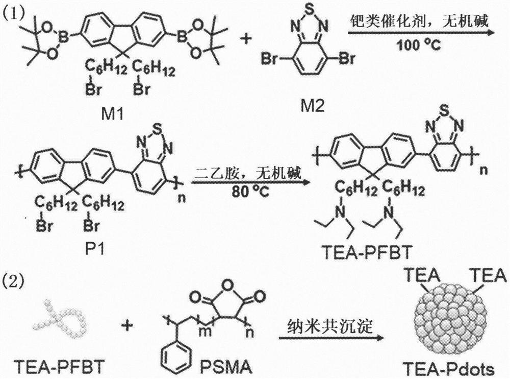
[0017] Example 1: Combining figure 2 , to synthesize polymer dots coupled with triethylamine.
[0018] M1 (100mg), M2 (59.7mg), tetrakis(triphenylphosphine) palladium (17.3mg) and potassium carbonate (207.3mg) were mixed in toluene / water (4mL / 1mL) and placed under argon atmosphere In a Shrek tube, after stirring at 100°C for 72 hours, precipitate twice in methanol to obtain a light orange solid P1. Then P1 (100mg) was mixed with diethylamine (1.17g) and potassium carbonate (221.1mg) in tetrahydrofuran / dimethylformamide (16mL: 4mL), placed in a Shrek tube under an argon atmosphere, 80 After stirring for 96 h at °C, TEA-PFBT was precipitated twice in methanol to obtain TEA-PFBT as an orange solid.
[0019] TEA-PFBT (100μL, 1mg mL -1 THF solution) and PSMA (20μL, 1mg mL -1 THF solution) was added into 1.88 mL THF, ultrasonically degassed for 20 minutes, then the mixture was quickly injected into 1 mL of water, and ultrasonic treatment was continued for 4 minutes. After conc...
Embodiment 2
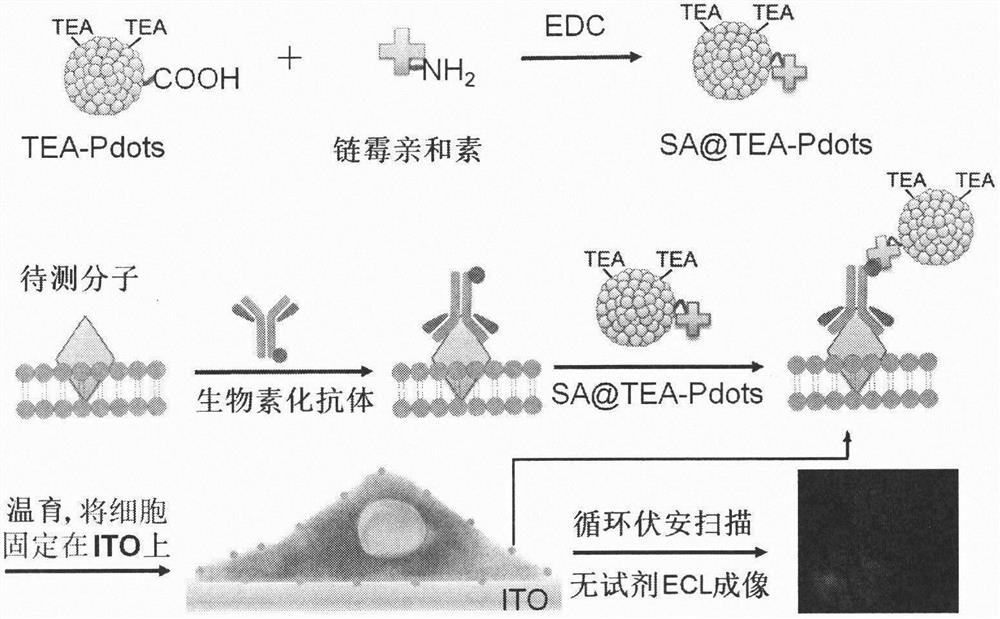
[0020] Example 2: Combining figure 1 , TEA-Pdots were coupled to streptavidin.
[0021] TEA-Pdots (1mL, 50μg mL -1 ), 4-hydroxyethylpiperazineethanesulfonic acid (20μL, 1M) and polyethylene glycol (20μL, 5wt%, M w =3350) after mixing, adjust the pH of the solution to about 7.0. Subsequently, freshly prepared EDC (20 μL, 5 mg mL -1 ) and streptavidin (20 μL, 1 mg mL -1 ) were sequentially added to the mixture, and stirred at room temperature for 5 hours. Then, bovine serum albumin (20 μL, 10 wt%) was added to the solution and stirring was continued for 30 minutes to prevent non-specific adsorption. Finally, streptavidin-coupled TEA-Pots (SA@TEA-Pots) were isolated by ultrafiltration three times with Triton X-100 (0.5 wt%).
Embodiment 3
[0022] Example 3: Binding figure 1 , reagent-free ECL imaging detection of analyte molecules on the surface of living cells using SA@TEA-Pots
[0023] at 5% CO 2 In a humid atmosphere at 37 °C, SK-BR-3 cells were incubated with 10% fetal bovine serum and 100 μg mL -1 Penicillin-streptomycin McCoy's 5A culture medium to obtain a concentration of 5 × 10 5 -2×10 6 cells / mL of cell dispersion. Take 200μL of this dispersion liquid and biotinylated antibody (1μL, 1mg mL -1 ) in the dark at room temperature for 45 minutes with rotation and shaking, and then washed three times with phosphate buffered saline (PBS). Subsequently, cells were incubated with 200 μL SA@TEA-Pots for another 45 minutes in the dark at room temperature, washed three times with PBS and transferred to 2.5cm×2.5cm ITO conductive slides at 37°C, 5% CO 2 Cells were immobilized on the surface of ITO conductive slides by incubating in medium for 8 hours.
[0024] The ECL imaging process was performed on a self-...
PUM
 Abstract
Description
Claims
Application Information
 - R&D Engineer
- R&D Manager
- IP Professional
- Industry Leading Data Capabilities
- Powerful AI technology
- Patent DNA Extraction
Browse by: Latest US Patents, China's latest patents, Technical Efficacy Thesaurus, Application Domain, Technology Topic, Popular Technical Reports.
© 2024 PatSnap. All rights reserved.Legal|Privacy policy|Modern Slavery Act Transparency Statement|Sitemap|About US| Contact US: help@patsnap.com