Supercharge Your Innovation With Domain-Expert AI Agents!

X-ray fluorescence and fluorescence bimodal imaging probe based on synchrotron radiation light source as well as preparation method and application of X-ray fluorescence and fluorescence bimodal imaging probe

A dual-mode imaging and fluorescence imaging technology, which is applied in the preparation of X-ray contrast agents, preparations for in vivo tests, and pharmaceutical formulations, to achieve fluorescence imaging and synchrotron radiation X-ray fluorescence imaging, with mild reaction conditions and simple operation Effect

Active Publication Date: 2022-04-22
SHANGHAI ADVANCED RES INST CHINESE ACADEMY OF SCI
View PDF1 Cites 0 Cited by
  • Summary
  • Abstract
  • Description
  • Claims
  • Application Information

AI Technical Summary

Problems solved by technology

However, there is currently a lack of synchrotron radiation brain imaging probes targeting certain neurons to study the structure and function of neurons. If a dual-modality imaging probe with high specificity X-ray imaging and fluorescence can be developed, both Advantages of imaging to better understand and understand brain structure and function

Method used


Image

  • X-ray fluorescence and fluorescence bimodal imaging probe based on synchrotron radiation light source as well as preparation method and application of X-ray fluorescence and fluorescence bimodal imaging probe
  • X-ray fluorescence and fluorescence bimodal imaging probe based on synchrotron radiation light source as well as preparation method and application of X-ray fluorescence and fluorescence bimodal imaging probe
  • X-ray fluorescence and fluorescence bimodal imaging probe based on synchrotron radiation light source as well as preparation method and application of X-ray fluorescence and fluorescence bimodal imaging probe

Examples


Embodiment 1

[0043] Example 1 Preparation of Phycoerythrin B-Immunoglobulin G Complex

[0044] Take 5.2 mg of Phycoerythrin B, 1 μL of triethylamine, and 10 μL of isobutyl chloroformate, dissolve it in 360 μL of 1,4-dioxane, and react at -10°C for 1 hour before use. Dissolve 0.622 mg of immunoglobulin G in 240 μL of PBS buffer and 60 μL of carbonate buffer for use, add 4 μL of Phycoerythrin B reaction solution and react overnight at 4°C. Remove the organic reaction solvent through a dialysis bag, dialyze in PBS solution for 48 hours, replace the PBS solution every 12 hours, centrifuge at 3000 rpm to remove large particles formed due to aggregation, use a sephadex G25 desalting column to achieve efficient desalination, and remove it through a 10000 ultrafiltration tube For particles below 10kDa, small molecular substances are removed to obtain a 150kDa complex of phycoerythrin B-immunoglobulin G.

[0045] In this example, the molar ratio of immunoglobulin G to phycoerythrin B is 1:15.

Embodiment 2

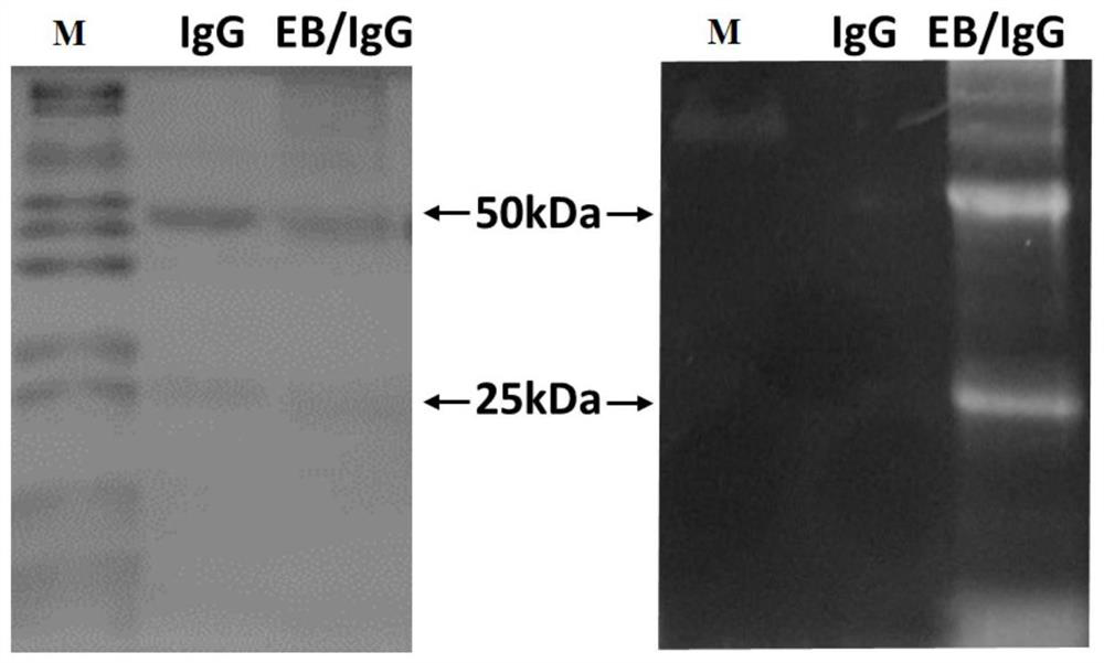
[0046] Example 2 Identification of Phycoerythrin B-Immunoglobulin G Complex

[0047] Take immunoglobulin G and phycoerythrin B-immunoglobulin G, boil with bromophenol blue loading buffer at 95°C for 10 minutes to denature, and after cooling, go through sodium dodecyl sulfate-polyacrylamide gel electrophoresis to treat bromophenol The blue reaches the bottom and stops, stained in the rapid Coomassie brilliant blue staining solution for 30-60min, decolorized using Syngene GBOX chemiluminescent imager and BlueLight Gel Imager to shoot the glue.

[0048] Such as figure 1 As shown, the band of immunoglobulin G connected with phycoerythrin B is close to that of immunoglobulin G under white light conditions, while under the irradiation of ultraviolet light with a wavelength of 340 nm, the light chain of phycoerythrin B-immunoglobulin G complex and The heavy chain band showed yellow-green fluorescence, indicating that Phycoerythrin B was successfully attached to IgG. It should be un...

Embodiment 3

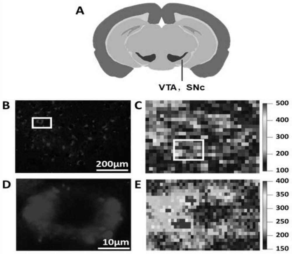
[0050] The preparation of embodiment 3 tissue sample

[0051] The ventricle was perfused with 4% paraformaldehyde and dehydrated with sucrose, and a 30 μm thick section was cut out using a Leica CM1950 microtome. The tissue section sample was placed at the bottom of a 24-well plate, and incubated with the primary antibody of the target neuron at 4°C for 18 hours. ~ 24 hours, and then co-incubated with the phycoerythrin B-immunoglobulin G complex in Example 1 for 2 hours at room temperature, sucked off the complex solution and washed, pasted the tissue on the mylar membrane, at room temperature Air dry under dark conditions.


PUM


Abstract

The invention discloses an X-ray fluorescence and fluorescence bimodal imaging probe based on a synchrotron radiation light source as well as a preparation method and application of the X-ray fluorescence and fluorescence bimodal imaging probe. The bimodal imaging probe is a phycoerythrin B-immune globulin G compound and is formed by covalently coupling tyrosine amino of immune globulin G and activated carboxyl of phycoerythrin B. The phycoerythrin B-immune globulin G compound has a fluorescence signal and an X-ray fluorescence signal at the same time. Aiming at the problem that a specific synchrotron radiation X-ray fluorescence imaging probe is not available in the prior art, the invention creatively provides the phycoerythrin B-immune globulin G compound and the preparation method thereof for the first time, and provides a target neuron bimodal imaging method. The invention has important significance and application value for neuron imaging and research of synchrotron radiation light sources in the field of biomedicine.

Description

technical field [0001] The invention relates to the technical field of biological imaging, and more specifically relates to a dual-mode imaging probe based on X-ray fluorescence and fluorescence of a synchrotron radiation light source, a preparation method and an application. Background technique [0002] The development of brain imaging technology and methods plays a very important role in studying the fine structure of the brain and understanding how the brain works. Fluorescence imaging is widely used because of its convenience and submicron resolution, but due to the scattering and absorption of tissues, its Imaging depth and coverage are limited, while X-rays have a good penetration depth, can provide excellent energy resolution, and allow fine spectral identification, which enables highly specific imaging of biological targets. In particular, synchrotron radiation X-ray imaging technology has become an important means of biomedical imaging. However, there is currently...

Claims


Application Information

Patent Timeline
Patent Type & Authority Applications(China)
IPC IPC(8): A61K49/04A61K49/00
CPCA61K49/04A61K49/0058A61K49/0043A61K49/0002Y02A50/30
Inventor 王丽华樊春海孙艳红闫美玲诸颖
Owner SHANGHAI ADVANCED RES INST CHINESE ACADEMY OF SCI
Features
  • R&D
  • Intellectual Property
  • Life Sciences
  • Materials
  • Tech Scout
  • Unparalleled Data Quality
  • Higher Quality Content
  • 60% Fewer Hallucinations
Learn More