Reagent strip for determining antibody and antigen of swine circular virus II
A porcine circovirus and antibody detection technology, which is applied in the field of rapid diagnostic equipment for livestock diseases, can solve the problems of complex test operation and long time consumption, and achieve broad market prospects, simple and fast operation, and good antigenicity
- Summary
- Abstract
- Description
- Claims
- Application Information
AI Technical Summary
Problems solved by technology
Method used
Image
Examples
preparation example Construction
[0026] (4) Preparation of gold standard glass wool
[0027] Prepare gold sol by sodium citrate reduction method, that is, add 2-4ml 0.5-2% trisodium citrate solution to boiling 50-100ml 0.01-0.05% chloroauric acid aqueous solution to obtain colloidal gold with a diameter of about 15nm. With 0.1mol / L K 2 CO 3 Adjust the pH of the colloidal gold to 8.5-9.5, and add the anti-PCV2 dCap monoclonal antibody or PCV2 dCap recombinant protein to be labeled into the gold sol at pH 8.5-9.5 at a labeling ratio of 1:1000-1:1300. After labeling for 10 minutes, add 20% PEG10000 to a final concentration of 0.05%, centrifuged at 1500-3000 r / min at 4°C for 20 min to remove unbound gold particles, centrifuged at 12,000 r / min at 4°C for 1 h, discarded the supernatant, and obtained the preliminary purified gold-labeled protein mixture, then used propylene glucose Glycan S-400 column chromatography, separation and purification of gold-labeled protein, to obtain colloidal gold-labeled anti-PCV2 dC...
Embodiment 1
[0036] Example 1 PCV2 antigen detection test strip
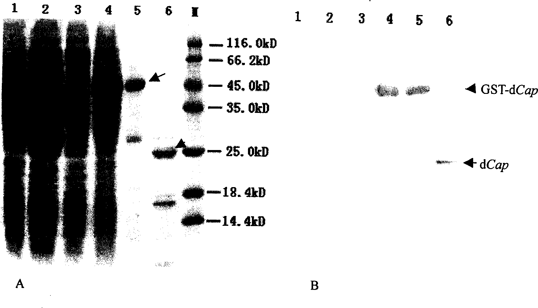
[0037] see figure 1 , figure 2 , prepare recombinant PCV2 dCap protein by (1) method step in the embodiment, prepare anti-PCV2 dCap monoclonal antibody mAb1 and mAb2 by (2) method step in the embodiment, prepare goat anti-mouse by (3) method step in the embodiment IgG polyclonal antibody, prepare gold-labeled glass wool according to the method steps in (4) in the embodiment, and finally, assemble various components into PCV2 antigen detection test strips, among the figures, 1 is the support layer, made of plastic sheet strips , 2 is the fiber layer at the sample end, made of glass wool, 3 is the fiber layer that absorbs the gold-labeled anti-PCV2 dCap monoclonal antibody, that is, the glass wool that absorbs the gold-labeled protein, referred to as the gold-labeled antibody cotton, and 4 is the cellulose membrane layer, the present embodiment adopts nitrocellulose membrane, and 5 is a water-absorbing material layer, that ...
Embodiment 2
[0039] Example 2 PCV2 antibody detection test strip
[0040] see figure 1 , figure 2, preparation of recombinant PCV2 dCap protein, anti-PCV2 dCap monoclonal antibody mAb1, the same as the examples, not repeated. Prepare goat anti-pig IgG polyclonal antibody by (3) method step in the embodiment, prepare gold-labeled glass wool by (4) method step in the embodiment, finally, various components are assembled into PCV2 antibody detection test strip, Fig. Among them, 1, 2, 4, 5, 8, 9, and 10 are the same as in Embodiment 3, and will not be repeated. 3,6,7 are different from the third embodiment. In this example, 3 is the glass wool that adsorbs the gold-labeled PCV2 dCap recombinant protein, 6 is the control blot of the anti-PCV2 dCap monoclonal antibody mAb1, and 7 is the detection blot of the goat anti-pig IgG polyclonal antibody.
[0041] When PCV2 antibody detection test strips are used to detect the PCV2 antibody level in pig serum, the blood of the pig to be tested is co...
PUM
 Abstract
Description
Claims
Application Information
 - R&D
- Intellectual Property
- Life Sciences
- Materials
- Tech Scout
- Unparalleled Data Quality
- Higher Quality Content
- 60% Fewer Hallucinations
Browse by: Latest US Patents, China's latest patents, Technical Efficacy Thesaurus, Application Domain, Technology Topic, Popular Technical Reports.
© 2025 PatSnap. All rights reserved.Legal|Privacy policy|Modern Slavery Act Transparency Statement|Sitemap|About US| Contact US: help@patsnap.com


