
Methods for producing protein domains and analyzing three dimensional structures of proteins by using said domains

a technology of three-dimensional structure and protein, which is applied in the field of producing protein domains and analyzing three-dimensional structure of proteins by using said domains, can solve the problems of difficult to obtain proteins which are toxic to host cells and/or degrade easily, difficult to obtain proteins, and difficult to maintain the integrity of three-dimensional structure and functions in vivo. achieve the effect of rapid and easy method

Inactive Publication Date: 2002-10-03
RIKEN
View PDF0 Cites 18 Cited by
  • Summary
  • Abstract
  • Description
  • Claims
  • Application Information

AI Technical Summary

Benefits of technology

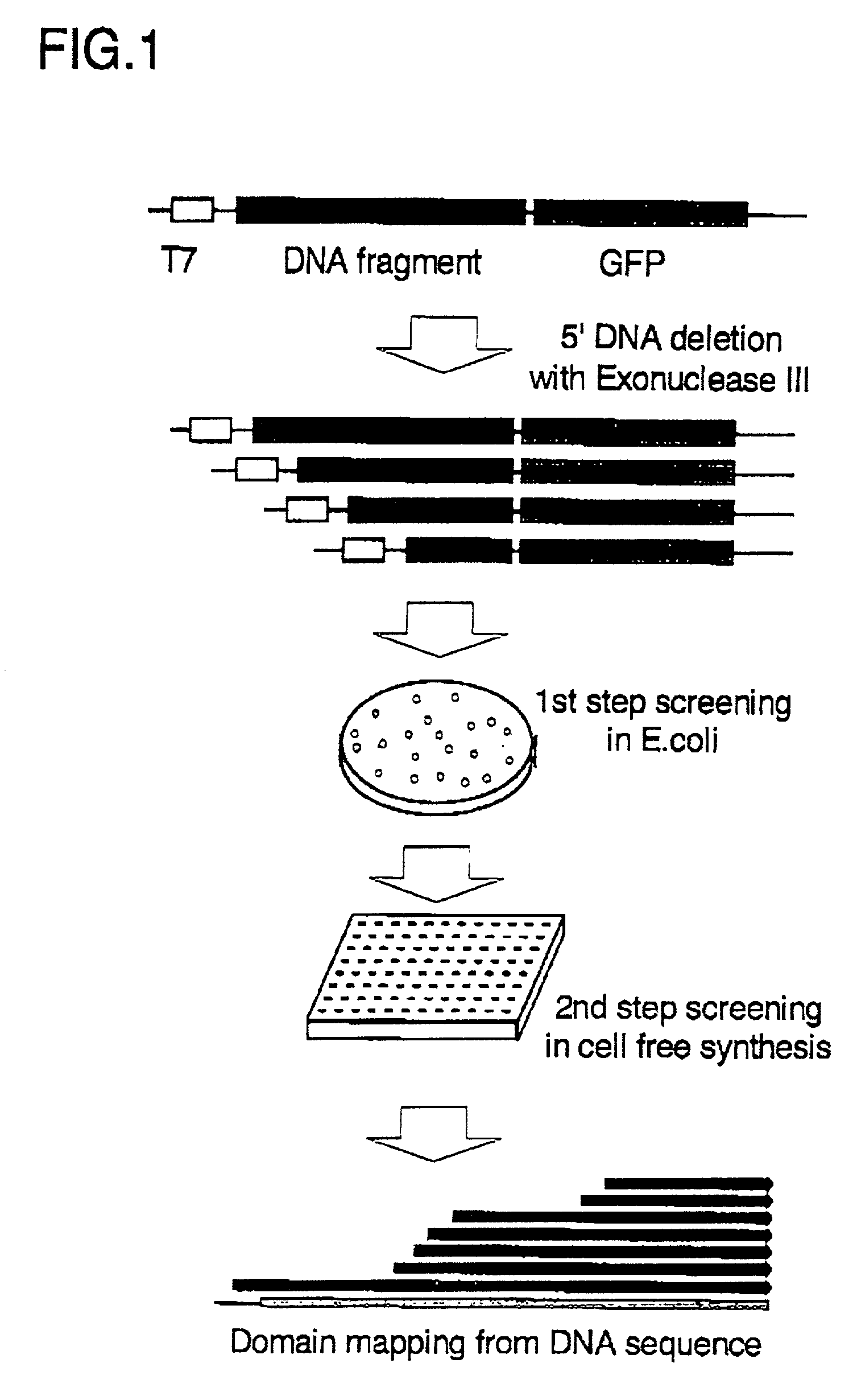
[0010] It is an object of the present invention to provide a rapid and easy method to select highly soluble protein domains suitable for three dimensional structural analysis.
[0039] There are several methods to fragment DNA and construct a DNA library of various lengths. We can classify them in two typical methods, out-vector fragmentation and in-vector fragmentation. In the out-vector fragmentation method, various lengths of DNA fragments are made at first, and then the fragments are cloned into an expression vector. In the in-vector fragmentation method, the DNA previously cloned into an expression vector is deleted from the fixed places in the vector to make various lengths of DNA fragments. With regard to the methods of the present invention, both of the above methods can be used, however, the method of in-vector fragmentation is more preferable. When a target DNA is randomly fragmented out of vector, deleted fragments should be re-ligated to the expression vector. This re-ligation leads the library to incline toward shorter fragments, for small fragments tend to be easily ligated. The ligation efficiency of blunt-end fragments is low compared to in-vector self-ligation. Because there is no definite method for random fragments with blunt ends to be ligated in the selected direction the number of effective fragments in the library, ligated in the right direction to a functional protein, automatically decreases to half. On the other hand, when DNA is fragmented in vector, the homogeneity of the deletion library is ensured, and self-ligations make it possible to efficiently make correctly ligated clones.
[0045] In the present invention, it is especially preferable to use Green fluorescent protein (GFP). GFP is a fluorescent protein isolated from the Pacific jellyfish, Aequorea victoria and the like. Its role is to transduce, by energy transfer, the blue chemiluminescence of another protein, aequorin which is a luminescent protein co-localized with GFP, into green fluorescent light (Prasher, D., et al., Gene 11, 229-233. (1992)). In contrast to said aequorin and luciferase, GFP does not require the specific fluorophore, and forms a fluorophore (chromophore) automatically on its own apoprotein. For this reason, to use as indices of gene expressions, GFP has been expressed in bacteria, yeast, slime mold, plants, insects, and in mammalian cells using the cloned GFP cDNA. Furthermore, mutations have been introduced into the GFP gene by various methods to make various GFP variants. It has been reported that, among the variants, some show stronger fluorescent intensities than the wild type GFP, and others which radiate fluorescence at the different wave lengths (Ehrig, T., O'kane, D., and Prendergast, F. FEBS Lett., 367, 163-166. (1995); Crameri, A., Whitehom, E., Tate, E., and Stemmer, W., Nature Biotech., 14, 315-319. (1996)). In this invention, these GFPs or the variants can be used as a functional protein to construct rapid and easy screening systems.
[0053] Domains of proteins selected in this manner can be synthesized efficiently through cell-free systems. In cell-free systems, because PCR products can be used directly as templates for expressions, simultaneous treatments of multiple samples and additions of various expression tags can be easily done.

Problems solved by technology

In the existing methods of protein synthesis, genetic engineering methods in which full length genes cloned from cDNA or a genomic library are introduced into living cells such as E. coli have been widely used, however, it is difficult to obtain proteins which are toxic to host cells and / or degrade easily because of instability.
It is also difficult to obtain proteins which aggregate easily in host cells as soluble proteins.
In the case where these proteins are expressed in an intact form in heterogeneous cells, they are likely to be insoluble, and it is difficult to maintain their intact three dimensional structures and functions in vivo.
However, in the case of particular genes, the soluble protein products cannot be obtained by any of these methods.
In general, these specialized approaches geared toward evaluating individual proteins have not been sufficient as methods for high-throughput analyses of three dimensional structures of many proteins, as well as for systematic understandings of structures and functions of the proteins.

Method used


Image

  • Methods for producing protein domains and analyzing three dimensional structures of proteins by using said domains
  • Methods for producing protein domains and analyzing three dimensional structures of proteins by using said domains
  • Methods for producing protein domains and analyzing three dimensional structures of proteins by using said domains

Examples


example 1

Construction of Plasmids and Introduction of Mutations

[0060] pGFPuv (Clontech), used as the reporter gene, was mutated at three sites using a site-directed mutagenesis kit (Stratagene). Replacement of phenylalanine at the 64.sup.th residue to leucine (F64L) and serine at the 65.sup.th residue to threonine (S65T) are the mutations to obtain red-shifted excitation peak and fluorescence about 35 times more intensely than wild type GFP when excited at 488 nm. These mutations also display improved solubility and the more efficient folding of proteins (Cormack, B. P., Valdivia, R. H., Falkow, S., Gene 173, 33-38. (1996)). The other mutation is a silent mutation to terminate the NdeI restriction site.

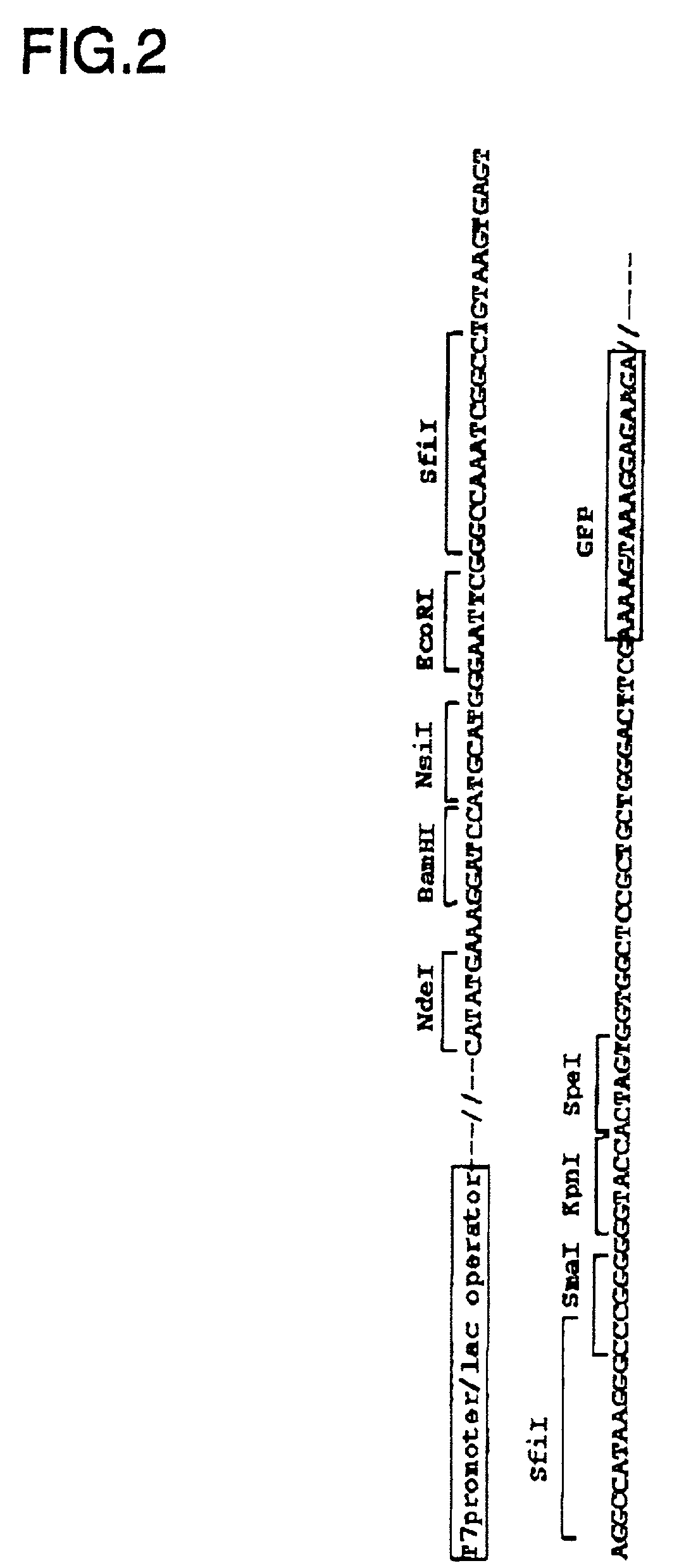
[0061] To produce GFP-fusion proteins ligated to the C-terminus of proteins coded on the inserted DNA, an expression vector has been constructed comprising a T7 promoter, the DNA insertion site, the mutant GFP gene, and a T7 terminator sequence. One plasmid constructed in this way, pGFPsfiI, h...

example 2

Construction of Deletion Library

[0062] Grb2 cDNA (without stop codon) was ligated to the SfiI site of the plasmid pGFPsfiI in the correct direction and frame to C-terminal GFP. Then, after digesting the N-terminal side of the inserted DNA with EcoRI or a NsiI, the inserted DNA was deleted with Exonuclease III from the 5' end, the region of single stranded DNA was digested with Mung-bean nuclease. Finally, the blunt ends were made using DNA polymerase, Klenow fragment. The lengths of deleted DNA were selected by electrophoresis and the size selected DNA was self-ligated for transformation of E. coli JM109(DE3) strain (Promega) to prepare the deletion library containing Grb2 cDNA.

example 3

Protein Expression

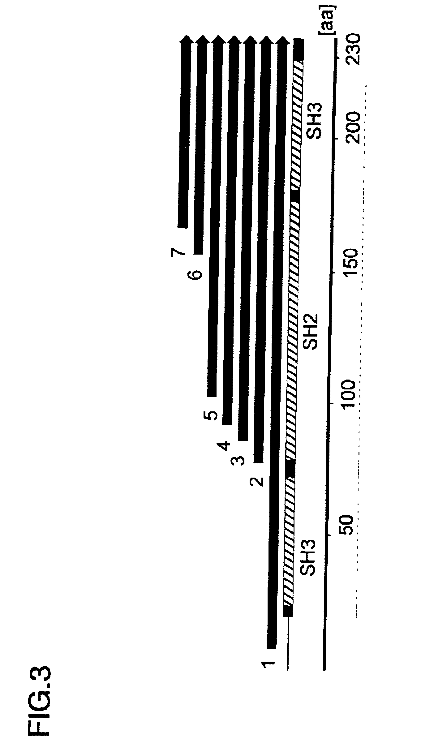
[0063] For the first screening step, the deletion library of GFP-fusion vectors prepared in Example 2 was transformed in JM109(DE3) (Promega) and cultured at 37.degree. C. over night. The fluorescence of the derived colonies was observed by excitation with a Dark Reader (BM Science) at 420-500 nm. When roughly observed with the excitation of the blue light (420-500nm), the obtained colonies could be classified into 3 categories on the basis of fluorescent intensity: strong, medium, and none. Among them, each 8 colonies of those emitting strong fluorescence, those emitting medium fluorescence, and those emitting no fluorescence were selected. And, base sequences of the total 24 clones were determined to identify the deletion sites. Most of the clones emitting strong fluorescence had relatively short (shorter than 35 amino acid residues) protein fragments of Grb2. This result suggests that the folding properness of the short protein fragment, with several tens of ami...


PUM

PropertyMeasurementUnit
Structureaaaaaaaaaa
Solubility (mass)aaaaaaaaaa
Fluorescenceaaaaaaaaaa

Abstract

There is provided a method for producing a soluble protein domain comprising: (a) preparing two or more DNA fragments by partially digesting a DNA coding for a protein; (b) expressing the protein which is coded on each of said DNA fragments, as a fusion protein with a functional protein; (c) selecting the fusion protein exhibiting said function among two or more fusion proteins synthesized in step (b); and, (d) synthesizing the soluble protein domain which is coded on said DNA fragment in a cell-free system, wherein said soluble protein domain is included in said fusion protein selected in step (c). By using this method, it can be easy and efficient to analyze the three dimensional structure of proteins of many clones.

Description

[0001] This application claims priority to Japanese Patent Application No. 2001-062703 filed on Mar. 6, 2001.[0002] The present invention relates to a method for producing a soluble protein domain and for analyzing a three dimensional structure of a protein. More specifically, the present invention relates to a method for selecting a basic unit of protein which is called a "fold" or "domain," followed by synthesis of said selected protein domain using cell-free systems, to a protein domain synthesized by this method, and to a method for analyzing the three dimensional structure of proteins by using said protein domain.[0003] Recently, DNA sequences of various species have been determined rapidly, and "structural genomics" is recognized as an important research area. With respect to a large number of genes selected from a mass of information about genomic sequences, structural genomics aims the systematic determination of three dimensional structures of proteins coded on each gene, a...

Claims


Application Information

Patent Timeline
IPC IPC(8): G01N23/20C07K14/00C07K14/72C07K19/00C12N1/21C12N15/09C12P21/02C12R1/19G01N24/08G01N33/68
CPCC07K2299/00C07K14/72
Inventor SEKI, EIKOKIGAWA, TAKANORIYOKOYAMA, SHIGEYUKI
Owner RIKEN
Learn More