Detection of mycobacteria

a technology of mycobacteria and detection method, which is applied in the field of detection of mycobacteria, can solve the problems of limited antigen detection assays for direct diagnosis of tuberculosis, low detection thresholds, and inability to identify subjects with latent infection, and achieve the effect of low detection thresholds

Inactive Publication Date: 2003-02-20
WALLIS ROBERT S
View PDF1 Cites 3 Cited by
  • Summary
  • Abstract
  • Description
  • Claims
  • Application Information

AI Technical Summary

Benefits of technology

[0107] In the method of the present invention, a sandwich ELISA (enzyme-linked immuosorbent assay) with a monoclonal antibody for capture and a secondary antibody for detection of bound monoclonal antibody is used. In a preferred embodiment, the signal from the assay is amplified using an recycling enzymatic method, which increases the signal over 500 fold. Special measures are included in order to reduce background noise. The assay offers numerous advantages as compared to existing methods for tuberculosis diagnosis. First, body fluids remote from the site of infection can be tested using the method of the present invention. The test can be used with infected body fluids, as well as urine and blood. Although urine and blood usually do not contain M. tuberculosis, these sites may contain secreted products of the infection. Detection of these secreted products provides a clear advantage in attempts to diagnose a localized infection such as is usually the case in tuberculosis. It is a particularly important advantage in diagnosis of tuberculosis in children, from whom quality sputum specimens are difficult to collect and are generally not available.
[0112] The present invention provides numerous advantages over the methods previously and currently in use to detect infection and / or disease with M. tuberculosis or MAC. For example, all of the previously reported tuberculosis antigen detection assays were applied only to infected fluids or to early cultures, the assays were considerably less sensitive than this assay, and in addition, relied upon an animal antiserum developed against a whole organism of another mycobacterial species (BCG) for detection, resulting in poor specificity. Furthermore, none of the previously described assays were applied to urine and blood samples.
[0114] Also, unlike the previously described methods, the present invention can be used for early monitoring of anti-tuberculosis therapy, providing a useful tool in predicting which individuals might experience treatment failure with a particular regimen. Thus, the present invention can also be used in the rapid evaluation of new anti-tuberculosis therapies.
[0115] Importantly for clinical diagnosis and public health tuberculosis clinics, the method of the present invention is rapid, requiring only 1-2 days to complete, as opposed to a minimum of 2-4 weeks for culture. Furthermore, the present invention can be performed during a single visit to the clinic, unlike the routine skin testing methods which require a return visit in order to observe and record the skin test results. Also importantly, the present invention provides an inexpensive method that is within the capabilities of existing clinical and public health mycobacteria laboratories. This is generally not true for methods that require specialized equipment and training (e.g., the polymerase chain reaction [PCR]). The test is very flexible, permitting the detection of various antigens or alpha antigen epitopes. The assay can be used to detect all secreted mycobacterial antigens by substituting a non-specific binding method for the capture antibody.
[0117] In contrast with the previous studies, the present invention provides a more sensitive method for the reliable detection of infection and / or disease due to M. tuberculosis or MAC, based on the detection of mycobacterial products, rather than the host response to the mycobacteria.
[0142] The present invention provides compositions and methods for the rapid detection of mycobacterial antigens in such samples as blood, serum, urine, sputum, and culture filtrates. Therefore, this invention provides significant advantages in the diagnosis, monitoring, and treatment of patients with infections and / or disease with such organisms as M. tuberculosis and MAC.

Problems solved by technology

Previous reports of antigen detection assays for direct diagnosis of tuberculosis have been limited to examination of fluids obtained from the site of clinical disease, such as cerebrospinal fluid, sputum, or bronchoalveolar lavage fluid.
In addition, the lower detection thresholds were only achieved in those assay which used a polyclonal antiserum for capture.
None of these assays were able to identify subjects with latent infection.

Method used


Image

  • Detection of mycobacteria
  • Detection of mycobacteria
  • Detection of mycobacteria

Examples


example 2

Production of Murine Monoclonal Antibody

[0154] The monoclonal antibody designated as TBC27 was produced for use in the ELISA method of the present invention. This monoclonal was developed by immunization with M. tuberculosis alpha antigen obtained using the methods of Example 1. Six week-old female Balb / c mice (Charles River, Mass.) were immunized with 50 .mu.g alpha antigen in incomplete Freund's adjuvant (DIFCO, Detroit, Mich.). Twice, animals were boosted with additional 50 .mu.g of antigen at two week intervals, and sacrificed 2 weeks after the last boost. Spleen cells were fused to SP2 / 0 Ag 14 myeloma cells (ATCC) using polyethylene glycol (Sigma), and were propagated in Dulbecco's modified Eagle' medium (Sigma) with added hypoxanthine, aminopterin, and thymidine. Supernatants of wells with visible growth were tested for antibody by ELISA. The wells of Immulon 2 plates (PGC) were sensitized overnight (i.e., 12-18 hours), with 50 .mu.l M. tuberculosis filtrate (10 .mu.g / ml), and...

example 3

Detection of Alpha Antigen in M. tuberculosis Culture Filtrates

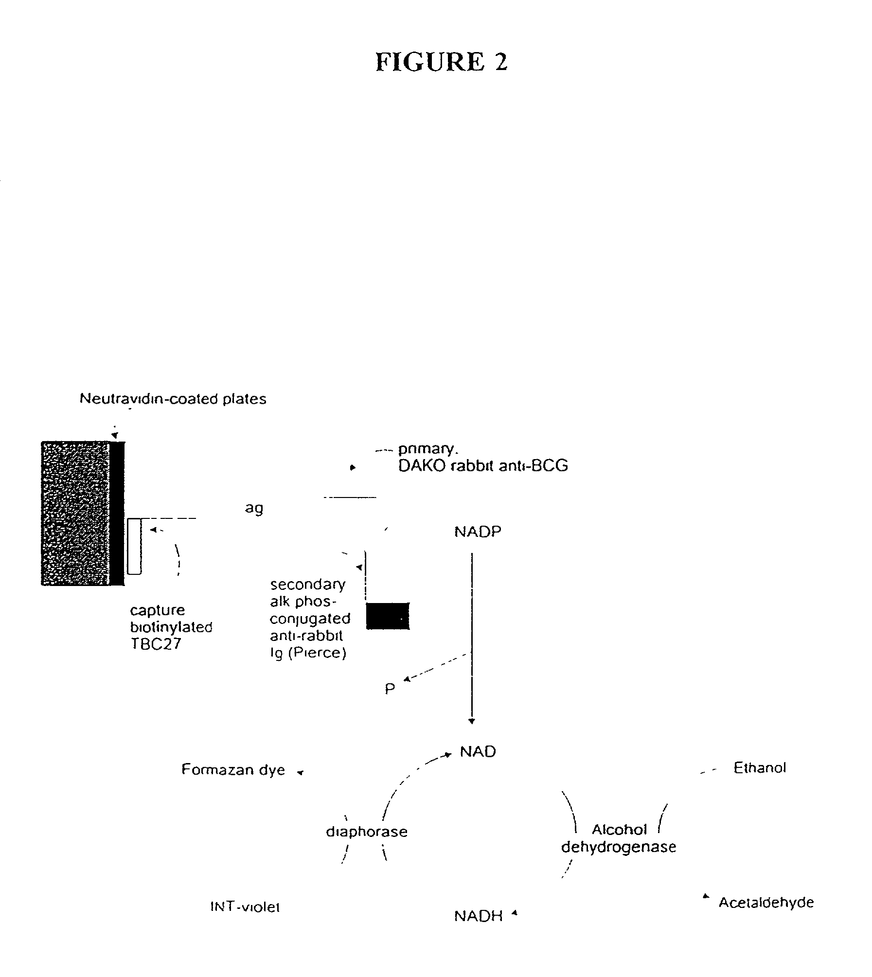
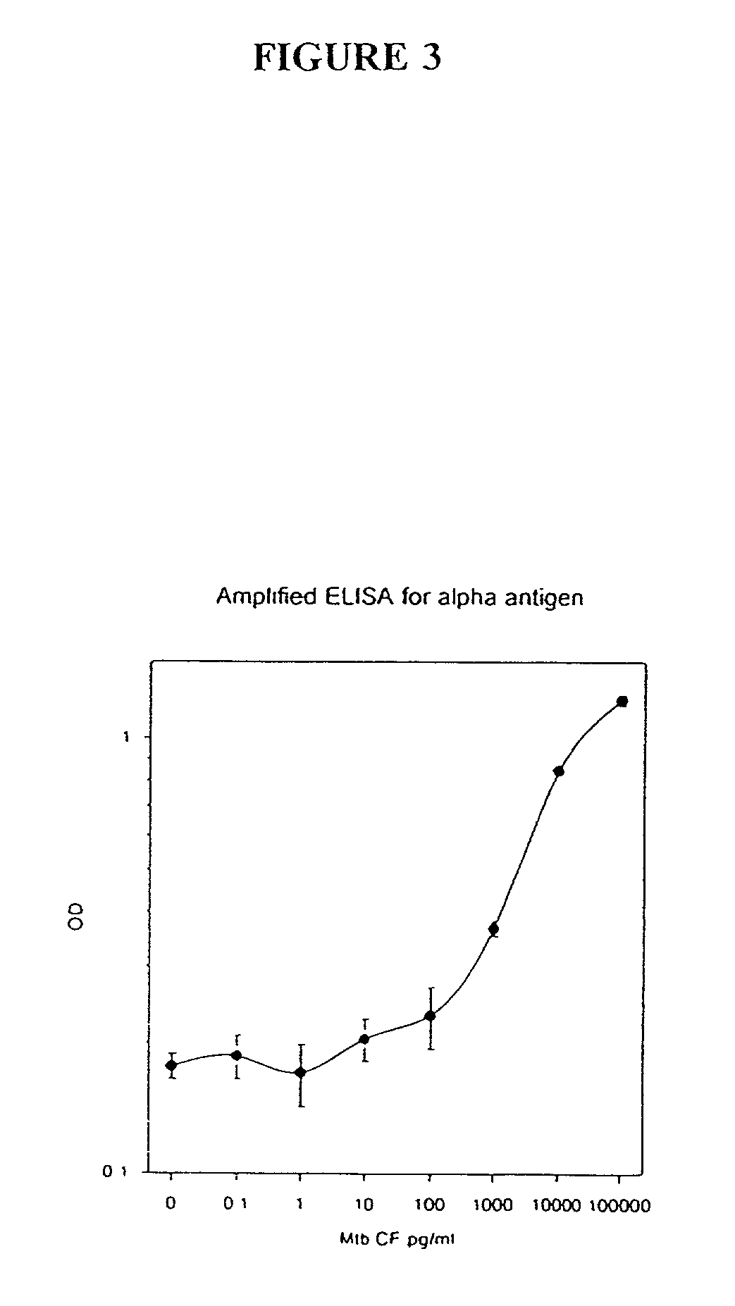
[0156] In this Example, an ELISA method (C. J. Stanley et al., "Enzyme amplification can enhance both the speed and the sensitivity of immunoassays," J. Immunol. Meth., 83:89-95 [1985]; and C. H. Self, "Enzyme amplification--a general method applied to provide an immunoassisted assay for placental alkaline phosphatase," J. Immunol. Meth., 76:389-93 [1985]) that substantially increases the speed and sensitivity of ELISA, was extensively modified for use for the detection of alpha antigen, including the addition of another step. In this method, an alkaline phosphatase conjugate was used to convert NADP to NAD, which then entered a regenerating cycle leading to production of a colored formazan dye. A schematic of the reaction is shown in FIG. 2. Its use in the alpha antigen assay has resulted in over a 1000 fold reduction in the lower limit of detection of the assay (i.e., from 20 ng / ml, to from 1 to 10 pg / ml). The samples ...

example 4

Recovery of M. tuberculosis Alpha Antigen in Spiked Samples

[0162] In this Example, the same methods as described in Example 3 were used for testing of serum or urine, with the exception that M. tuberculosis culture filtrates were added to normal pooled normal human serum or urine, as described above. This experiment was conducted in order to determine whether alpha antigen from M. tuberculosis filtrate could be detected in such samples. The results of this experiment are shown in FIG. 4. As can be seen from FIG. 4, the sensitivity of the assay was not substantially affected by either diluent (i.e., normal serum or urine). However, the study suggests that separate diluents for standards be used, depending upon the type of clinical sample.


PUM

PropertyMeasurementUnit
volumeaaaaaaaaaa
volumeaaaaaaaaaa
poweraaaaaaaaaa

Abstract

The present invention is directed to methods and compositions for the detection of infection and disease due to members of the genus Mycobacterium. In particular, the present invention is well-suited to the detection and identification of patients with disease or infection due to M. tuberculosis or MAC.

Description

[0001] The present invention relates to the detection and diagnosis of infection and disease due to the mycobacteria, especially Mycobacterium tuberculosis and other mycobacteria commonly associated with disease in immunocompromised patients including those with acquired immunodeficiency syndrome (AIDS). The present invention is suited for rapid screening of large populations for the presence of M. tuberculosis carriers, as well as diagnosing and monitoring disease or infection in patients who present at healthcare or public health facilities.[0002] Organisms within the genus Mycobacterium include obligate parasites, saprophytes, and opportunistic pathogens. Most species are free-living in soil and water, but for species such as M. tuberculosis and M. leprae, the causative agents of tuberculosis and leprosy respectively, the major ecological niche is the tissue of humans and other warm-blooded animals.[0003] Despite the fact that most mycobacteria do not cause disease, a relatively ...

Claims


Application Information

Patent Timeline
Patent Type & Authority Applications(United States)
IPC IPC(8): G01N33/569
CPCG01N33/5695
Inventor WALLIS, ROBERT S.
Owner WALLIS ROBERT S
Why Eureka
Try Eureka