
Method of purifiying nucleic acid using nonwoven fabric and detection method

a nucleic acid and nonwoven fabric technology, applied in the direction of nucleic acid reduction, microorganisms, organic chemistry, etc., can solve the problems of requiring centrifugation, requiring more time and trouble, and requiring a large amount of time and trouble, so as to achieve a rapid and simple method of preparing nucleic acids, simple and high yield

Inactive Publication Date: 2005-02-17
ASAHI KASEI KK
View PDF8 Cites 28 Cited by
  • Summary
  • Abstract
  • Description
  • Claims
  • Application Information

AI Technical Summary

Benefits of technology

[0021] It is an object of the present invention to provide a method of purifying cellular nucleic acids from a sample containing blood cells or the like in a simpler and higher yield manner than prior art techniques. It is another object of the invention to provide a rapid and simple method of preparing nucleic acids which can be used for conventional nucleic acid amplification techniques or nucleic acid sequence analysis techniques.

Problems solved by technology

Although this method is simpler than the phenol method, it has a disadvantage of entailing numerous steps and requiring a centrifugation procedure.
Furthermore, because it uses a chaotropic agent or ethanol which strongly inhibit PCR and other enzyme reactions, it has a disadvantage of a necessity of thoroughly removing such substances through complicated and time-consuming procedures.
However the method described for purification of the nucleic acid after cell lysis is a prior art method, and a new method is not disclosed therein.
Such a method of preparing nucleic acid from peripheral blood leukocytes using cell-adsorbing fiber aggregates as leukocyte separation filters has been known, but these methods had a disadvantage that after the disclosed filtering method employed up to the steps of leukocyte separation or nucleic acid extraction, the nucleic acid must be purified by existing nucleic acid purification methods, thereby complicating the procedure and requiring more time and trouble.
It is stated that characteristic features of the filters suitable for this method are that it is impossible to capture a purified DNA when it passes through the filters, and that when lysed cells are passed through filters, the DNA yield is reduced by 80% and thus it is not practical.
Such methods wherein purification is carried out after adsorbing the cells onto the filter also have a drawback that the filter must be selected according to the type of cell.
Otherwise, the viscosity is too high to allow filtration with the filter.
This method has a drawback that an alcohol or a chaotropic agent must be added during adsorption in order to increase the yield of the nucleic acid.
Nucleic acid preparation methods are commonly known, but elution is time consuming and requires setting of suitable elution conditions.
These methods have drawbacks because they require labor for preparation of the dry solid medium and are limited in the volumes of samples that can be added to the dry solid medium, thereby reducing the density of nucleic acid that can be immobilized on the medium, while organic solvents such as phenol or alcohol must be used to remove the protein including hemoglobin, and the procedure is thus complicated and time consuming.
The drawbacks of this method, however, include a necessity of using a chaotropic agent and organic solvent, the need to completely remove the chaotropic agent and ethanol, which strongly inhibit the PCR reaction, and the resulting complex and time consuming operations.
In addition, because the filter is normally used for blotting, it is not suited for the purpose of purifying nucleic acid from biological substances such as blood.
The disadvantages of this method are that because the cells must be bound first to the solid, the solid must therefore be selected to match the type of cell, and that there is a restriction on the flow rate for filtration, because the cells must also be bound without disrupting them.

Method used


Image

  • Method of purifiying nucleic acid using nonwoven fabric and detection method
  • Method of purifiying nucleic acid using nonwoven fabric and detection method
  • Method of purifiying nucleic acid using nonwoven fabric and detection method

Examples


example 1

[0201] Purification of Nucleic Acid from Fresh Human Blood with Nonwoven Fabric

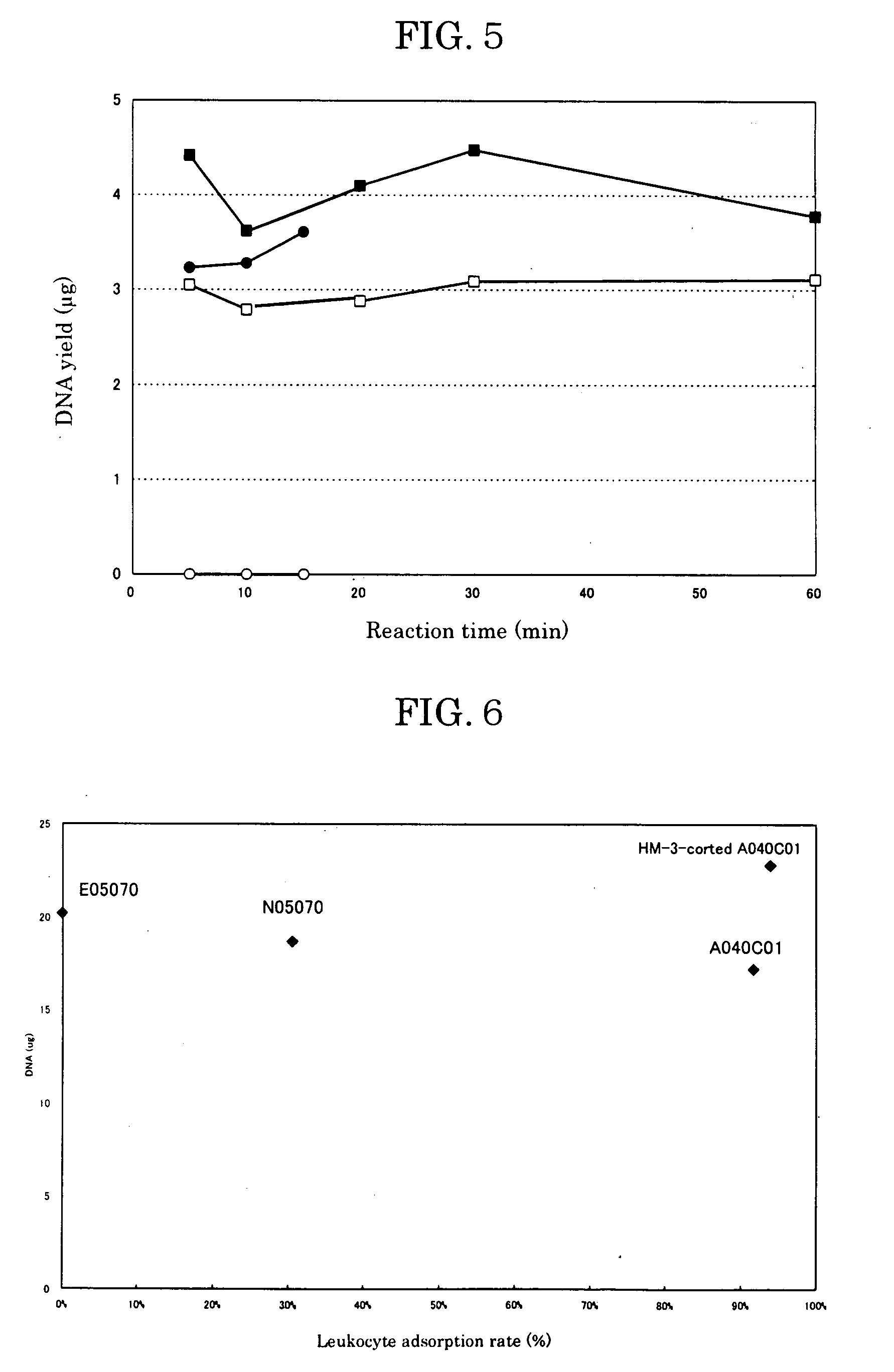
[0202] Blood was taken from a healthy donor and heparin sodium (Shimizu Pharmaceutical) was added as an anticoagulant at 10 units per milliliter of blood. The leukocyte count of the blood was measured with a flow cytometer (FACS Calibur, Becton Dickinson) using a LeucoCOUNT Kit (Becton Dickinson). The leukocyte count in 0.25 ml of blood was 1.16×10⁶. The nonwoven fabric used was a product by Asahi Kasei Corp. The nonwoven fabric was cut into 12 mm-diameter disks, four of which were stacked and set in a filter holder (SWINNEX, MILLIPORE), with a 10 ml glass syringe set upstream and a suction pump set downstream in connection with the filter holder. The nonwoven fabric was initially washed with 3 ml of Digestion Buffer (10 mM Tris, pH 8; 100 mM NaCl; 25 mM EDTA; 0.5% SDS).

[0203] Next, 0.05 μg of Proteinase K (PCR-Grade, Roche) was added to 0.25 ml of the human blood, and after further adding 0.25 ml of 2×...

example 2

[0206] Analysis of Purified Human Nucleic Acid

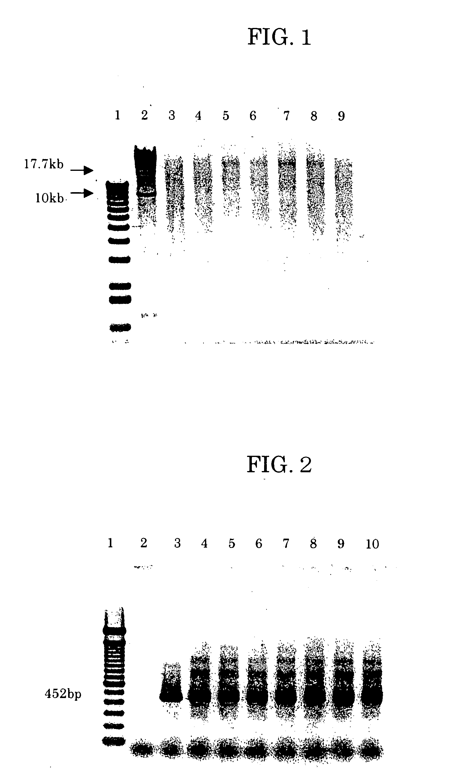
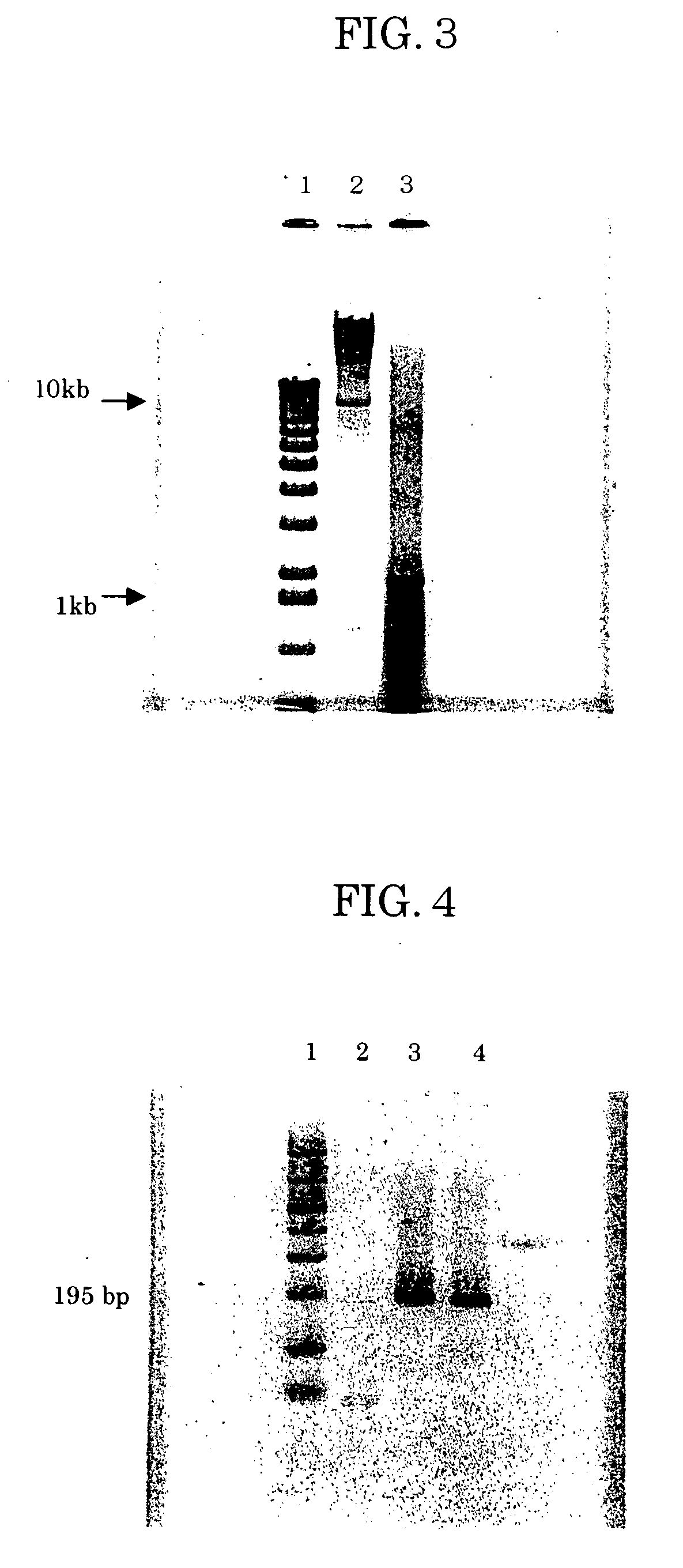
[0207] The purified nucleic acid obtained in Example 1 was subjected to 0.7% agarose electrophoresis and the sizes were confirmed. After adding 1.5 μl of 10×Loading Buffer (1% SDS, 50% glycerol, 0.05% Bromophenol Blue, TaKaRa) to 10 μl of the effluent of Example 1 and thoroughly mixing, the total amount was subjected to 0.7% agarose electrophoresis. After electrophoresis in a Mupid Minigel Migration Tank (Advance) at 50 V for 90 minutes, the gel was stained with ethidium bromide and photographed with a BioImage Gel Print 2000i / VGA. As shown in FIG. 1, the purified nucleic acid contained nucleic acid fragments of various sizes of from several kilobases to several tens of kilobases.

[0208] It was then confirmed that the purified DNA obtained in Example 1 could be used as a PCR template. A glyceraldehyde 3-Phosphate Dehydrogenase (G3PDH) 0.45 kb Control Amplimer Set by Clontech (Cat. No. 5405-3) was used for primers. The effluent of Exampl...

example 3

[0210] Purification of E. coli Nucleic Acid

[0211] HM-3-coated A040C01 (Asahi Kasei) was cut into 12 mm-diameter disks, four of which were stacked and set in a filter holder (SWINNEX, MILLIPORE), with a 10 ml glass syringe set upstream and a suction pump set downstream from the filter holder. The nonwoven fabric disks were initially washed with 3 ml of Digestion Buffer (10 mM Tris, pH 8; 100 mM NaCl; 25 mM EDTA; 0.5% SDS)

[0212] After adding 50 μl of E. coli DH5 glycerol stock to 3 ml of LB medium (1 g Tryptone Peptone (DIFCO); 0.5 g Yeast Extract (DIFCO); 1 g NaCl; 200 μl 1N NaOH; 100 ml distilled water), the mixture was cultured at 37° C. for 4.5 hours to obtain a culture solution with A600=1.56. The E. coli density based on absorbance was considered to be approximately 6.2×10⁹ cells / ml. A 1.6 ml portion of the culture solution (1010 E. coli cells) was taken and centrifuged at 15,000 rpm for 1 minute. The cell precipitate was suspended in 0.25 ml of LB medium. After adding 0.05 μg...


PUM

PropertyMeasurementUnit
temperatureaaaaaaaaaa
pHaaaaaaaaaa
pore sizeaaaaaaaaaa

Abstract

A method of separating and purifying nucleic acids from samples containing cells, such as blood and culture solutions. According to the method of the invention, a cell extract obtained by cell disruption is adsorbed by a filter made of a nonwoven fabric and the nucleic acid is eluted after washing the filter. Alkaline conditions of pH 12 or higher may be employed for elution of the nucleic acid, or the filter-adsorbed nucleic acid may be eluted by treatment with active oxygen or by using a surfactant. Nucleic acids separated and purified by the method of the invention can be used in nucleic acid amplification and nucleic acid sequence analysis techniques.

Description

TECHNICAL FIELD [0001] The present invention relates to a simple method of preparing nucleic acids at a high purity from cells using a nonwoven fabric, and to a preparation kit therefor. The invention further relates to a method of amplifying nucleic acid from a nonwoven fabric which has adsorbed the nucleic acid, to a method of detecting a nucleic acid sequence from a nonwoven fabric which has adsorbed the nucleic acid, and to a method and kit for preparation of the nonwoven fabric which has adsorbed the nucleic acid, to be used in the aforementioned method. BACKGROUND ART [0002] Nucleic acids, including DNA, are generally prepared from cells by treating a sample containing the cells with SDS or Proteinase K and then denaturing and removing the protein with phenol to purify the nucleic acid (Molecular Cloning 2nd Edition, 9.16-9.23, Cold Spring Harbor Laboratory Press, 1989). Because of the trouble and time required for this procedure, however, simpler methods are desired. [0003] A...

Claims


Application Information

Patent Timeline
Patent Type & Authority Applications(United States)
IPC IPC(8): C12N15/10
CPCC12N15/1017C12Q1/6806C12Q2523/308C12N15/09D04H13/00
Inventor KANNO, KIMIYOSHIODA, NAOZUMIARITOMI, MASAHARUSATO, AKIKO
Owner ASAHI KASEI KK
Learn More