Detection of cell membrane-associated proteins using membrane fragments displayed on encoded microparticle arrays

a technology of cell membrane and array, which is applied in the field of detection of cell membrane-associated proteins using membrane fragments displayed on encoded microparticle arrays, can solve the problems of predicting a high risk of graft rejection, adverse events or toxicities, certain drugs, etc., and achieves the effect of small size and increased assay accuracy

Inactive Publication Date: 2005-03-17
BIOARRAY SOLUTIONS
View PDF2 Cites 23 Cited by
  • Summary
  • Abstract
  • Description
  • Claims
  • Application Information

AI Technical Summary

Benefits of technology

To determine the percentage of reactive antibodies, membrane fragment arrays displaying a representative spectrum of antigens are contacted with patient sera. Following completion of the labeling step, the percentage of bead types within the array scoring positive is determined. In contrast to conventional methods of using allo-antibody typing trays which require placement of multiple aliquots of patient serum into each of multiple wells which contain cells from different individual cell lines, the READ™ format used herein completes the entire analysis on a single small aliquot of serum by performing a fully “multiplexed” analysis in a single reaction using a randem encoded array of membrane fragments. To increase the accuracy of the assay, antibodies purified from human serum could be used in the assay. The array optionally may contain additional molecular receptors of interest, such as a subarray of encoded bead-displayed auto-antigens. The entire array is small in size. In contrast to flow cytometric methods of analysis of the prior art, no separate step of analysis is required.

Problems solved by technology

If certain receptors are over-expressed, certain drugs may cause adverse events or toxicity.
Conversely, if certain receptors are under-expressed or completely absent, certain drugs may not be effective, and signal transduction may not occur.
The presence in a prospective transplant recipient of antibodies against donor HLA alleles, also known as a “donor-specific cross-match,” is predictive of a high risk of graft rejection.
In this “panel-reactive antibody” (PRA) testing, serum reactivity against a high percentage of HLA alleles present in a normal human population is predictive of a high risk of graft rejection.
This method is labor intensive, requires viable cells, may be nonspecific and requires a subjective evaluation of the cells.
In such cases, the incidence of false-negative results was higher for ELISA testing than for panel studies.
Moreover, when using flow cytometry with lymphocytes, use of ten or more different lymphocytes tends to produce confusing signals.
As a consequence, studies using lymphocytes have been limited to presenting a small panel of HLA antigens that do not adequately reflect the distribution of HLA antigens in a normal human population.
The methods of Sumitran-Karuppan are limited, however, because the pooling of antigens causes sensitivity to certain rare HLA antigens.
Moreover, the method is not capable of quantifying the relative amounts of PRA.

Method used


Image

  • Detection of cell membrane-associated proteins using membrane fragments displayed on encoded microparticle arrays
  • Detection of cell membrane-associated proteins using membrane fragments displayed on encoded microparticle arrays
  • Detection of cell membrane-associated proteins using membrane fragments displayed on encoded microparticle arrays

Examples


example i

Extraction of Membrane Proteins from Lymphocytes

Membranes were extracted from human peripheral blood lymphocytes (“PBLs”) and from human spleen cell preparations using the following procedure. First, the cell samples were placed in separate tubes and spun down at 14,000 g for 2 minutes. Next, the supernatant was collected and aliquots were suspended in 50 μl of 50% glycerol in 1×PBS. All the samples were frozen at −86° C. for later use.

A protease inhibitor cocktail (Signia P8340) was prepared as 100 times concentrated stock solution, and added to the following homogenization buffer, which was used to disrupt the cell membrane solution under conditions preserving their integrity. The cocktail had the formula: 250 mM sucrose 10 mM HEPES 1 mM EDTA 1 mM PMSF

Protease inhibitor cocktail (the foregoing was brought up to 10 ml with H2O).

50 μl of ice cold homogenization buffer was added to the cell pellets. A mortar and pestle was used to grind the cell pellets, and the pestle wa...

example ii

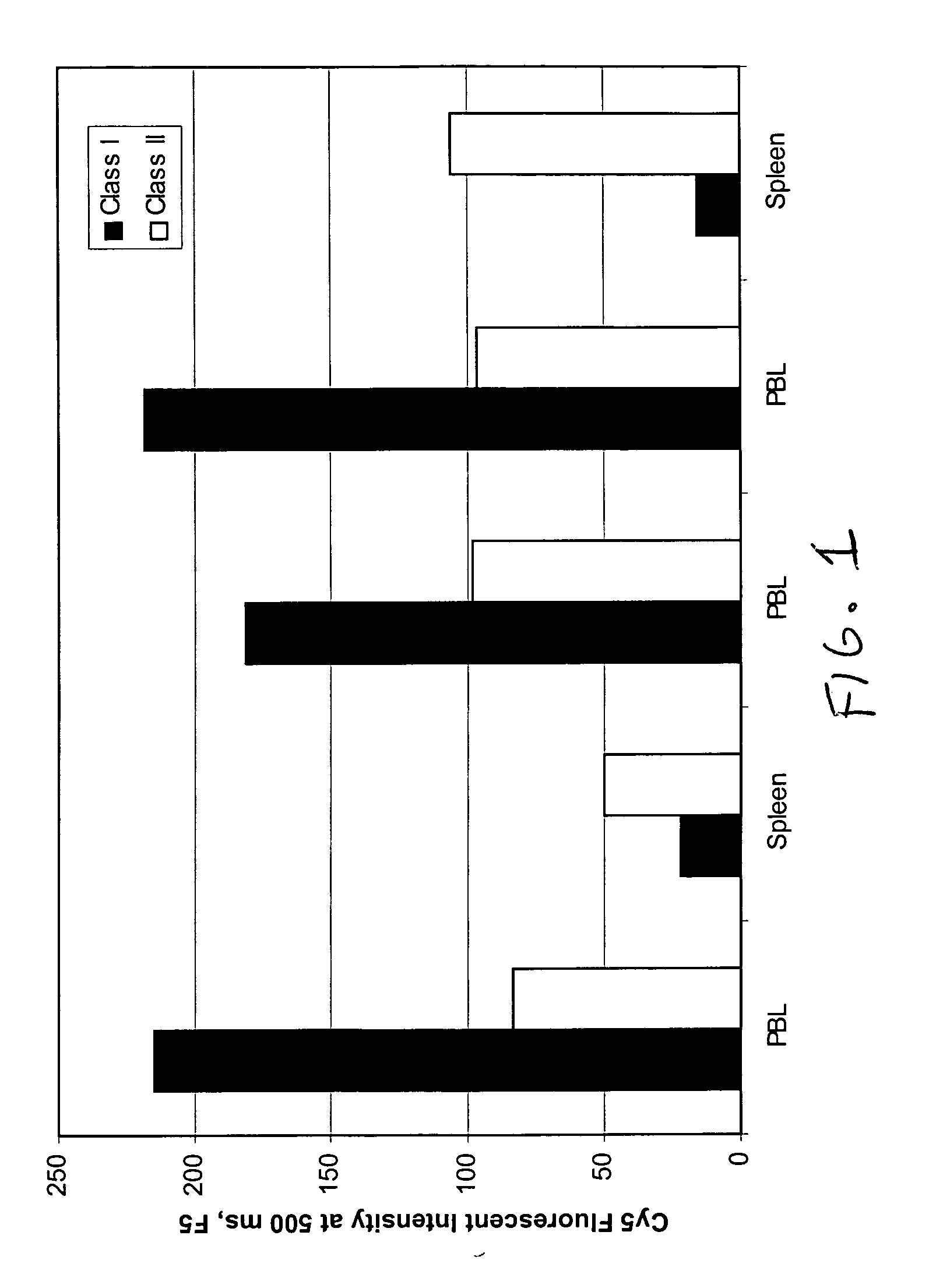
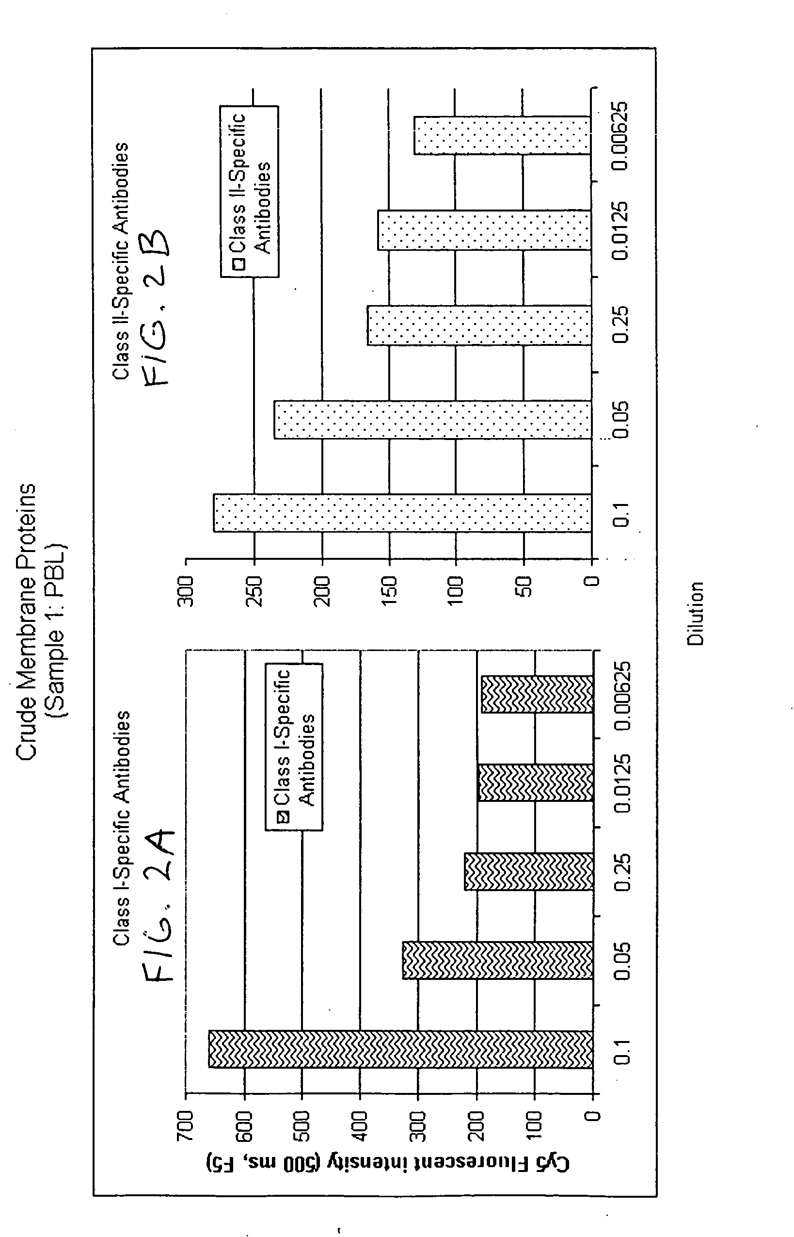
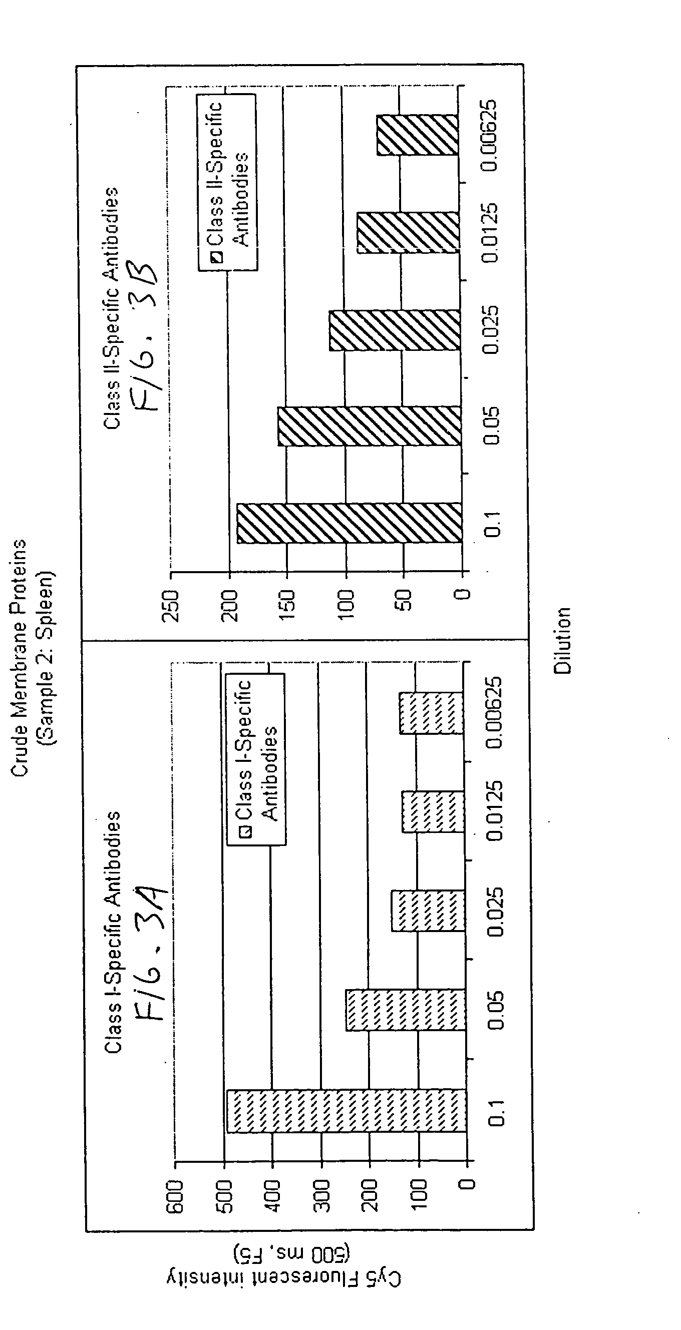
Determination of Relative Abundance of Class I and Class II HLA in Different Cell Lines

Preparing Encoded Bead Arrays: Membrane preparations in PBS-CHAPS were extracted from different cell lines and were affixed to encoded beads of 3.2 micron diameter by placing 5 μl of a 1% suspension of such beads into each tube containing an entire preparation. Beads were collected, then resuspended in 100 μl of storage buffer containing 1% Bovine Serum Albumin with protease inhibitors. Beads coated with different membrane protein were pooled into one tube for assembly of bead arrays on chips (“BeadChips”). Overlapping pools of antigens are formed by including in the array membrane fragments from a sufficiently large number of cell lines so as to represent a sampling of antigens found in a normal population. Such an array permits the determination of a relative percentage of PRA simply by evaluating the percentage of bead types scoring positive in the assay.

Assay: Positive control sera reactiv...

example iii

Covalent Attachment of Proteins to Encoded Microparticles

Antibodies were covalently attached to tosyl-activated microparticles by the following method, which was used to attach anti-cytokine monoclonal antibodies to such microparticles. A similar method can be used to attach fragments, including Fab. Five hundred microlitres of PBST (Phosphate Buffered Saline (PBS), 1% vol / vol Tween-20, pH 7.2) were placed in a 1.5 mL Eppendorf tube, and fifty microliters of a suspension containing 1% w / w microparticles (0.5 mg beads) were added and mixed by vortexing. Beads were first collected by centrifuging for 3 minutes at 10,000 rpm and discarding the supernatant. Next, beads were washed once in 1 mL of PBST and once in 1 mL of PBS using centrifugation in each step as described above. Beads were resuspended in 500 μL of PBS, pH=7.2. A designated amount of specific proteins was added to each suspension at a concentration of 400 μg protein per mg beads. The coupling reaction was allowed to pro...


PUM

PropertyMeasurementUnit
sizesaaaaaaaaaa
sizesaaaaaaaaaa
sizesaaaaaaaaaa

Abstract

Disclosed are methods relating to cell membrane fragments associated with microbeads, so that the characteristics of the cells the fragments originated from can be determined. The fragments can be oriented with what was the outer surface of the cell membrane facing outwardly, so that the antigens associated with the membrane can be contacted with ligands (including antibodies) to antigens in the membranes which would be accessible to antibodies in vivo. The system is useful, inter alia, for detection of panel reactive antibodies in donor serum, as well as detection of other cell membrane antigens; or quantitation of particular cell membrane antigens.

Description

BACKGROUND Determining the type and relative proportion of an individuals' cell surface or membrane-associated proteins is medically useful because over-expression, under-expression or complete lack of certain receptors or transmembrane channels frequently is indicative of disease or state of disease. If certain receptors are over-expressed, certain drugs may cause adverse events or toxicity. Conversely, if certain receptors are under-expressed or completely absent, certain drugs may not be effective, and signal transduction may not occur. Human leukocyte antigens (HLA) represent a class of cell surface proteins (also referred to herein as transplantation antigens), whose great variability from one individual to another forms the molecular basis for the immune system's ability to distinguish “self” from “non-self” cells and tissues. Individuals sensitized to HLA, for example in the course of pregnancy, or as a result of blood transfusion or organ transplatation, develop allo-antib...

Claims


Application Information

Patent Timeline
Patent Type & Authority Applications(United States)
IPC IPC(8): A61BC12M1/34C12Q1/68G01N33/53G01N33/543G01N33/564G01N33/567G01N33/569G01N33/68
CPCG01N33/5432G01N33/564Y10T436/110833G01N33/6854Y10S435/971G01N33/56972
Inventor YANG, JIACHENGTAN, ENQING
Owner BIOARRAY SOLUTIONS
Why Eureka
Try Eureka