Method of isolating epithelial cells, method of preconditioning cells, and methods of preparing bioartificial skin and dermis with the epithelial cells and preconditioned cells

a technology of which is applied in the field of isolating epithelial cells and preconditioning cells, can solve the problems of difficult identification of these two types of cells from each other, skin tissue or internal organ damage, and the expected non-separation of stem cells

Inactive Publication Date: 2005-07-28
KOREA ATOMIC ENERGY RES INST
View PDF1 Cites 23 Cited by
  • Summary
  • Abstract
  • Description
  • Claims
  • Application Information

AI Technical Summary

Benefits of technology

[0056] The present inventors have verified the effect of in vitro preconditioning on a va...

Problems solved by technology

It is difficult to identify these two types of cells from each other.
Therefore, stem cells are expected not to be easily separated, compared to other cells.
Skin tissue or internal organ may be partially damaged by burn, traumatic injury, ulcer, etc.
Green's method provides a sufficiently high cell yield for research purpose cell isolation, but not enough for cell isolation for tissue engineering-based industrial use.
Disadvantageously, however, inactivation of thermolysin or dispase cannot be controlled in the 2-step enzyme treatment method.
These two enzymes are known to retain its function in an enzyme-substrat...

Method used


Image

  • Method of isolating epithelial cells, method of preconditioning cells, and methods of preparing bioartificial skin and dermis with the epithelial cells and preconditioned cells
  • Method of isolating epithelial cells, method of preconditioning cells, and methods of preparing bioartificial skin and dermis with the epithelial cells and preconditioned cells
  • Method of isolating epithelial cells, method of preconditioning cells, and methods of preparing bioartificial skin and dermis with the epithelial cells and preconditioned cells

Examples


example 1

Cell Isolation and Culture

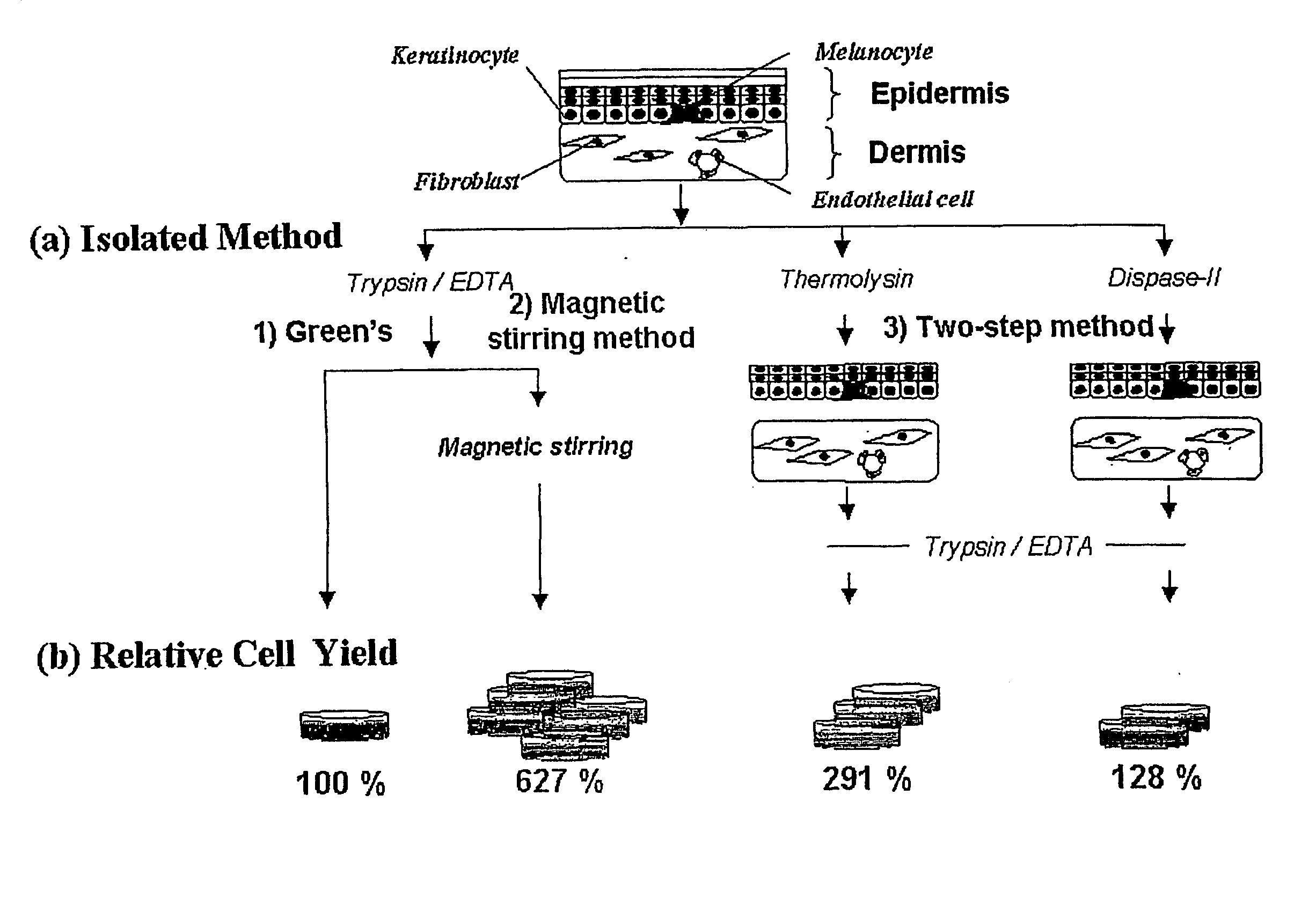
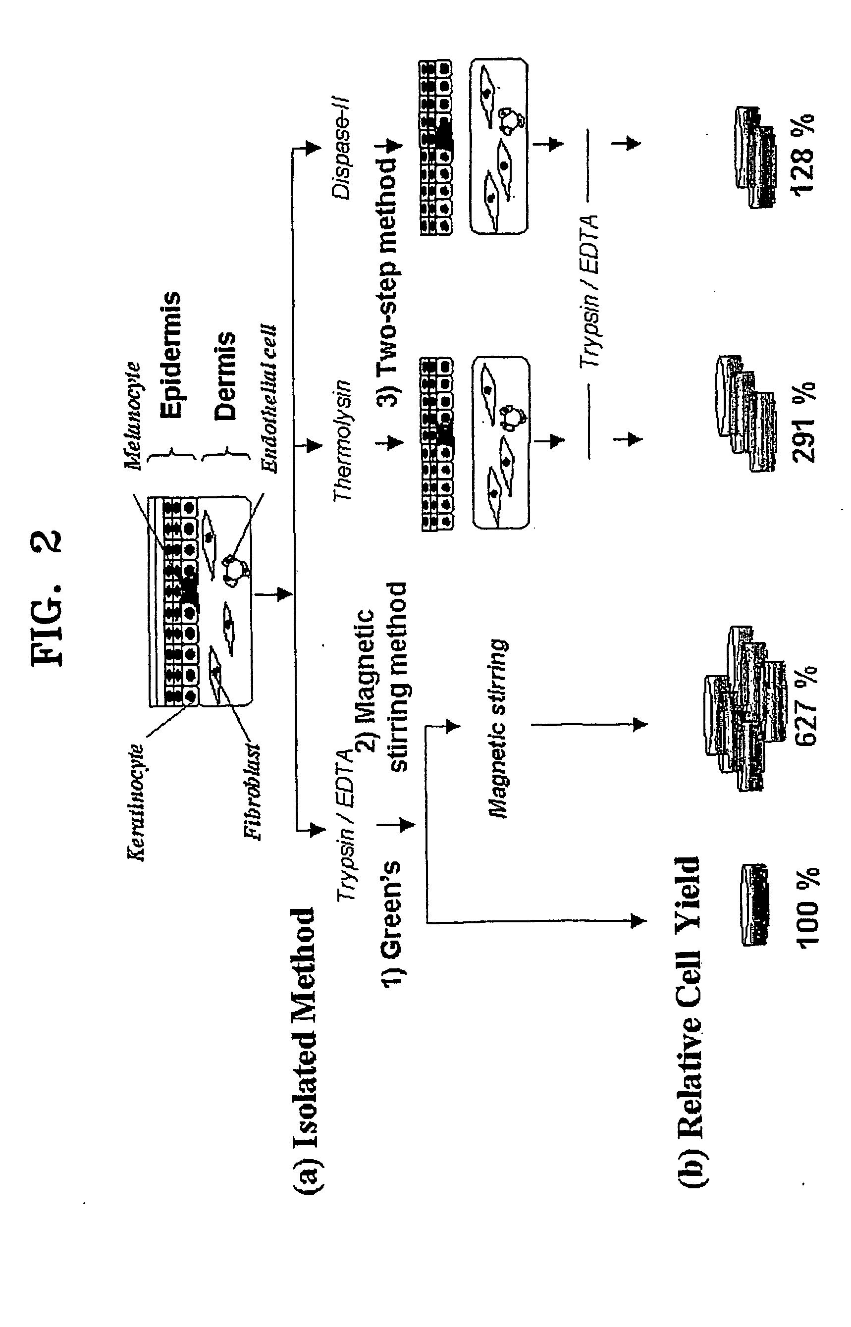
[0086] Primary keratinocytes were isolated from adult human foreskins obtained by circumcision. The adult human foreskins were placed in an epidermal minimal medium (hereinafter, E-medium) containing 1% penicillin, streptomycin, and 250 ng / ml Fungizone (Cat. No. 15240-062, Gibco) at 4° C. before cell isolation. Primary keratinocytes were isolated not later than 24 hours from circumcision.

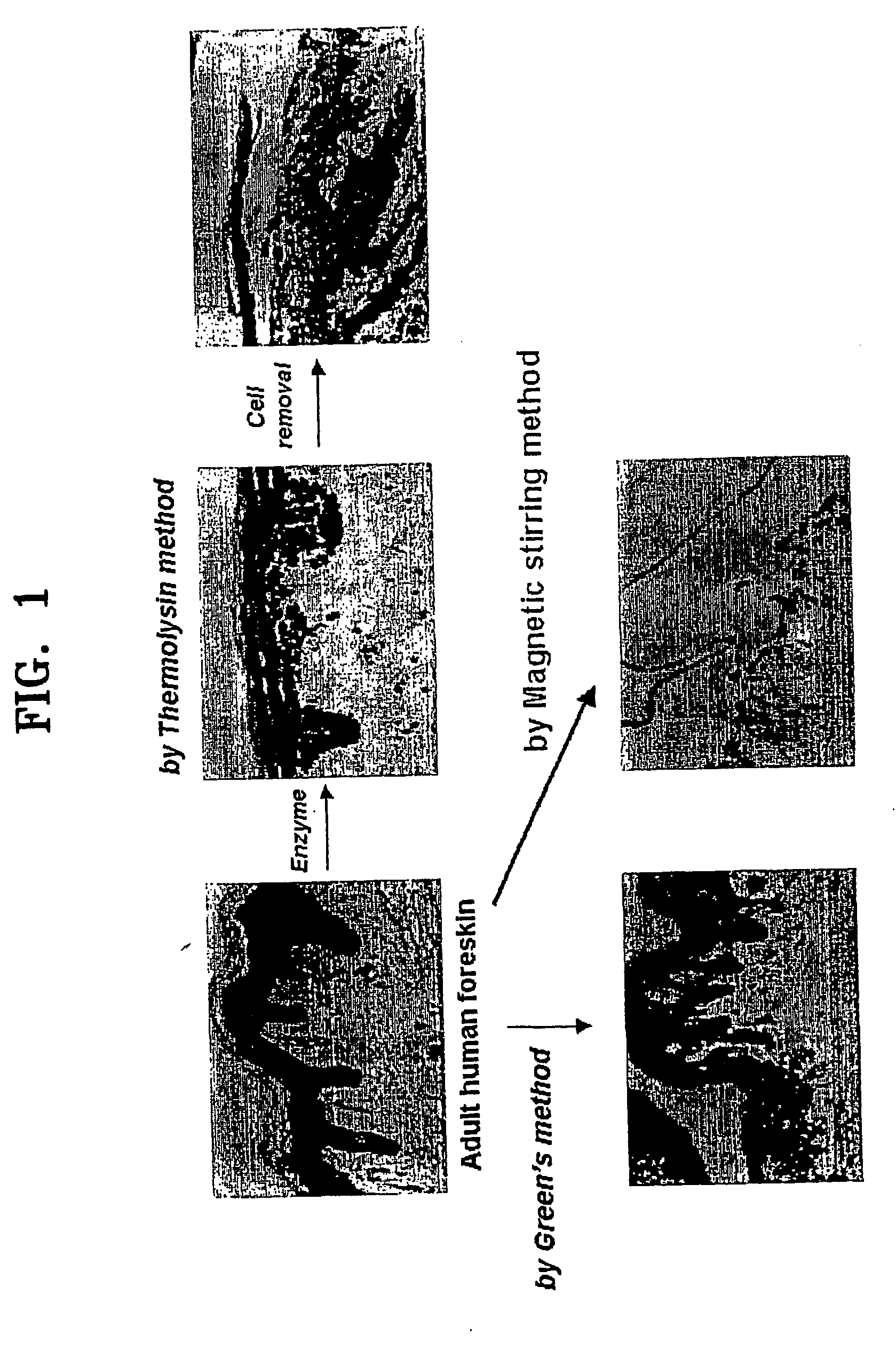
[0087] The foreskin sample was washed at least 8 times in a phosphate buffered saline (PBS) solution containing 5% penicillin / streptomycin. Subcutaneous tissue was mostly removed from the dermis of the foreskin sample with a pair of sterile surgical scissors, and the remaining portion was cut into tissue fragments not larger than 1-2 mm2.

[0088] Cell isolation was carried out by four methods, (i) magnetic stirring method according to the present invention, and conventional methods including (ii) Green's method, (iii) thermolysin method, and (iv) dispase method, based upon...

example 2

Fluorescence Activated Cell Sorting (FACS)

[0098] Levels of β1-integrin expression in cells isolated in Example 1 according to the four methods were compared by FACS to measure the percentage of β1-integrin bright cells in the isolated cells, which could be predominantly expressed with β1-integrin known as a stem cell marker. The cells isolated by the respective four methods were incubated along with β1-integrin antibodies (Chemicon) and followed with fluorescein isothiocyanate (FITC)-conjugated goat anti-mouse antibodies on ice for 45 minutes. The cells were washed in phosphate buffered saline (PBS) containing 5% bovine serum albumin (BSA). At the end of staining, cells were resuspended in a medium at a density of 1×10⁶ cells / ml and sorted using a FACStarPlus (Beckton Dickinson). At least 10,000 cells were analyzed by flow cytometry in each experiment. The results of each experiment was calibrated using fluorescent native antibodies and isotype control antibodies (refer to Effect 4...

example 3

Immunostaining

[0099] Keratinocytes isolated in Example 1 were cultured on coverslips and fixed for 10 minutes at 4° C. in a 1:1 mixture of ethanol and methanol. To identify whether the isolated and cultured cells exclusively consisted of keratinocytes, the fixed cells were stained with pan-cytokeratin antibodies acting as an epithelial cell marker (refer to FIG. 8 and Effect 2). In addition, the fixed cells were stained with α2 integrin antibodies (chemicon) to determine whether the isolated and cultured cells showed basal cell characteristics (refer to FIG. 8 and Effect 4), and with involucrin antibodies to determine the number of differentiating cells (refer to FIG. 8 and. Effect 5). The β1 integrin and α2 integrin antibodies used were mouse monoclonal antibodies, and the pan-cytokeratin (Novocastra) and Involucrin (Biomedical Technologies, a keratonicyte differentiation indicator) antibodies used were rabbit polyclonial antibodies. Cell incubation in the presence of primary anti...


PUM


Abstract

A method of isolating epithelial cells from a human skin tissue or internal organ tissue using trypsin and ethylene-diamine tetraacetic acid (EDTA) simultaneously with the application of magnetic stirring, a method of preconditioning isolated biological cells by the application of physical stimulus, i.e., strain, are provided. Epithelial cells can be isolated by the method with increased yield, colony forming efficiency (CFE), and colony size. Also, the increased percentage of stem cells in isolated cells is advantageous in therapeutic tissue implantation by autologous or allogeneic transplantation. In skin cells preconditioned by the application of strain, cell division is facilitated, and the secretion of extracellular matrix components and growth factors and the activity of matrix metalloproteinases (MMPs) are improved. When preconditioned cells are implanted by autologous or allogeneic transplantation to heal a damaged tissue, the improved cell adhesion, mobility, and viability provides a biological adjustment effect against a variety of stresses or physical stimuli which the cells would undergo after implantation, with improved capability of integration into host tissue, thereby markedly improving the probability of success in skin grafting.

Description

TECHNICAL FIELD [0001] The present invention relates to a method of isolating epithelial cells and a method of preconditioning cells, and more particularly, to a method of isolating epithelial cells from skin or internal organs using trypsin and ethylenediamine tetraacetic acid (EDTA) simultaneously with magnetic stirring, and a method of in vitro preconditioning isolated skin cells with the application of physical stimuli during cell culture. BACKGROUND ART [0002] Human skin tissue is roughly divided into three parts: the epidermis on top, the dermis underneath, and the subcutaneous tissue. The epidermis consists of epithelial cells stratified from the basement membrane between the epidermis and the dermis, and melanocytes and Langerhans Cells. The dermis consists of fibroblasts and extracellular matrix secreted by the fibroblasts. [0003] Skin epithelial cells have different cell ages and degrees of differentiation for each cell layer. This is because stem cells in the basal layer ...

Claims


Application Information

Patent Timeline
IPC IPC(8): A61K35/12A61L27/00C12N5/071
CPCA61K35/12C12N5/0629C12N5/0698C12N2509/10C12N2502/1323C12N2502/28C12N2509/00C12N2502/094
Inventor SON, YOUNG-SOOKPARK, HYUN-SOOKKIM, CHUN-HOKANG, HYUN-JUKIM, CHANG-HWANKIM, YOUN-YOUNGCHOI, YOUNG-JULEE, SU-HYUNGIN, YONG-JAE
Owner KOREA ATOMIC ENERGY RES INST
Why Eureka
Try Eureka