High throughput screening assays utilizing affinity binding of green fluorescent protein

a green fluorescent protein and screening assay technology, applied in the field of biotechnology research and development, can solve the problems of reducing the cost of drug development, affecting the quality of drug development, so as to improve the quantitation, enhance the signal-to-noise ratio, and improve the effect of quantitation

Inactive Publication Date: 2006-08-24
WARD WILLIAM W +1
View PDF7 Cites 0 Cited by
  • Summary
  • Abstract
  • Description
  • Claims
  • Application Information

AI Technical Summary

Benefits of technology

[0027] Reducing the interfering compounds and concentrating the sample allow for a decrease in noise and allow the use of higher intensity excitation wavelength irradiation and generation of higher signals. This results in greatly increased signal-to-noise ratios. In a preferred embodiment, the signal-to-noise ratio is increased about one to many orders of magnitude.
[0031] In one embodiment, the method uses a high intensity or ultra-high intensity light source to irradiate the trapped and concentrated GFP. Such light sources are known in the art, for example argon lasers. The removal of interfering compounds allows for the light intensity to be greatly increased without risk of elevated noise due to scattering or autofluorescence. Under such conditions, where the GFP is trapped and concentrated, with the exclusion of scattering and autofluorescent contaminants, the true GFP signal is proportional to the intensity of the light at the excitation wavelength.
[0032] In a preferred embodiment, the method is automated for quantifying a plurality of assays. The automation of the method is by methods such as are known in the art for automatically processing the assays—handling and transporting samples and reactants, performing incubations, mixing, removal or addition of components, quantifying reactants, recording data appropriately. In a preferred embodiment, the automation is developed as part of a high throughput screening system. In one embodiment, the assay is miniaturized to allow smaller volumes and faster manipulation of samples, with lower consumption of reactants. Miniaturization also facilitates significantly increasing the light intensity for the excitation wavelength.
[0035] In one embodiment of this method, the increased sensitivity of the assay allows for the detection of more subtle differences among the various biological regulatory machinery and structural components which are involved in the nucleic acid expression system.
[0039] Yet another aspect of the invention features combinations of the aforementioned elements into a HTS assay system with greatly enhanced signal-to-noise ratios, greater sensitivity, and improved quantitation relative to existing assays. Other and further features and advantages of the invention will become apparent from the following description.

Problems solved by technology

Despite its importance to the industry and, indirectly, to the public, drug screening is often a bottleneck in the development of new drugs to alleviate conditions ranging from the common cold to cancer.
Traditional methods have been either labor intensive, time-consuming, or too expensive.
In addition, many potentially valuable therapeutic agents may be missed because of inadequate screening assays.
Pressure to reduce the cost of drugs also forces the cost of drug development down.
However all these advances have created a situation where the potential ability to rapidly and cost effectively screen chemical compounds for ‘activity’ on a multitude of theoretical targets has outstripped the basic biological strategies and principles of assay development.
Such methods do allow for rapid screening, however they often suffer from problems such as high background levels, or low levels of signal—this a particularly problematic situation, especially when the signal-to-noise ratio is low because it can result it both false positives and false negatives.
In addition to the problems of high background and low signal which plague many assay systems, assays which are based on radiolabeled compounds pose additional hazards to those who work with them and are a waste disposal problem.
Considering the expense associated with drug screening and the cost of moving a screened compound forward to the next phase of drug development, the cost of falsely identifying a potential compound as useful is significant.
Of potentially even greater cost to both the pharmaceutical company and to the public is the cost of a false negative.
In addition to the billions of potential dollars in lost revenue, and lives not saved, it may cause the researchers to miss an entire class of compounds which may have been useful to treat other conditions.
Most of the applications have been restricted to microscopic examination of transformed cells.
One problem which has plagued assays using the green fluorescent proteins to date is that of ‘noise’, most particularly in non-microscopic assays.
Despite the signal created by the emission of the GFP, there are numerous sources of background fluorescence, autofluorescence, scatter and other interference in the assays in which it has been used.
Another problem common in such assays is low signal strength.
In many cases the intensity of the light used for excitation is limited by the anticipated noise.
The spectral properties of the GFPs used to date are also somewhat limiting, in that large amounts of expression are often needed to overcome detection limits.
In summary, the GFP reporter assays that have been attempted for high throughput screening have tended to suffer from low signal-to-noise ratios.

Method used


Image

  • High throughput screening assays utilizing affinity binding of green fluorescent protein
  • High throughput screening assays utilizing affinity binding of green fluorescent protein
  • High throughput screening assays utilizing affinity binding of green fluorescent protein

Examples


example 1

The Limits of Detection

[0091] Assume spherical bacterial cells (or e.g. trapping particle, bead, and the like), 1 μM in diameter. Such cells expressing GFP are easily detected by fluorescence microscopy.

[0092] Given the basic formula for volume (V) of a sphere:

V=4 / 3πr3, where r=radius of the sphere:

V=(4 / 3)⁢(3.14)⁢(0.5⁢ ⁢μ⁢ ⁢M)3=(4)⁢(0.125)⁢10-12⁢cm3⁡(Rounding)=0.5⨯10-12⁢cm3

[0093] If the 1 μM sphere were all (100%) GFP, of density (ρ)=1.3 g / cm3 then the amount (mass (m), in grams, g) of GFP readily visualized by fluorescence microscopy is:

V×ρ=m

(0.5×10⁻¹² cm3)(1.3 g / cm3)=0.65 pg [0094] Converting to moles (MWGFP=27,000)

(0.65×10⁻¹² g)(1 mole / 27,000 g)=2.4×10⁻¹⁷ moles [0095] And using Avagadro's Number to reduce moles to number of molecules:

(2.4×10⁻¹⁷ moles)(6.022×10²³ molecules / mole)=14.4×10⁶ molecules

However, recalling that this number is based on the unrealistic assumption that the hypothetical 1 μM sphere (e.g. bacterial cell or trapping particle) consisted 100% of GFP, a ...

example 2

Detection Limit on Conventional Fluorometers

[0110] Calibration curves were performed on three commercial fluorometers optimized for GFP detection, using GFP expressed in E. coli cells. The fluorometers include a Turner 110 filter fluorometer, a Hoefer TKO 100 fluorometer, and a computer-operated Thermo Lab Systems MFX fluorometric microplate reader.

[0111] The detection limit for the wild-type GFP on the Turner 110 was 5 pmoles per assay. To minimize scatter, the fluorometer was set to the 10× slit setting and E. coli cells were at OD660 of about 0.25—i.e. the determined limit is for nonturbid samples only. The sensitivity for the Hoefer TKO 100 was determined to be 12 pmoles per assay. This detection limit was essentially unaffected by scatter caused by the E. coli cells. The Thermo Labs MFX was determined to be capable of detecting GFP down to 10 pmoles per assay, a result also virtually not influenced by scatter.

[0112] By comparison, when the GFP from cells in a 200 μl micropla...

example 3

Trapping GFP by Hydrophobic Interaction

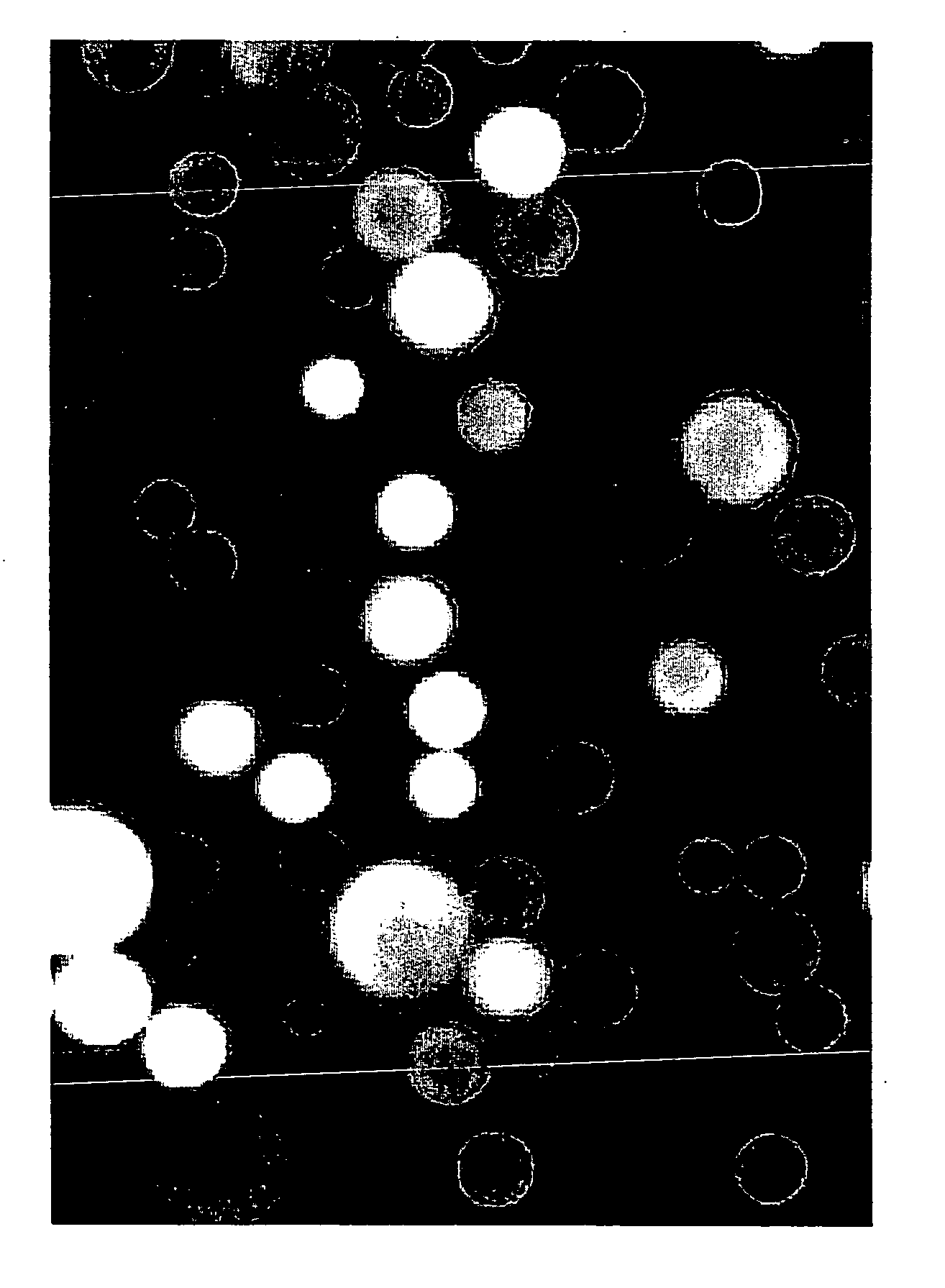
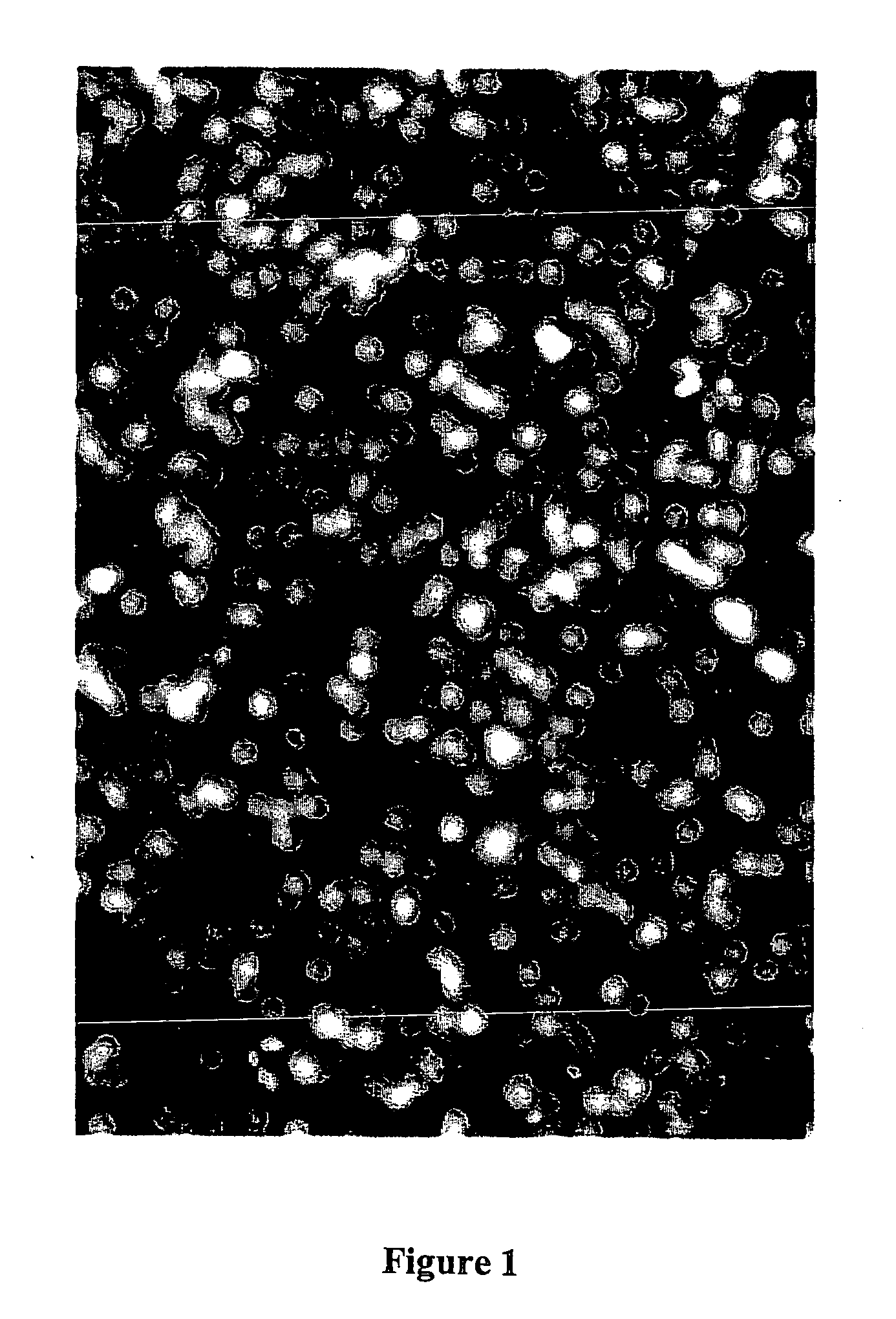
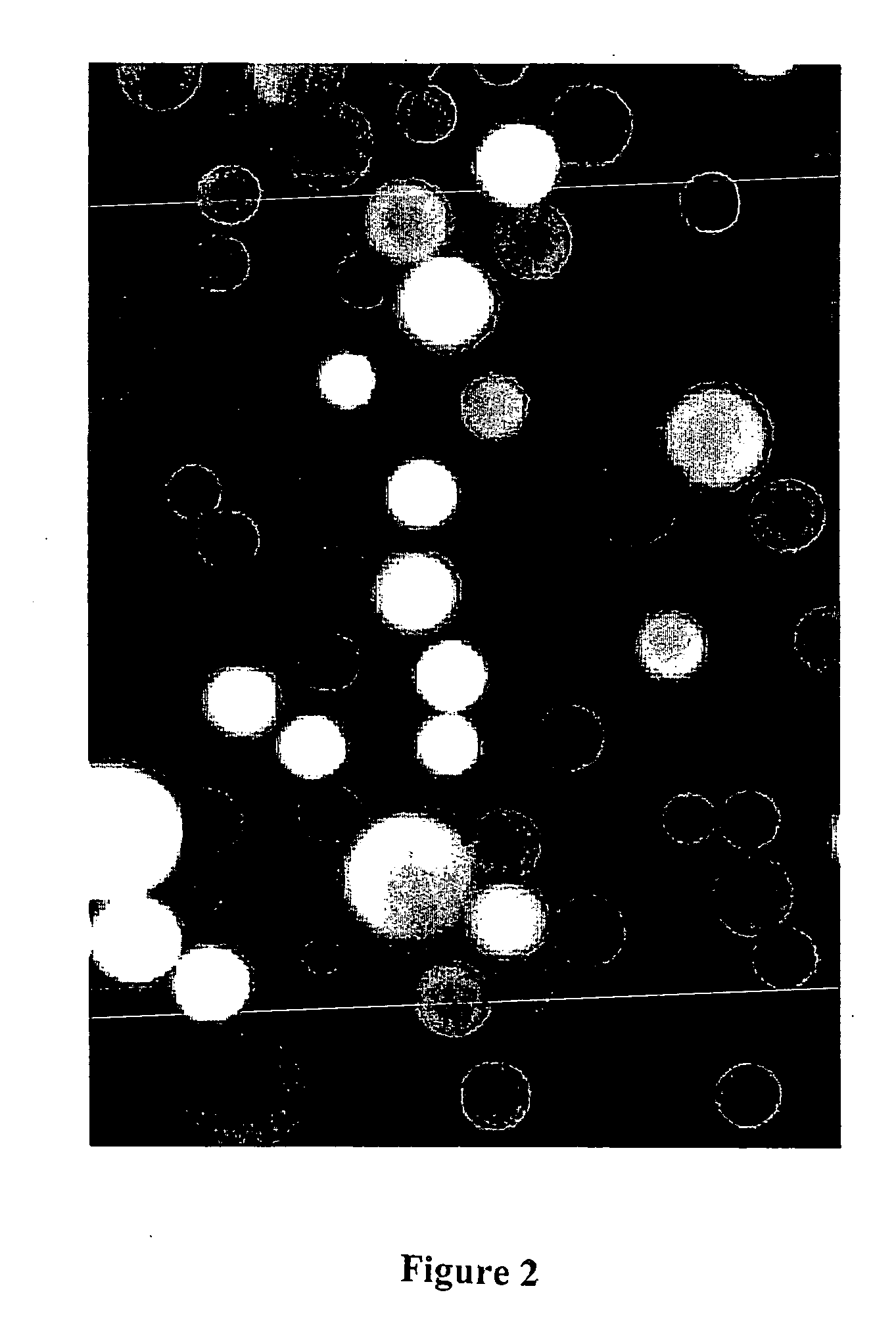
[0114] C4 derivatized silica beads, 5 uM in diameter, (reversed phase HPLC beads from BioRad product number 125-0134) were used to trap GFP by hydrophobic interaction. The C4 (n-butyl)-derivitized silica based beads were dispersed in methanol and then added to an aqueous solution of wild-type recombinant GFP. The GFP bound immediately to the beads by hydrophobic interaction, producing fluorescently labeled beads so intense in their fluorescence that, despite their tiny size, they can be easily viewed, individually, by the unaided eye on the surface of a hand held long wavelength (365 nm) UV lamp. In this case, the beads were viewed with a BH-2 Olympus fluorescence microscope using a high pressure mercury arc lamp and a blue excitation filter selected for optimal excitation of fluorescein. Results are shown in FIG. 1.

[0115] In all micrographs presented in this patent application, the ocular lens was 10×. In this particular view (see FIG. 1), t...


PUM

PropertyMeasurementUnit
areaaaaaaaaaaa
areaaaaaaaaaaa
excitation wavelengthaaaaaaaaaa

Abstract

Novel methods of detecting fluorescent proteins are described. The methods result in vastly improved signal-to-noise ratios in assays measuring fluorescence of a fluorescent protein specifically by employing a unique trapping step to microconcentrate the fluorescent protein and by using improved optical techniques. The trapping step may be a chemical or physical process or a combination thereof leading to substantial microconcentration of the fluorescent protein with concomitant removal of contaminants or interfering compounds. The methods are readily adaptable to high throughput screening and can be engineered for use with a wide variety of assays currently using microplate readers. Green fluorescent protein and fluorescent coral proteins are among preferred fluorescent proteins for the methods.

Description

CROSS REFERENCE TO RELATED APPLICATION [0001] This application claims benefit of U.S. Provisional Application No. 60 / 295,184, filed Jun. 1, 2001, the entirety of which is incorporated by reference herein.FIELD OF THE INVENTION [0002] This invention relates to the field of pharmaceutical and biotechnology research and development. Specifically, this invention provides methods and devices for rapid screening of compounds with potential as therapeutic agents or research tools. BACKGROUND OF THE INVENTION [0003] Various scientific and scholarly articles are referred to in parentheses throughout the specification. These articles are incorporated by reference to describe the state of the art to which this invention pertains. In addition, any sequences referred to by Accession Number of a publicly accessible database are incorporated by reference herein. [0004] The screening of potential candidates for therapeutic agents is critical to maintaining a full pipeline of products for the pharma...

Claims


Application Information

Patent Timeline
Patent Type & Authority Applications(United States)
IPC IPC(8): G01N33/53G06F19/00G01N33/58
CPCG01N33/582
Inventor WARD, WILLIAM W.THOMSON, CATHERINE
Owner WARD WILLIAM W
Why Eureka
Try Eureka