
Method of diagnosing wood decay and decay diagnostic agent

Inactive Publication Date: 2006-09-07
SDS BIOTECH CO LTD
View PDF4 Cites 3 Cited by
  • Summary
  • Abstract
  • Description
  • Claims
  • Application Information

AI Technical Summary

Benefits of technology

[0013] The present inventors have obtained the results that an antibody obtained by sensitizing an animal with an antigen which is a protein having a molecular weight of 1,000 to 100,000 obtained by culturing a naturally occurring wood-destroying fungus such as Fomitopsis palustris is widely reactive not only with extract of wood decayed by the wood-destroying fungi used to obtain the protein but also extract of wood decayed by other wood-destroying fungi while the antibody shows no significant reactivity with extract of wood where fungi other than wood-destroying fungi are growing, and thus completed the invention.
[0014] That is, the present invention provides a method for diagnosing wood decay, an agent used in the method of wood-decay diagnosis, a simple a method for diagnosing decay in wood and a test kit used in the method for diagnosing decay in wood.
[0044] Diagnosis of wood-decay degree is conducted by contacting extract obtained from the test subject sample wood (the wood to be examined) with the above-obtained antibody. Examples of test subject sample wood include a cut piece of wood, a drilled piece of wood, sawdust and wood powder, which are taken out of the wood to be examined. Extraction is conducted by immersing these samples into the following extraction solvent. The extraction solvent is an aqueous solvent preferably prepared by adding to water at least one kind of surfactant such as nonionic surfactant, saccharides, osmotic pressure adjusting agent such as inorganic salts or pH adjuster such as inorganic salts or organic salts. The surfactant is not particularly limited, however, preferred is a surfactant with less protein modification. Examples thereof include Triton-X series (e.g. Triton X-100). Examples of buffer liquid include PBS (phosphate buffered saline), and the pH value is in a range of 4 to 10, preferably from 6 to 9. Extraction time may be determined arbitrarily, and preferred time range is 1 minute to 1 hour. For the purpose of improving extraction efficiency, it is useful to use ultrasonic treatment or homogenizer treatment in combination.
[0057] That is, a plate made of a resin such as polystyrene, polyethyrene or polypropylene on which a groove of 0.5 to 10 cm in width, 1 to 50 cm in length and 0.1 to 10 cm in depth, preferably 1 to 5 cm in width, 5 to 20 cm in length and 0.5 to 2 cm in depth is provide is prepared. The form of the groove may be either a bottomed form (like a container) or a bottomless form (hereinafter the plate is referred to as “device”). The membrane-like material (hereinafter abbreviated as “membrane”) of cellulose or modified cellulose such as nitrocellulose, glass, nylon, polyethylene, polyester or PVDF is cut in a form to fit in the groove of the device, and placed in the groove. By allowing at least a part of the membrane except for positions to be spotted to be in contact with the inner surface of the groove in this step, the operations thereafter can be smoothly conducted.
[0059] In this case, spotting is conducted by dropping the extract on the membrane with appropriate intervals between spots and for easy observation of color changes in spotted portions, the form of the groove in the device may be designed according to the spotted positions. For example, scale lines or diamond marks may be provided on the plate surface between the grooves in the device, so that spotting can be accelerated with optimum intervals according to the predetermined number of spots, and spaces for writing down the respective sample numbers to identify each spot. By doing so, spotted positions in the membrane and correspondence of one spot with one sample can be clearly indicated, whereby multiple samples can be precisely examined at the same time by using a single device. Moreover, it is preferable to prepare a standard spot on the plate or membrane by using a sample wood whose decay degree is known. That is, presence or absence of decay caused by wood-destroying fungi can be judged from the color change of each spot and the decay degree can be quantitatively evaluated by comparison in absorption (or comparison in contrasting density when visually observed by naked eye) between the spots of test subject sample woods and the standard spot thus prepared in advance. Such a standard spot can be provided by spotting a standard sample obtained from a wood whose decay degree is already known by decrease in weight or the like (e.g. wood of weight loss rate of 1%, 3%, 5%, 7% or 10%). A single standard spot may be provided or multiple standard spots with different decay degrees may be provided.
[0062] According to the present invention, presence or absence of wood decay caused by wood-destroying fungi can be easily determined in a short period of time. In the method of the present invention, which can determine wood decay specifically caused by wood-destroying fungi and does not include response to fungi present on the wood surface and metabolites therefrom, possibility of wrong diagnosis is extremely low. Furthermore, the agent for detecting wood-destroying fungi, which is obtained by sensitizing an animal with a single species of wood-destroying fungus, can be easily prepared and therefore production of the agent is easy and uniform products can be supplied easily.

Problems solved by technology

Wood is an excellent material and used in a wide range of fields, mainly as a building material, however wood has a disadvantage that it is susceptible to the breakdown of materials by microbial action (biodeterioration).
In most cases of the latter damage, where the damage is caused by growth of wood-destroying fungi inside the wood material, the damage cannot be judged by visual observation only.
However, with respect to method (1), wood decay at an early stage cannot be detected by examination by percussion or touch, and determination by (1) is ambiguous.
With respect to method (2), it takes long to obtain results of culturing fungi and such a method requires expert technique.
With respect to method (3), although a PILODYN, which drives a steel pin by a spring into wood and measures the degree of decay from the penetration depth, the determination depends on density and quality of the sample wood and therefore, quantitative results cannot be obtained and decay cannot be detected at an early stage.
With respect to method (4), decay inspection by sonic wave or by acoustic emission (AE) not only uses a considerably large apparatus and is expensive, but also requires expert knowledge and technique.
Thus, all the conventional decay-detection approaches have disadvantages and none of them is widely used.
Moreover, the document describes that various fungi responded to control antiserum (antiserum not inoculated with fungi) Generally, many kinds of fungi including mold fungi are present on the surface of a wood material suspected of being decayed and therefore, the method involving responses from those fungi present on the surface is not effective as a method for detecting wood-destroying fungi.
However, this method, which requires culturing multiple kinds of fungi, is cumbersome.
Accordingly, the method possesses low reliability.
Postia placenta is a wood-destroying fungus which does not inhabit in Japan and since there is a control measure to prevent the fungus from entering in the country, the fungus is not available in Japan.
However, the document does not include description about Fomitopsis palustris, Trametes versicolor and Serpula lachrymans, which are representative types of wood-destroying fungi in Europe, USA and Japan and therefore, the method cannot be said to be useful in detection of overall kinds of typical wood-destroying fungi.
Thus, in conventional detection methods using antigen-antibody reaction, the object to detect various kinds of wood-destroying fungi simultaneously without detecting fungi other than wood-destroying fungi cannot be attained.

Method used


Image

  • Method of diagnosing wood decay and decay diagnostic agent

Examples


example 1

Preparation of Antibody

(1) Preparation of Antigen

[0065] 50 ml of liquid medium (pH 5.5) which contains 1% cellobiose, 0.2% NH4NO3, 0.2% KH2PO4, 0.05% MgSO4.7H2O, 0.01% CaCl2.2H2O, 0.57 ppm H3BO4, 0.036 ppm MnCl2.4H2O, 0.31 ppm ZnSO4.7H2O, 0.039 ppm CuSO4.5H2O, 0.018 ppm (NH4)6Mo7O24.4H2O, 0.015 ppm FeSO4.7H2O, 1 ppm thiamine hydrochloride, 0.5% peptone and 0.05% yeast extract was added into a 500 ml flask for culture, the flask was plugged with cotton and sterilized in an autoclave at 121° C. for 30 minutes.

[0066]Fomitopsis palustris (Berk. Et Curt.) Gilbn. & Ryv. FFPRI 0507 was inoculated in this medium, statically cultured at 27° C. for 2 weeks and the obtained cultured solution was filtered by a glass filter, to thereby obtain cultured filtrate. The cultured filtrate was filtered by an ultrafilter membrane (Ultrafree-15, Biomax 100, a membrane-attached unit by Millipore Corporation), to thereby obtain a fraction having a molecular weight of 100,000 or less. This fraction was ...

example 2

Diagnosis by Using ELISA Method

(1) Preparation of Decayed Wood Chips and Extract from the Decayed Wood Chips

[0068] As culture substrates, 10 pieces of cedar splint wood and 20 ml of pure water were placed in a 500 ml-volume Erlenmeyer flask, and after plugged with cotton, the medium was sterilized in an autoclave at 121° C. for 30 minutes.

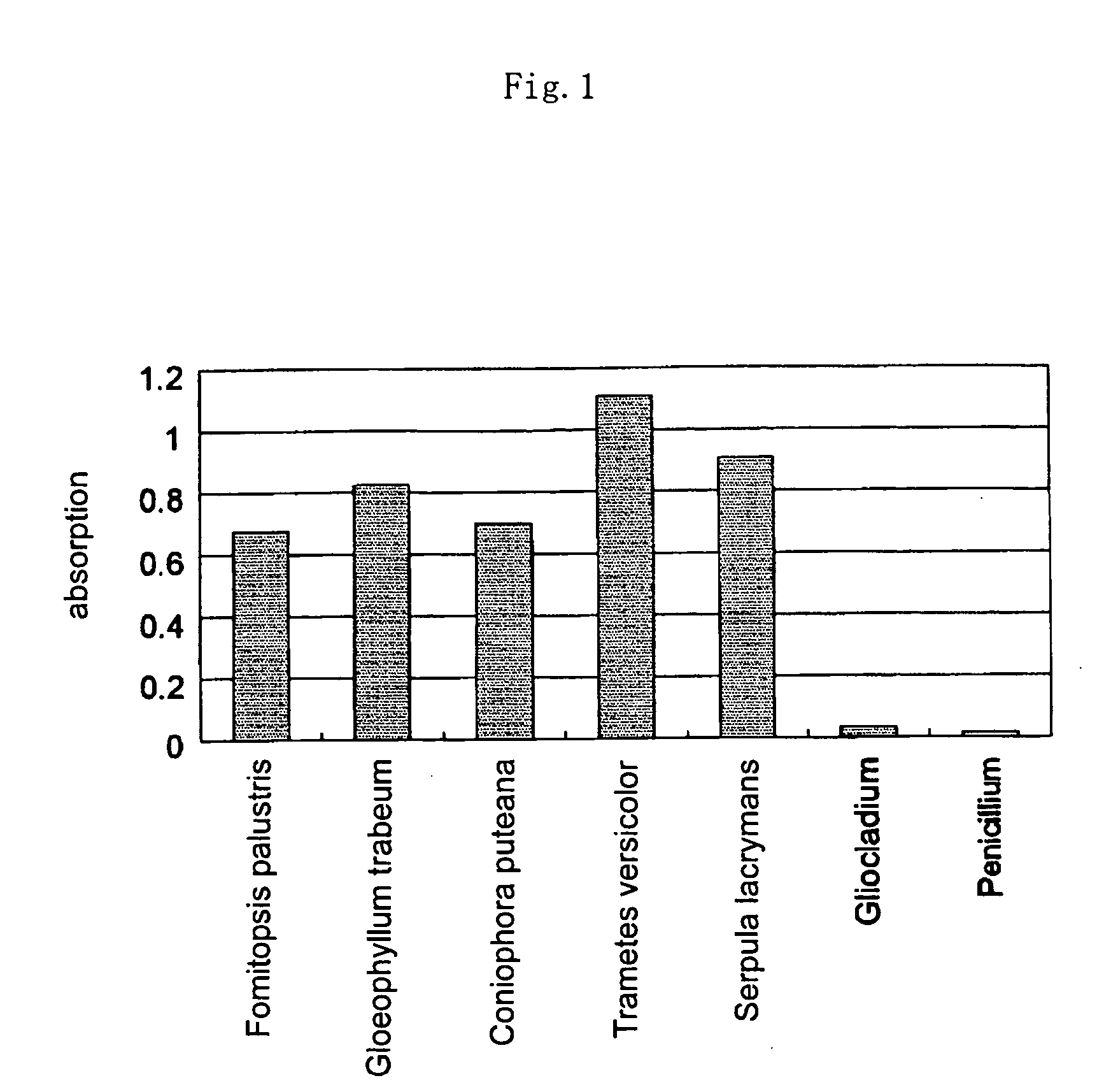
[0069] In this way, 7 of such an Erlenmeyer flask were prepared and strains of 7 kinds of fungi, Fomitopsis palustris, Gloeophyllum trabeum, Coniophora puteana, Serpula lacrymans, Trametes versicolor, which are wood-destroying fungi, Penicillium funiculosum (which is used in JIS fungus resistance test) which is a wood surface contaminant fungus and Gliocladium virens Miller (which is used in JIS fungus resistance test, former name: Trichoderma viride) were respectively inoculated, and kept at 24° C. for 4 weeks.

[0070] After 4 weeks, from thus decayed wood pieces, small chip-like wood samples were taken our by using a manual drill. To the sampl...

example 3

Determination of Wood Decay by Dot-Blot Method

[0076] The wood extract was prepared in the same manner as in Example 2, and 10 μl of the extract from each of the test subject wood samples and a control were spotted to a nitrocellulose membrane (0.451 μl of nitrocellulose membrane, product of BIO-RAD Laboratories, Inc., hereinafter simply abbreviated to as “membrane”). This membrane was dried for 10 minutes. The membrane was washed twice with PBS which contained 0.05% Tween 20 (hereinafter, this operation is referred to as “washing”). Then, the membrane was immersed in a blocking buffer liquid (PBS which contained 1% BSA and 0.05% Tween 20), incubated at 37° C. for 10 minutes and the membrane was subjected to washing.

[0077] The antibody solution obtained in (2) of Example 1 was diluted with PBS, the membrane was immersed in the solution, and the membrane was incubated at 37° C. for 10 minutes. After washing the membrane, the membrane was immersed in alkaline phosphatase-conjugated g...


PUM

PropertyMeasurementUnit
Temperatureaaaaaaaaaa
Decay rateaaaaaaaaaa
Molecular weightaaaaaaaaaa

Abstract

The invention provides a simple method which enables precise diagnosis of wood decay at an early stage of decay and an agent used for the diagnosis method. The invention provides a method for diagnosing wood decay, wherein wood decay is judged by contacting extract of wood to be examined (test subject wood) with an antibody obtained by sensitizing an animal to an antigen which is a protein having a molecular weight of 1,000 to 100,000 obtained by culturing a naturally occurring wood-destroying fungus and an agent for the diagnosis method.

Description

TECHNICAL FILED [0001] The present invention relates to a method for diagnosing decay in wood and an agent for diagnosing wood decay used in the method. Further, the present invention relates to a simple method for diagnosing decay in wood and a test kit used in the method for diagnosing decay in wood. BACKGROUND ART [0002] Wood is an excellent material and used in a wide range of fields, mainly as a building material, however wood has a disadvantage that it is susceptible to the breakdown of materials by microbial action (biodeterioration). [0003] Biodeterioration of wood is roughly classified into feeding damage by insects such as termites and decay caused by microorganisms such as wood destroying fungi. The former damage can be judged by visual observation and / or simple test. In most cases of the latter damage, where the damage is caused by growth of wood-destroying fungi inside the wood material, the damage cannot be judged by visual observation only. Moreover, since growth of w...

Claims


Application Information

Patent Timeline
IPC IPC(8): G01N33/569C07K16/14G01N33/53G01N33/46G01N33/48G01N33/577
CPCG01N33/56911G01N33/56961G01N2333/375
Inventor TANAKA, KEIJITSUECHIGO, TAKASHIHIRAMOTO, MASAHIKO
Owner SDS BIOTECH CO LTD
Learn More