

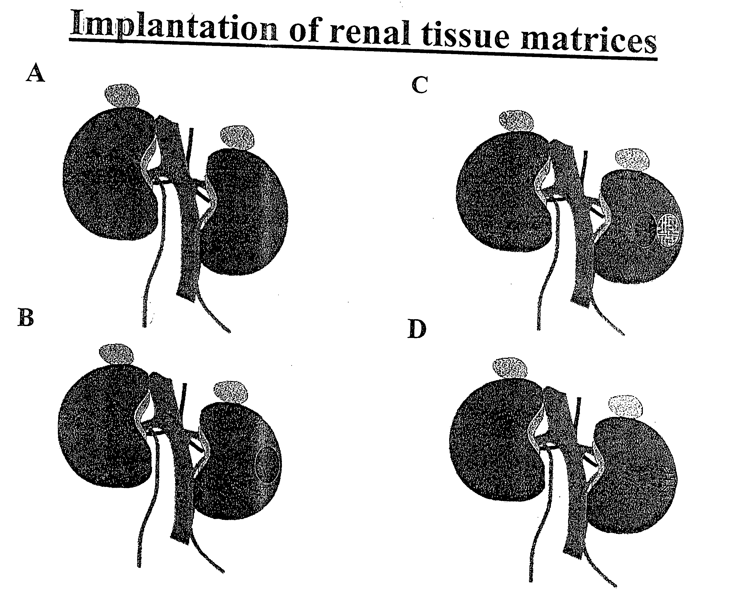
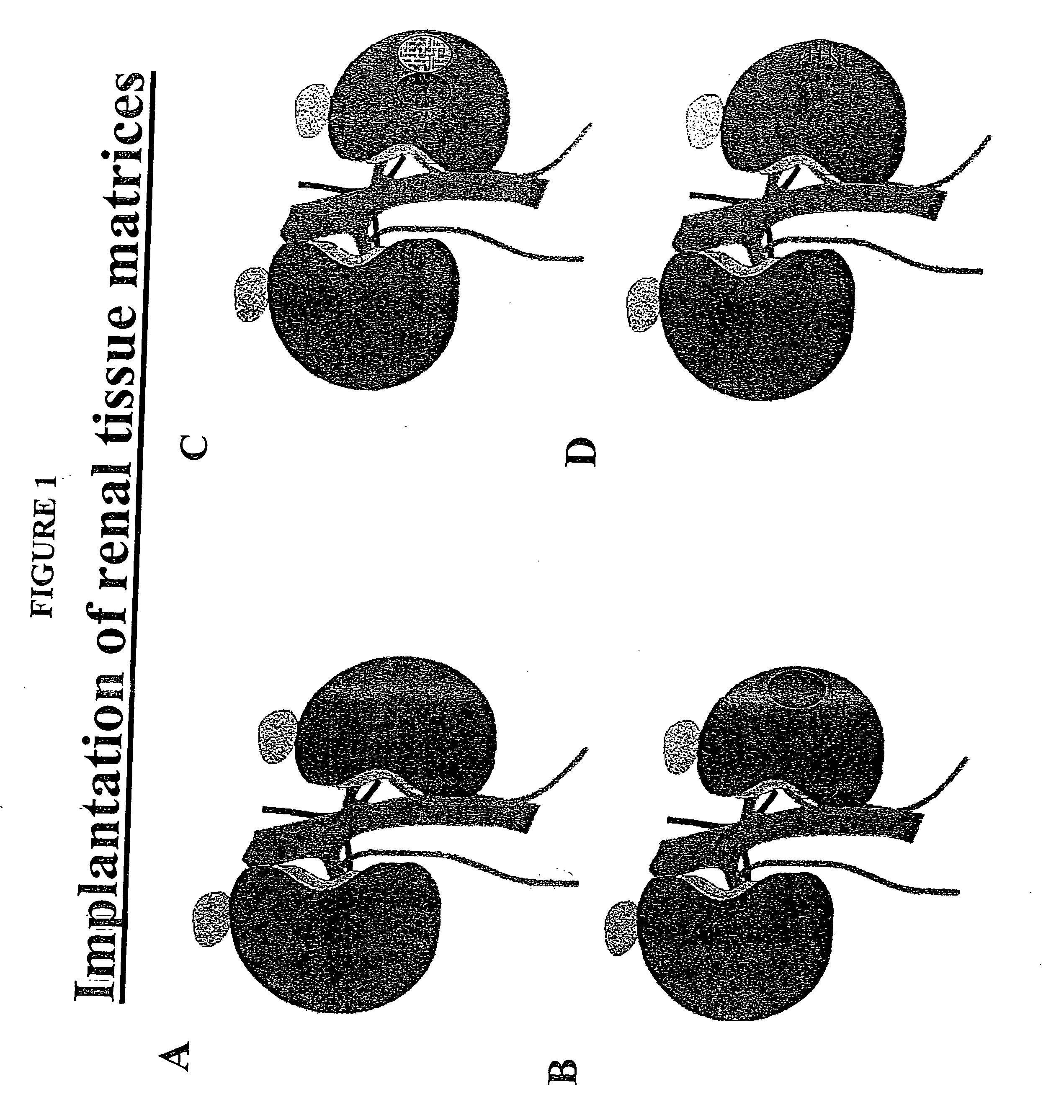
Augmentation of organ function
a technology for organs and functions, applied in the field of organ function augmentation, can solve the problems of reducing kidney function capacity, significant inconvenience for most patients, and significant percentage of any organ function los
- Summary
- Abstract
- Description
- Claims
- Application Information
AI Technical Summary
Benefits of technology
Problems solved by technology
Method used
Image

Examples
example 1
Isolation of Kidney Cells
[0129] Small kidneys, for example, from one week old C7 black mice, were decapsulated, dissected, minced and suspended in Dulbecco's Modified Eagles's Medium (DMEM; Sigma, St. Louis, Mo.) containing 15 mM Hepes, pH 7.4 and 0.5 μg / ml insulin, 1.0 mg / ml collagenase and 0.5 mg / ml dispase, a neutral protease from Bacillus polymyxal (Boehringer Mannheim, Indianapolis, Ind.).
[0130] Large kidneys, for example, swine kidneys, were arterially perfused at 37° C. for 10 minutes with calcium free Eagles minimum essential medium within three hours of extraction. The kidneys were then perfused with 0.5 mg / ml collagenase (Type IV, Sigma, St. Louis, Mo.) in the same buffer supplemented with 1.5 mM MgCl2 and 1.5 mM CaCl2. The kidneys were then decapsulated, dissected, minced and suspended in Dulbecco's Modified Eagles's Medium (DMEM; Sigma, St. Louis, Mo.) containing 15 mM Hepes, pH 7.4 and 0.5 μg / ml insulin, 1.0 mg / ml collagenase and 0.5 mg / ml dispase, a neutral protease ...
example 2
In vitro Culturing of Kidney Cells
(i) Isolation of Rat Tail Collagen
[0132] Tendon was stripped from rat tails and stored in 0.12 M acetic acid in deionized water in 50 ml tubes. After 16 hours at 4° C. overnight.
[0133] Dialysis bags were pretreated to ensure a uniform pore size and removal of heavy metals. Briefly, the dialysis bag is submerged in a solution of 2% sodium bicarbonate and 0.05% EDTA and boiled for ten minutes. Multiple rinses of distilled water was used to remove the sodium bicarbonate and 0.05% EDTA.
[0134] The 0.12 M acetic acid solution comprising rat tendons was placed in treated dialysis bags and dialyzed for two or three days to remove acetic acid. The dialysis solution was changed every 3 to 4 hours.
(ii) Coating Tissue Culture Plates:
[0135] The culture flasks, 75 cm2, were coated with a solution containing about 30 μg / ml collagen (Vitrogen or rat tail collagen), about 10 μg / ml human fibronectin (Sigma, St. Louis, Mo.) and about 10 μg / ml bovine serum albu...
example 3
Isolation and Culturing of Endothelial Cells
[0138] Endothelial cells, were isolated from a dissected vein. Perivenous heparin / papaverine solution (3 mg papaverine HCl diluted in 25 ml Hanks balanced salt solution (HBSS) containing 100 units of heparin (final conc. 4 / ml)), was used to improve endothelial cell preservation. A proximal silk loop was placed around the vein and secured with a tie. A small venotomy was made proximal to the tie and the tip of vein cannula was inserted and secured in place with a second tie. A second small venotomy was made beyond the proximal tie and the vein was gently flushed with Medium 199 / heparin solution Medium 199 (M-199) supplemented with 20% fetal bovine serum, ECGF (100 mg / ml), L-glutamine, heparin (Sigma, 17.5 / ml) and antibiotic-antimycotic), to remove blood and blood clots. Approximately 1 ml of a collagenase solution (0.2% Worthington type I collagenase dissolved in 98 ml of M-199, 1 ml of FBS, 1 ml of PSF, at 37° C. for 15-30 min, and filter...
PUM
 Abstract
Description
Claims
Application Information