Hyperspectral microscope for in vivo imaging of microstructures and cells in tissues

a hyperspectral microscope and tissue technology, applied in the field of medical diagnostics, can solve the problems of sampling error, time-consuming tissue sectioning, and requiring tissue removal from the patient, and achieve the effects of high signal sensitivity, enhanced image contrast and visibility, and high resolution

Inactive Publication Date: 2007-07-12
LAWRENCE LIVERMORE NAT SECURITY LLC
View PDF11 Cites 67 Cited by
  • Summary
  • Abstract
  • Description
  • Claims
  • Application Information

AI Technical Summary

Benefits of technology

[0011] Accordingly, the present invention is directed to a high resolution optical imaging method, that includes: illuminating at one or more incidence angles, one or more structures and cells of tissue components using one or more predetermined off-axis illumination sources; microscopically interrogating the illuminated structures and cells using one or more optical elements, wherein the optical elements further include long working distance objectives; collecting one or more spectral bands and/or a degree of polarization states resulting from the interrogation; and processing the collected one or more spectral bands and/or polarization states resulting from the one or more incidence illumination angles to enhance image contrast and visibility of the interrogated structures and cells.
[0012] Still another aspect of the present invention is directed to an optical apparatus that can provide high signal sensitivity while utilizing various optical imaging approaches. Such a system utilizes long working distance microscope objectives so as to enable off-axis illumination of predetermined tissue thereby allowing for excitation at any optical wavelength and so as to substantially eliminate spectral noise from the optical elements.
[0013] Accordingly, the present provides hyperspectral imaging methods and a simple less expensive microscope apparatus that enables real-time monitoring of microstructures...

Problems solved by technology

While this process has led to a much deeper understanding of cellular structure, tissue sectioning is time consuming, requires removal of tissue from the patient, and has inherent sampling error.
However, the most important limitation is the delay, due to processing, in providing the surgeon with clinically relevant information at the time of surgery.
These nonlinear imaging techniques offer unique diagnostic capabilities, however their implementation in a clinical environment requires the accommodation of complex instrumentation and specialized technical expertise.
In addition, the focuse...

Method used


Image

  • Hyperspectral microscope for in vivo imaging of microstructures and cells in tissues
  • Hyperspectral microscope for in vivo imaging of microstructures and cells in tissues
  • Hyperspectral microscope for in vivo imaging of microstructures and cells in tissues

Examples


Embodiment Construction

[0035] Referring now to the drawings, specific embodiments of the invention are shown. The detailed description of the specific embodiments, together with the general description of the invention, serves to explain the principles of the invention.

General Description

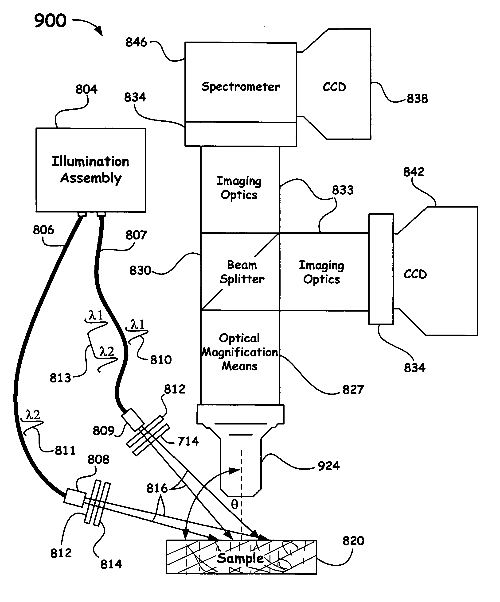
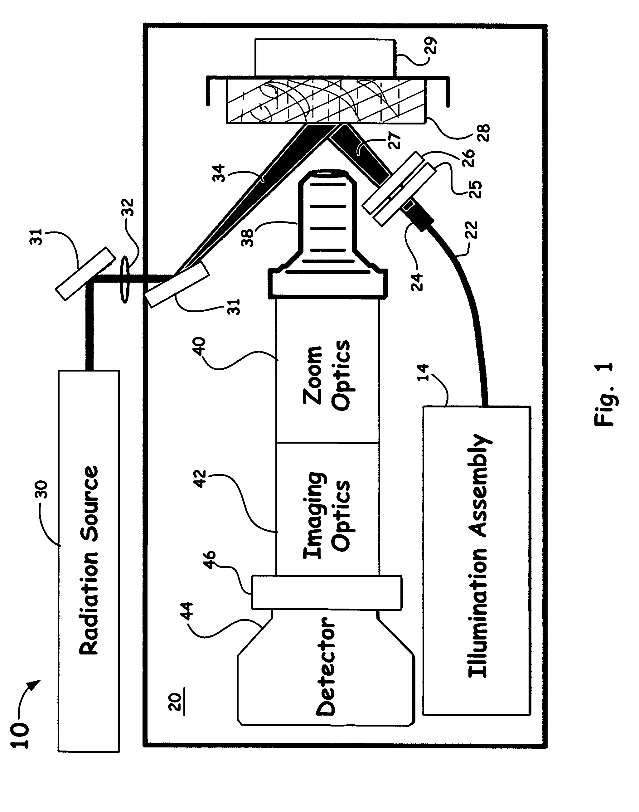
[0036] A real-time monitoring microscope system of the present invention (suitable for in vivo application in a clinical setting) provides: a) contrast between components of intact tissue with no processing to reveal histopathologic information, b) sufficiently high spatial resolution of down to about 0.5 μm to separate structures and components of interest and, c) fast image acquisition by utilizing pulsed sources for real time imaging of an object (tissue component or organ) that may be continuously moving due to heartbeat and blood flow.

[0037] The main strength of the microscope of the present invention for interrogation of microstructures and cells in tissues is the combined investigative hyperspectral / multimodal ...


PUM


Abstract

An optical hyperspectral/multimodal imaging method and apparatus is utilized to provide high signal sensitivity for implementation of various optical imaging approaches. Such a system utilizes long working distance microscope objectives so as to enable off-axis illumination of predetermined tissue thereby allowing for excitation at any optical wavelength, simplifies design, reduces required optical elements , significantly reduces spectral noise from the optical elements and allows for fast image acquisition enabling high quality imaging in-vivo. Such a technology provides a means of detecting disease at the single cell level such as cancer, precancer, ischemic, traumatic or other type of injury, infection, or other diseases or conditions causing alterations in cells and tissue micro structures.

Description

[0001] The United States Government has rights in this invention pursuant to Contract No. W-7405-ENG-48 between the United States Department of Energy and the University of California for the operation of Lawrence Livermore National Laboratory.BACKGROUND OF THE INVENTION [0002] 1. Field of the Invention [0003] The present invention relates to a medical diagnostic for investigating tissue components. More particularly, the present invention relates to a micro-scale instrument that utilizes autofluorescence emission and / or polarized elastic light scattering for real-time monitoring of microstructures and cells in tissues. [0004] 2. Description of Related Art [0005] The diagnostic gold-standard of histological evaluation of living tissues typically entails fixation, sectioning, and staining to obtain thin samples which exhibit high contrast under the microscope. While this process has led to a much deeper understanding of cellular structure, tissue sectioning is time consuming, require...

Claims


Application Information

Patent Timeline
IPC IPC(8): G06K9/00G02B21/00
CPCA61B5/0071A61B5/0075A61B5/0086A61B5/0088A61B5/0091G02B21/16G01N21/6458G01N21/6486G01N2021/4792G02B21/082A61B5/417
Inventor DEMOS, STAVROS G.
Owner LAWRENCE LIVERMORE NAT SECURITY LLC
Why Eureka
Try Eureka