Compositions and methods for convection enhanced delivery of high molecular weight neurotherapeutics

Inactive Publication Date: 2007-11-08
RGT UNIV OF CALIFORNIA
View PDF54 Cites 27 Cited by
  • Summary
  • Abstract
  • Description
  • Claims
  • Application Information

AI Technical Summary

Benefits of technology

[0012] The invention is directed to the therapeutic treatment of CNS disorders. The invention overcomes problems associated with many previous treatment regimens by employing local convection enhanced delivery. The invention additionally overcomes issues associated with local delivery, such as limited tissue distribution, unwanted binding site interaction and toxicity with the use of high molecular weight neurotherapeutics and, optionally, a facilitating agent. The invention stems in part from the finding that high molecular weight neurotherapeutics comprising active therapeutic agents may be convected in the CNS of large mammals and exhibit increased tissue distr

Problems solved by technology

Disorders of the central nervous system (CNS) often result in serious morbidity, death or impairment of mobility because of the lack of effective surgical or medical therapies.
Although potentially therapeutic compounds exist for treating many of these disorders, delivering effective doses of these agents selectively to target CNS tissue has remained a challenge.
Systemic toxicity and an inability to cross the blood brain barrier frequently compromise the efficacy of compounds that exhibit promising activity in vitro.
Additionally, many compounds that are capable of crossing the blood brain barrier exhibit non-uniform, inconsistent patterns of distribution as well as a frequent inability to effectively penetrate target tissue.
Further, compounds delivered intraventricularly have also exhibited non-uniform distribution and poor target tissue penetration.
Accordingly, the poor efficacy exhibited by therapeutic agents to date in respect of the treatment of CNS disorders may be due to administration and tissue distribution rather than the activity of agents per se.
However, there are a number of limitations associated with direct infusion and diffusion-based delivery of therapeutics to target CNS regions, the most critical being a low tissue distribution volume.
Without means for monitoring the distribution of infused neurotrophin, it is difficult to determine the therapeutic efficac

Method used


Image

  • Compositions and methods for convection enhanced delivery of high molecular weight neurotherapeutics
  • Compositions and methods for convection enhanced delivery of high molecular weight neurotherapeutics
  • Compositions and methods for convection enhanced delivery of high molecular weight neurotherapeutics

Examples


example 1

Gadolinium-Loaded Liposomes Allow for Real-Time Magnetic Resonance Imaging of Convection-Enhanced Delivery in the Primate Brain

[0181] The robust distribution of liposomes obtained in small rodent brains in the prior art did not guarantee similar results in the much larger primate CNS. Therefore, several key factors were further explored in the present studies. These included correlating the volume of infusion and volume of distribution in larger brains, developing a real-time imaging system, establishing the convectabilty of high molecular weight therapeutics in large mammal brain, and evaluating the accuracy of MRI monitoring for detection of liposome distribution.

[0182] Distribution of liposomes after convection-enhanced delivery detected by fluorescence labeling (data not shown): In order to test the feasibility of CED of liposomes in the non-human primate brain, liposomes (20 mM phospholipids) loaded with fluorescent dyes (either rhodamine or Dil-DS, the difference in fluoresc...

example 2

Real-Time Visualization and Characterization of Liposomal Delivery into the Monkey Brain by Magnetic Resonance Imaging

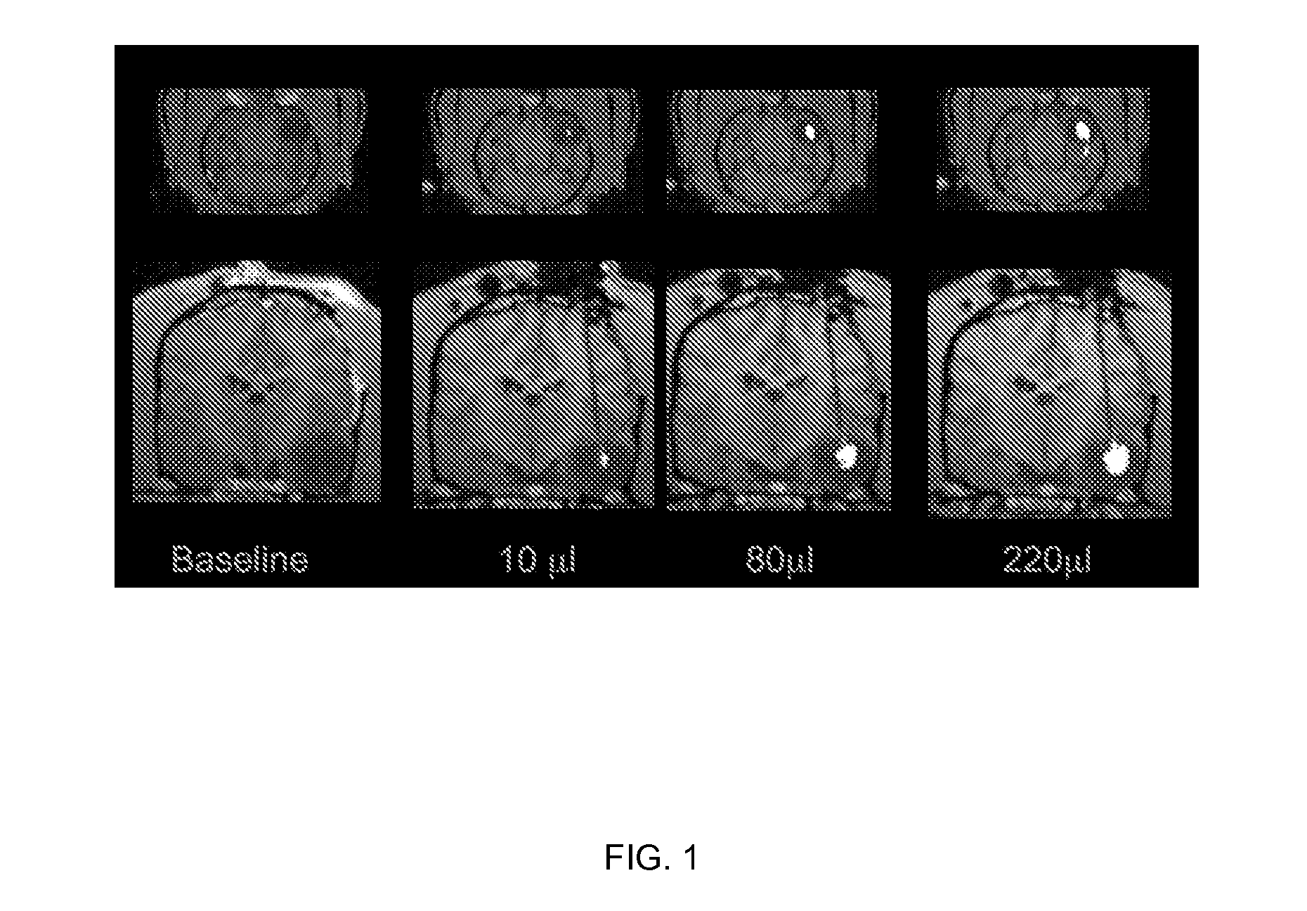
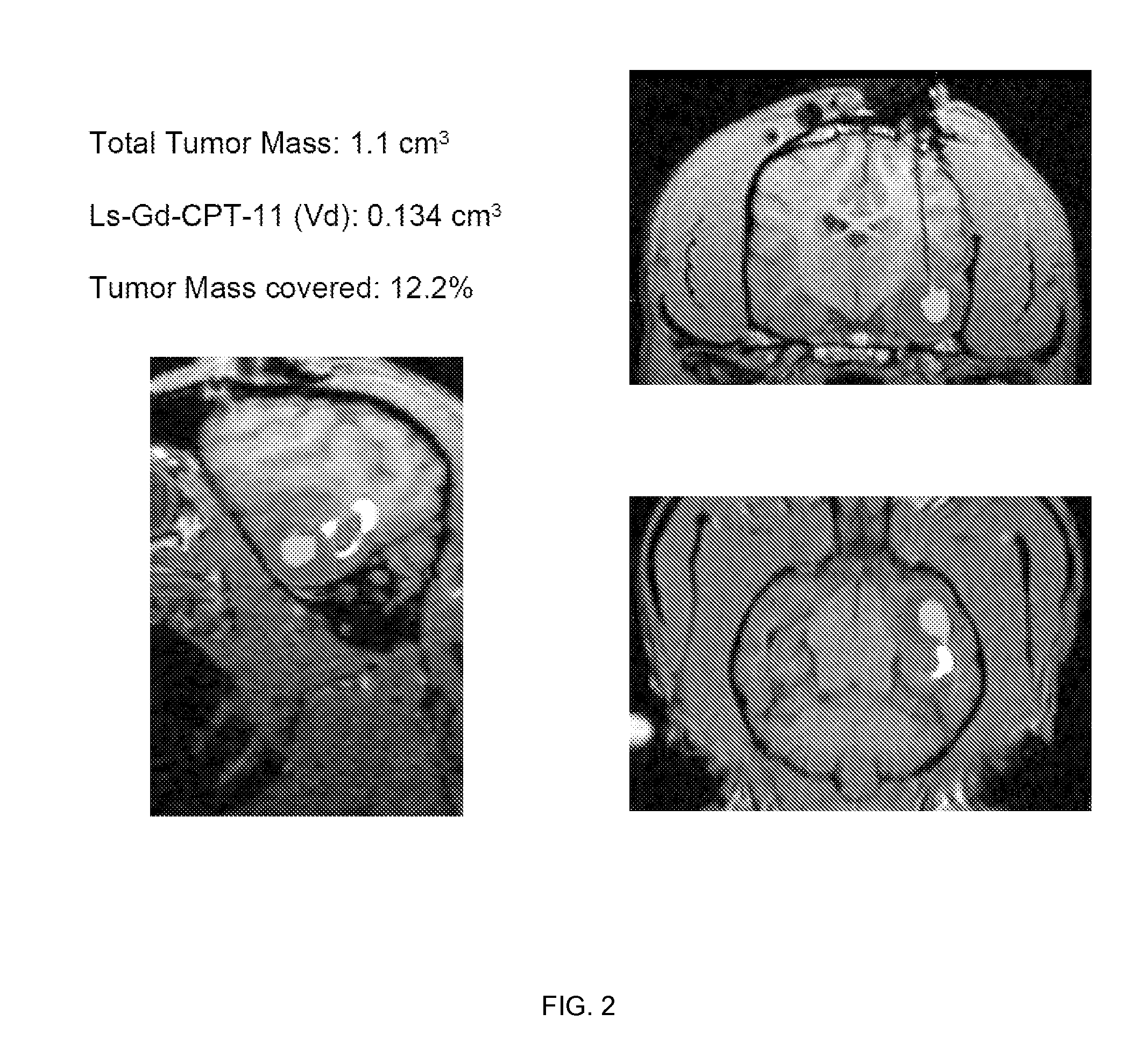
[0188] Magnet resonance imaging of Gadoteridol-loaded liposomes during CED in primate brain (data not shown): Infusion was started simultaneously in all three targeted regions (brainstem, putamen and corona radiata) of primate CNS, and placement of cannulas was verified before infusion pumps were turned on. After stabilization of animal vital signs, up to 700 μl of liposomes was convected with increasing rates of infusion. Robust and reflux-free Vd was achieved at all 3 sites. Brainstem infusion distributed rostrally towards mid-brain and caudal towards medulla oblongata. Some distribution into cerebellum via superior cerebellar peduncle was seen at 700 μl infusion. Liposomal distribution in corona radiata was primarily confined to white matter, and distributed into the non-infused contralateral hemisphere via corpus callosum above 500 μl infusion volume. Infusion i...

example 3

Effects of the Perivascular Space on Convection-Enhanced Delivery of Liposomes in Primate Putamen

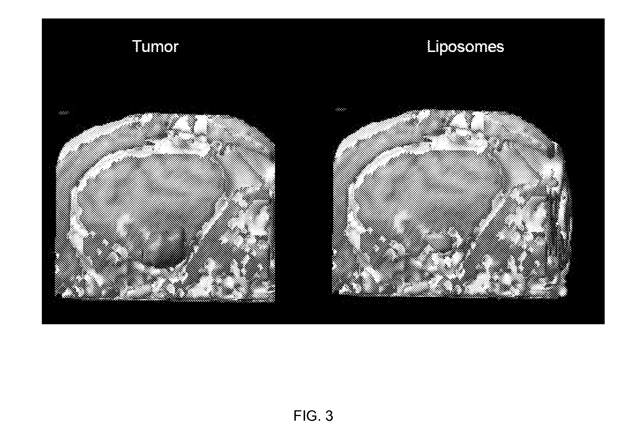
[0193] MRI monitored leakage out of non-human primate striatum after liposomal infusion (data not shown): We established a method to monitor in real time the infusion of liposomes loaded with a surrogate marker. We then used this system to infuse various anatomical structures in non-human primate brain including putamen. CED of up to 300 μl of liposomes was performed in non-human primate putamen, and subsequent distribution was monitored. Placement of cannula in primate putamen was verified for each animal by MRI prior infusion of liposomes. MRI was used to monitor CED of liposomes throughout the infusion procedure and reflux-free delivery was established to ensure optimal convection parameters. After starting the primate putamen infusion procedure, signal enhancement was detected in the perivascular space of the medial cerebral artery (MCA). At the lateral putamen border, lateral stria...


PUM

PropertyMeasurementUnit
Diameteraaaaaaaaaa
Atomic weightaaaaaaaaaa
Compositionaaaaaaaaaa

Abstract

A method of therapeutic treatment of CNS disorders using local convection enhanced delivery.

Description

CROSS-REFERENCE TO RELATED APPLICATIONS [0001] This application claims priority to U.S. provisional patent application Ser. No. 60 / 795,371 filed on Apr. 26, 2006, incorporated herein by reference in its entirety, and to U.S. provisional patent application Ser. No. 60 / 900,492 filed on Feb. 9, 2007, incorporated herein by reference in its entirety.STATEMENT REGARDING FEDERALLY SPONSORED RESEARCH OR DEVELOPMENT [0002] This invention was made with Government support under Grant No. P50 CA097257 awarded by the National Institutes of Health (NIH), and under Grant No. U54 NS045309 awarded by the National Institute of Neurological Disorders and Stroke (NINDS). The Government has certain rights in this invention.INCORPORATION-BY-REFERENCE OF MATERIAL SUBMITTED ON A COMPACT DISC [0003] Not Applicable NOTICE OF MATERIAL SUBJECT TO COPYRIGHT PROTECTION [0004] A portion of the material in this patent document is subject to copyright protection under the copyright laws of the United States and of...

Claims


Application Information

Patent Timeline
IPC IPC(8): A61K38/17A61K9/127
CPCA61K9/1271A61K9/1272A61K49/1812A61K31/4745A61K38/17A61K31/00A61P25/00A61P35/00
Inventor BANKIEWICZ, KRYSTOF S.KUNWAR, SANDEEP
Owner RGT UNIV OF CALIFORNIA
Why Eureka
Try Eureka