

Superparamagnetic Gadolinium Oxide Nanoscale Particles and Compositions Comprising Such Particles
a gadolinium oxide nanoscale and superparamagnetic technology, applied in the field of superparamagnetic gadolinium oxide nanoparticles, can solve the problems of signal loss, impede tissue fine structure delineation, weak signal intensity enhancement of such agents,
- Summary
- Abstract
- Description
- Claims
- Application Information
AI Technical Summary
Benefits of technology
Problems solved by technology
Method used
Image

Examples
example 1
Synthesis of Gd2O3 Nanocrystals Coated with Diethylene Glycol (DEG)
[0026] Nanocrystalline gadolinium oxide was synthesized by the polyol method, as described previously in Feldmann C. Polyol-mediated synthesis of nanoscale functional materials. Adv. Funct. Mater. 2003; 13: 101-107; Bazzi R et al., Synthesis and luminescent properties of sub-5-nm lanthanide oxides nanoparticles, Journal of Luminescence. 2003; 102-103: 445-450; and Söderlind, F., et al., Synthesis and characterization of Gd2O3 nanocrystals functionalized by organic acids, J. Colloid Interface Sci., 288: 140-148 (2005).
[0027] Gd(NO3)36H2O (2 mmol), solid NaOH (2.5 mmol) and de-ionized water (a few drops) was dissolved in 15 ml diethylene glycol ((HOCH2CH2)2O, DEG) and the mixture is heated to 140° C. When the reactants are completely dissolved, the temperature is raised to 180° C. and held constant for 4 h, yielding a dark yellow colloid. The colloid is diluted with deionized water to adjust the gadolonia concentrati...
example 2
Synthesis of Gd2O3 Nanocrystals Coated with Other Agents, or Alternative Synthesis
[0028] Gd(NO3)36H2O (2 mmol) and NaOH (6 mmol) were dissolved in two separate beakers, each containing 10 ml of DEG. The two solutions were mixed, heated to about 210° C., and held at that temperature for 30 minutes under stirring. To the hot solution oleic acid in DEG (1.6 mmol in 5 ml) was added yielding a brownish syrup. After washing and centrifuging several times in methanol, an off-white powder was collected. Oleic acid was replaced by, respectively, citric acid, 16-hydroxyhexadecanoic acid, 16-aminohexadecanoic acid, or hexadecylamine. In all cases, 1.6 mmol acid / amine in 5 ml DEG were used.
[0029] Gd2O3 nanocrystals can also be prepared with a rather different method, suitably called a combustion method [W. Zhang, et al., “Optical properties of nanocrystalline Y2O3:Eu depending on its odd structure”, J. Colloid and Interface Sc., 262 (2003) 588-593], was performed in the following way. Equal v...
example 3
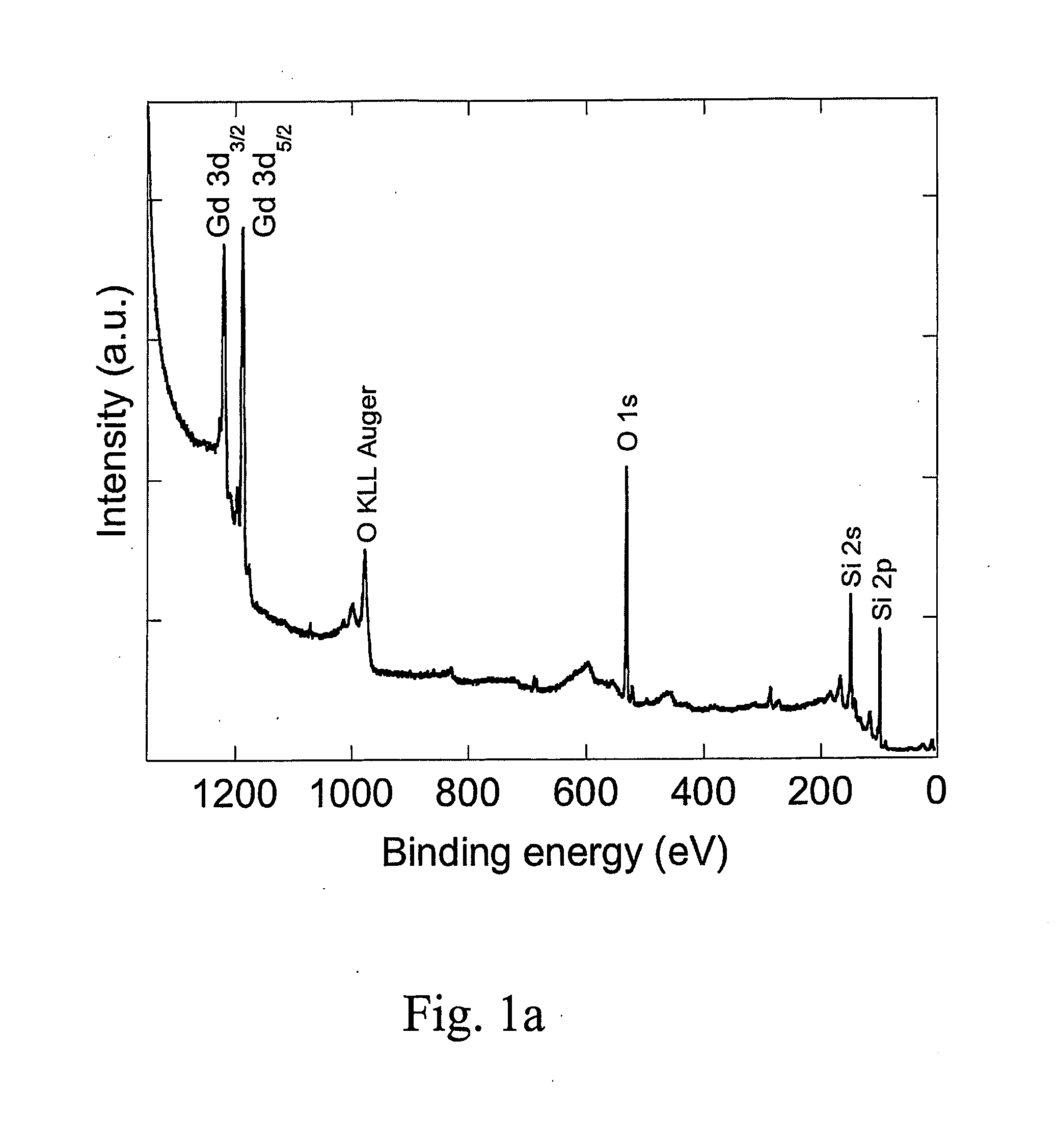
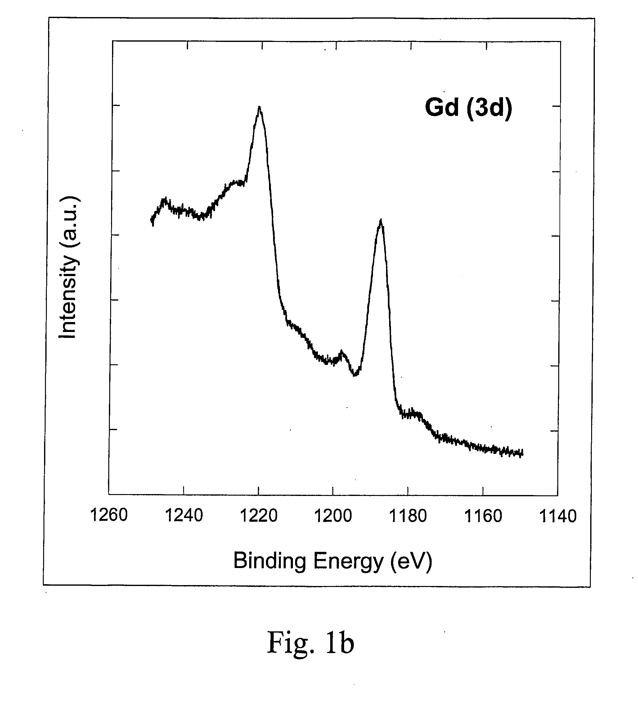
Characterisation with X-Ray Photoelectron Spectroscopy (XPS)
[0030] In order to confirm that correct Gd2O3 nanocrystals were prepared by studying composition and binding energy of the particles, the XPS spectra were recorded on a VG instrument using unmonochromatized Al Kα photons (1486.6 eV) and a CLAM2 analyzer. The power of the X-ray gun was 300 W. The spectra were based upon photoelectrons with a takeoff angle of 30° relative to the normal of the substrate surface. The pressure in the analysis chamber was 3*10−10 mbar and the temperature 297 K during the measurements. The VGX900 data analysis software was used to analyze the peak position. To clean the silicon (SiOx) substrates, the surfaces were first washed with a 6:1:1 mixture of MilliQ water: HCl (37%): H2O2 (28%) for 5-10 minutes at 80° C. followed by a 5:1:1 mixture of MilliQ water:NH3 (25%):H2O2 (28%) for 5-10 minutes at 80° C. The silicon surfaces were after each washing step carefully rinsed with MilliQ water. Gadoliniu...
PUM
 Abstract
Description
Claims
Application Information