Method of Analyzing Biosample by Laser Ablation and Apparatus Therefor

a biosample and laser ablation technology, applied in the direction of analysis using chemical indicators, particle separator tube details, instruments, etc., can solve the problems of mainly done qualitative analysis, long analysis time required, complicated staining and color development process, etc., to avoid swaying the analytical result, the time required for analysis can be shortened, and the result is highly reliable

Inactive Publication Date: 2008-01-24
RIKEN +1
View PDF2 Cites 24 Cited by
  • Summary
  • Abstract
  • Description
  • Claims
  • Application Information

AI Technical Summary

Benefits of technology

[0065]The present invention exerts an excellent effect that time required for analysis can be shortened significantly comparing to conventional art.
[0066]Further, the present invention exerts an excellent effect that highly reliable result can be obtained avoiding sway of analytical result by observers.
[0067]Further, the present invention exerts an excellent effect that one-time analysis of a multiplicity of genes or the like can be performed on a single biosample, workload and time efficiencies can be improved, analysis of a plurality of genes or the like can be performed under conditions completely free from any difference in background attributed from biosample.
[0068]Further, the present invention exert

Problems solved by technology

However, in the above-described conventional various method of analyzing biosample, there existed a problem that processes required for staining and color development was complicated and time required in analysis became long time.
Further, the above-described conventional various methods of analyzing a biosample, a man needs to analyze the biotissue section or the like after processing by observing it under a microscope, there was a problem that a qualitative analysis was mainly

Method used


Image

  • Method of Analyzing Biosample by Laser Ablation and Apparatus Therefor
  • Method of Analyzing Biosample by Laser Ablation and Apparatus Therefor
  • Method of Analyzing Biosample by Laser Ablation and Apparatus Therefor

Examples


example 1

[0116]In the following, description will be made for a method of fabricating a biotissue section, which is formed by slicing a mouse brain, as an example of fabricating a biosample.

1. Preparation of Mouse Brain

[0117]Animal: As a model animal, 10 to 11 week-old male wild-type CD-1 mice and C57BL / 6J purchased from Oriental Yeast Co., Ltd. were used.

[0118]Fixative as a reagent when preparing a mouse brain is as shown in the following table 1.

TABLE 1Reagent: fixativeNeutral fixative: 1 L 4% PFA (para form aldehyde) liquid (pH 7.5)PFA40gSucrose40gNa2HPO4•12H2O11.4gNaH2PO4•2H2O3.3gAcidic fixative: Original-Bouin liquid (pH 3.5 to 4.0)Saturated picric acid300mlFormalin100mlAcetic acid20ml

[0119]By using the above-described reagent, a paraffin section and a frozen section of the mouse brain were fabricated as biotissue sections by the following method.

[0120]

[0121]1) Fabricating a paraffin section

[0122]After anesthetizing a mouse by ether to eliminate pain response, an abdominal cavity was op...

example 2

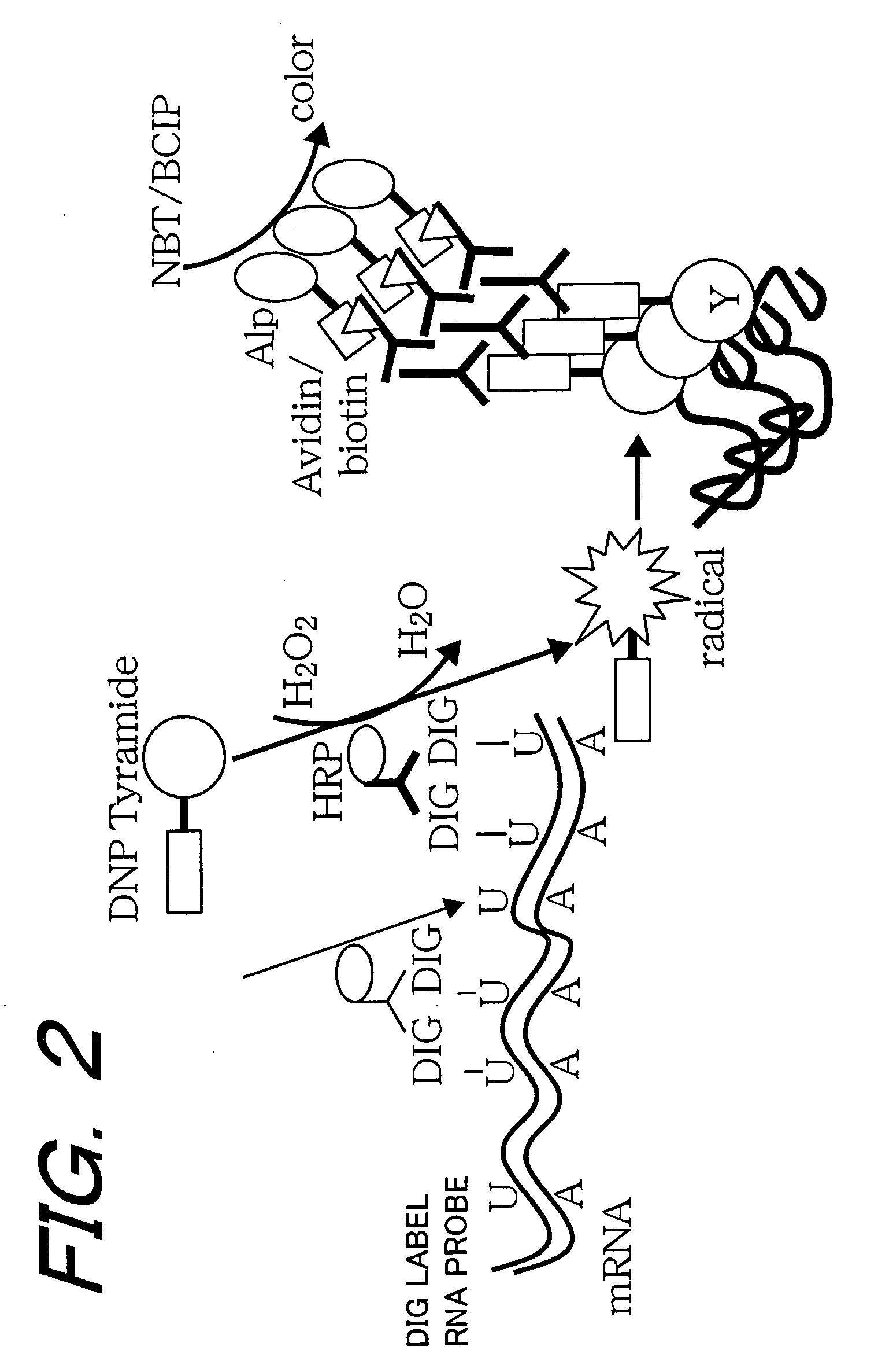
[0174]Next, description will be made for detection using a Pt-labeled RNA probe.

[0175]The expression of the gene of microtubule-associated protein MAP2, which exists on dendrite in a large volume, in the mouse brain was investigated.

[0176]It is to be noted that the preparation of the mouse brain, the design of the primer, and the purification of plasmid were performed in the same manner as “Example 1”.

1. Fabricating a Probe

[0177]1) Fabricating Pt-labeled RNA probe

[0178]A template DNA was created by the reaction liquid and reaction condition shown in FIG. 5. Further, an RNA probe was fabricated.

[0179]By using the PCR product and the primer containing the recognition sequence (*T7, SP6 adaptor) of T7 and SP6 polymerase, PCR was performed again. The reaction liquid composition and the reaction condition are as shown in Table 6. Electrophoresis was applied to the reaction product in 2% agarose gel and confirmed.

*T7 adaptor:GAGCGCGCGTAATACGACTCACTATAGGGCSP6 adaptor:TTGTGCGGCCATTTAGGTGACA...

modified example

[0191]It is to be noted that the above-described embodiments can be modified as shown in (1) to (12) below.

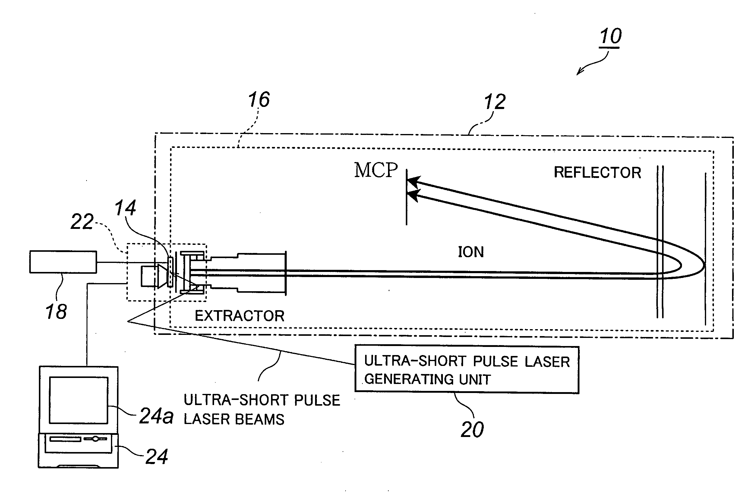
(1) In the above-described embodiments, the time-of-flight mass spectrometer that performs mass spectrometry by measuring the time of flight of atoms was used as a mass spectrometer, and mass spectrometry of a plurality of atoms can be performed simultaneously by one-time laser irradiation when the time-of-flight mass spectrometer is used. Further, even in the case where the ion cyclotron Fourier transform mass spectrometer is used as the mass spectrometer, mass spectrometry of a plurality of atoms can be performed simultaneously.

(2) In the above-described embodiments, description was made for mass spectrometry as an analysis method of molecule, but it goes without saying that the invention is not limited to this and the present invention may be used for analysis other than mass spectrometry.

(3) In the above-described embodiments, the rotational inlet terminal 18 that rotates t...


PUM

PropertyMeasurementUnit
Temperatureaaaaaaaaaa

Abstract

A method of analyzing a biosample that enables substantial shortening of time required for analysis, further enabling obtaining highly reliable results through means for avoiding sway of analytical results depending on observers, and that enables one-time analysis of a multiplicity of genes, etc. on a single biosample to thereby enhance workload and time efficiencies, and that enables analysis of multiple genes, etc. under conditions completely free from any difference in background attributed to biosamples. There is provided a method comprising irradiating a biosample as an analyte with ultra-short pulse laser beams to thereby effect an ablation thereof so that molecules contained in the biosample are atomized into constituting elements, ionizing the constituting elements resulting from the atomization and analyzing the ionized constituting elements to thereby analyze analytical-target molecules of the biosample.

Description

TECHNICAL FIELD [0001]The present invention relates to a method of analyzing a biosample using laser ablation and an apparatus therefor, more particularly to a method of analyzing a biosample using laser ablation and an apparatus therefor, which are capable of significantly improving analysis efficiency comparing to a conventional ones. For example, the invention relates to a method of analyzing a biosample using laser ablation and an apparatus therefor, in which a biotissue section is used as the biosample and which are preferable for use in the mass spectrometry of nucleic acid in the biotissue section.BACKGROUND ART [0002]Conventionally, as a method of analyzing a biosample such as a biotissue section, various chemical staining method and methods utilizing the hybridization of a nucleic-acid probe (in situ hybridization method) have mainly been used.[0003]Herein, the chemical staining method is a method of observing a tissue, the morphology of a cell, and the existence and the lo...

Claims


Application Information

Patent Timeline
IPC IPC(8): G01N33/48B01L11/00G01N1/00G01N24/00B01L99/00C12N15/09C12Q1/68G01N27/62G01N27/64G01N33/483G01N33/50G01N33/543H01J49/10H01J49/40
CPCG01N33/54373Y10T436/25Y10T436/24H01J49/162
Inventor HAYASHIZAKI, YOSHIHIDEKAWAI, JUNHAYASHI, TOSHIZOKAMIYA, MAMORU
Owner RIKEN
Why Eureka
Try Eureka