
Methods of detecting one or more cancer markers

a cancer marker and detection method technology, applied in the field of detection methods of one or more cancer markers, can solve the problems of ineffectiveness, inapplicability of nucleic acid based methods, and inability to detect specific or accurately, etc., and achieve rapid and facile detection and/or diagnosis, high sensitivity and selectivity, and early detection

Inactive Publication Date: 2008-01-31
ANTARA BIOSCI
View PDF50 Cites 25 Cited by
  • Summary
  • Abstract
  • Description
  • Claims
  • Application Information

AI Technical Summary

Benefits of technology

[0012] The present invention provides for the early, rapid and facile detection and / or diagnosis of cancer through the detection of cancer markers in biologic fluids including, inter alia, urine and blood. In certain embodiments, the present invention solves the problem of multiplex detection for multiple cancer markers, while eliminating the need for nucleic acid isolation / amplification and the problems associated with non-specific nucleic acid hybridization. The non-specific hybridization and low sensitivity observed in the detection methods currently known in the art are overcome by the present invention which provides novel methods that exploit, in a synergistic manner, the high sensitivity and selectivity of antibody: antigen interaction and nucleic acid hybridization.
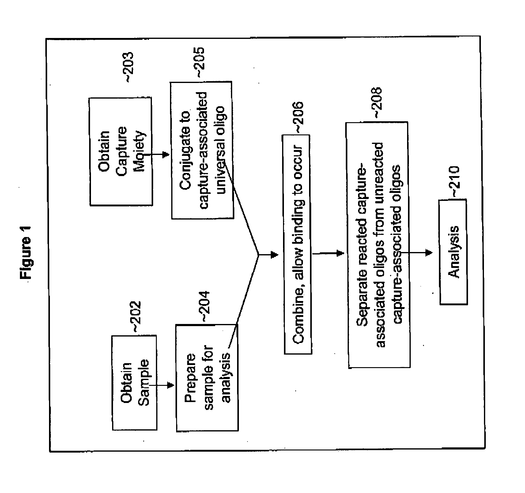
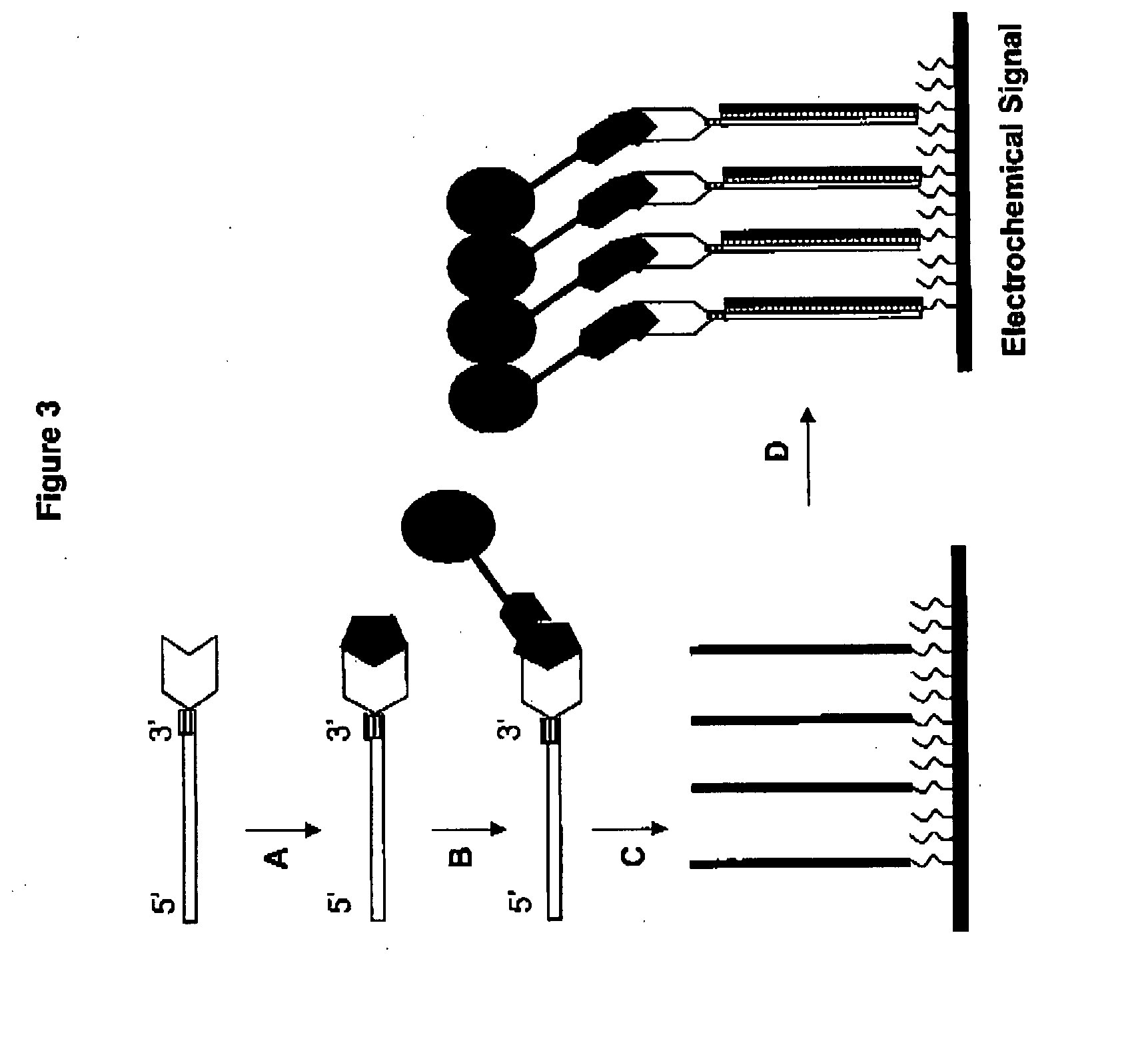
[0018] In a preferred embodiment, the complexed capture-associated oligo is subjected to one or more rounds of one or more types of amplification. In a particularly preferred embodiment, isothermal amplification can be employed to produce the polymerization products to increase the number of single stranded nucleic acid molecules available for binding (e.g., annealing to, hybridizing with, etc.) to the chip-associated oligo, thereby enhancing the signal created through a hybridization event. In these embodiments, the capture-associated oligo can be used as a template for linear amplification, with the capture-associated oligo being preferably designed to encode a complementary (or substantially complementary) sequence to a polymerase recognition sequence at its 3′ end following the complementary (or substantially complementary) region of the chip-associated oligo. Following interaction of the cancer marker with the capture moiety, the resultant complex is isolated (e.g., via immobilization with immobilized binding partners) and contacted with a “priming” oligonucleotide—an oligonucleotide that is complementary to, inter alia, the 5′ to 3′ polymerase recognition sequence to form a double-stranded polymerase recognition site. Following annealing of the priming oligonucleotide to the capture-associated oligo, an excess of mononucleotides and the appropriate polymerase(s) can be added under conditions that promote of otherwise facilitate polymerization and linear amplification of the capture-associated oligo to form polymerization products. This polymerization reaction is preferably performed under conditions that allow for the repeated use of the template strand (e.g., the capture-associated oligo) for multiple rounds of polymerization so as to result in multiple copies of the polymerization products being formed from each such complexed capture-associated oligo. In such embodiments, the chip-associated oligo will have the same sequence (or substantially similar sequence) as the capture-associated oligo, and both will be complementary (or substantially complementary) to the polymerization product(s). In a preferred embodiment, the polymerase recognition site created by this double-stranded region is a phage-encoded RNA polymerase recognition sequence (e.g., polymerase recognition sequences for T7, T3, SP6, and the like).

Problems solved by technology

Because many markers identified for various cancer states are present in trace amounts (low concentrations in biological samples), traditional antibody-based techniques fail to detect them specifically or accurately, if at all.
Nucleic acid based methods are not applicable and / or are ineffective for a variety of reasons including, inter alia, the common problem that the gene that encodes the marker in question may be identified or identifiable, but is not detectable or amplifiable.

Method used


Image

  • Methods of detecting one or more cancer markers
  • Methods of detecting one or more cancer markers
  • Methods of detecting one or more cancer markers

Examples


example i

Preparation of Monoclonal Antibodies

[0140] A peptide corresponding to amino acid residues in a desired cancer marker antigen is synthesized with a peptide synthesizer (Applied Biosystems) according to methods known in the art. The peptide emulsified with Freund's complete adjuvant is used as an immunogen. And administered to mice by footpad injection for primary immunization (day 0). The booster immunization is performed four times or more in total. The final immunization is carried out by the same procedure two days before the collection of lymph node cells. The lymph node cells collected from each immunized mouse and mouse myeloma cells are mixed at a ratio of 5:1. Hybridomas are prepared by cell fusion using polyethylene glycol 4000 or polyethylene glycol 1500 (GIBCO) as a fusing agent. The lymph node cells of the mouse are fused with mouse myeloma PAI cells (JCR No. B0113; Res. Disclosure Vol. 217, p. 155, 1982), and the resulting hybridomas are selected by culturing the fused ...

example ii

Preparation of DNA-Antibody Conjugates

[0143] A capture-associated universal oligonucleotide can be prepared on a solid support that has been treated with 3-amino-1,2-propanediol in order to introduce the 3′ amino group with an automated DNA synthesizer (e.g., 3400 DNA synthesizer, Applied Biosystems). Typical cleavage and purification steps are employed to obtain the modified universal oligonucleotide. The universal oligonucleotide is then incubated with N-succinimidyl 3-(2-pyridyldithio)propionate in PBS at a molar ratio between 1:30 to 1:35 for 30 minutes at room temperature. Dithiothreitol is typically added to this solution, resulting in a final concentration of 10 mM for 5 minutes. The universal oligonucleotide is then purified and recovered by applying this reaction mixture to a PBS equilibrated Sepharose column, washing the column several times, and eluting the universal oligonucleotide in a 0.6M NaCl phosphate buffer.

[0144] A monoclonal antibody is incubated with γ-maleimi...

example iii

Immobilization of Nucleic Acid Probe to a Platinum Electrode Surface

[0146] A platinum electrode is exposed to a high temperature to air-oxidize the surface of the electrode. The oxidized electrode is treated with cyanogen bromide (CNBr) to activate the oxide layer. The nucleic acid is attached to the electrode by contacting the electrode in a solution of single stranded nucleic acid. The single stranded nucleic acid is obtained by commonly employed means including, but not limited to, either standard oligonucleotide synthesis techniques or by thermal denaturation of a double stranded nucleic acid molecule.

[0147] Alternatively, a custom synthesized oligonucleotide containing a thiol group at the 5′ or the 3′ end is spotted on a gold electrode. This procedure involves placing approximately 100 nL of the probe solution containing the oligonucleotide probe (5 μmol / L), 400 mmol / L sodium chloride, and 0.1 mmol / L HCl, on the electrode and then keeping the electrode at room temperature fo...


PUM

PropertyMeasurementUnit
Electrical conductanceaaaaaaaaaa
Electric chargeaaaaaaaaaa

Abstract

The present invention provides a methods and compositions for early diagnosis of cancer by rapid and specific detection of one or more cancer markers in a sample.

Description

CROSS REFERENCE TO RELATED APPLICATIONS [0001] This application claims the benefit of U.S. Provisional Patent Application Ser. No. 60 / 830,129, filed Jul. 11, 2006, currently pending, which is herein incorporated by reference in its entirety for all purposes.FIELD OF THE INVENTION [0002] This invention relates to methods and compositions capable of rapid diagnosis of cancers as well as kits for performing such diagnosis. BACKGROUND OF THE INVENTION [0003] In the following discussion certain articles and methods will be described for background and introductory purposes. Nothing contained herein is to be construed as an “admission” of prior art. Applicant expressly reserves the right to demonstrate, where appropriate, that the articles and methods referenced herein do not constitute prior art under the applicable statutory provisions. [0004] Enzyme-linked immunosorbent assay (ELISA) is a widely used method for measuring the concentration of a particular molecule (e.g., a hormone or dr...

Claims


Application Information

Patent Timeline
IPC IPC(8): C12Q1/68
CPCC12Q1/6886
Inventor LABGOLD, MARC R.JOKHADZE, GEORGE G.
Owner ANTARA BIOSCI
Learn More