Method and arrangement for high-resolution microscope imaging or cutting in laser endoscopy

a laser endoscope and microscope technology, applied in the field of high-resolution microscope imaging or laser endoscopy, can solve the problems of hindering the transmission of femtosecond pulses, low excitation efficiency and detection efficiency, and no commercial light endoscope with focusing optics having a high numerical aperture. achieve the effect of precise imaging and/or micro-cutting

Active Publication Date: 2008-04-03
JENLAB
View PDF9 Cites 153 Cited by
  • Summary
  • Abstract
  • Description
  • Claims
  • Application Information

AI Technical Summary

Benefits of technology

[0018]It is the object of the invention to find a novel possibility for endoscopic application of multiphoton processes which permits a precise imaging and / or microcutting of materials, particularly biological materials, with an accuracy of under one millimeter by means of femtosecond laser radiation.

Problems solved by technology

Thus far, there have been no commercial light endoscopes with focusing optics having a high numerical aperture.
In addition, all commercial light endoscopes are based on optical materials and light guides which hinder the transmission of femtosecond pulses due to high dispersion.
However, the resolution, excitation efficiency and detection efficiency are low due to the low NA.
This would require cost-intensive, elaborate laser apparatus.
Further, there would be a high risk potential.
A cutting precision in the sub-100 μm range within the target could not be achieved because of collateral damage, including the formation of large cavity bubbles in a range greater than a cell dimension and uncontrolled auto-focusing (collateral damage correlates to the pulse energy).
All of the methods and arrangements mentioned above have the disadvantage that they cannot be applied for endoscopic use of multiphoton processes by means of the radiation of a femtosecond laser inside materials and within the body for precision image generation and / or cutting with an accuracy of under one millimeter.
At the present time, optical microscopic scanning methods are mostly used for external or near-surface examinations because flexible, miniaturized devices have not succeeded in scanning methods due to the absence of miniaturized scanners.

Method used


Image

  • Method and arrangement for high-resolution microscope imaging or cutting in laser endoscopy
  • Method and arrangement for high-resolution microscope imaging or cutting in laser endoscopy
  • Method and arrangement for high-resolution microscope imaging or cutting in laser endoscopy

Examples


Embodiment Construction

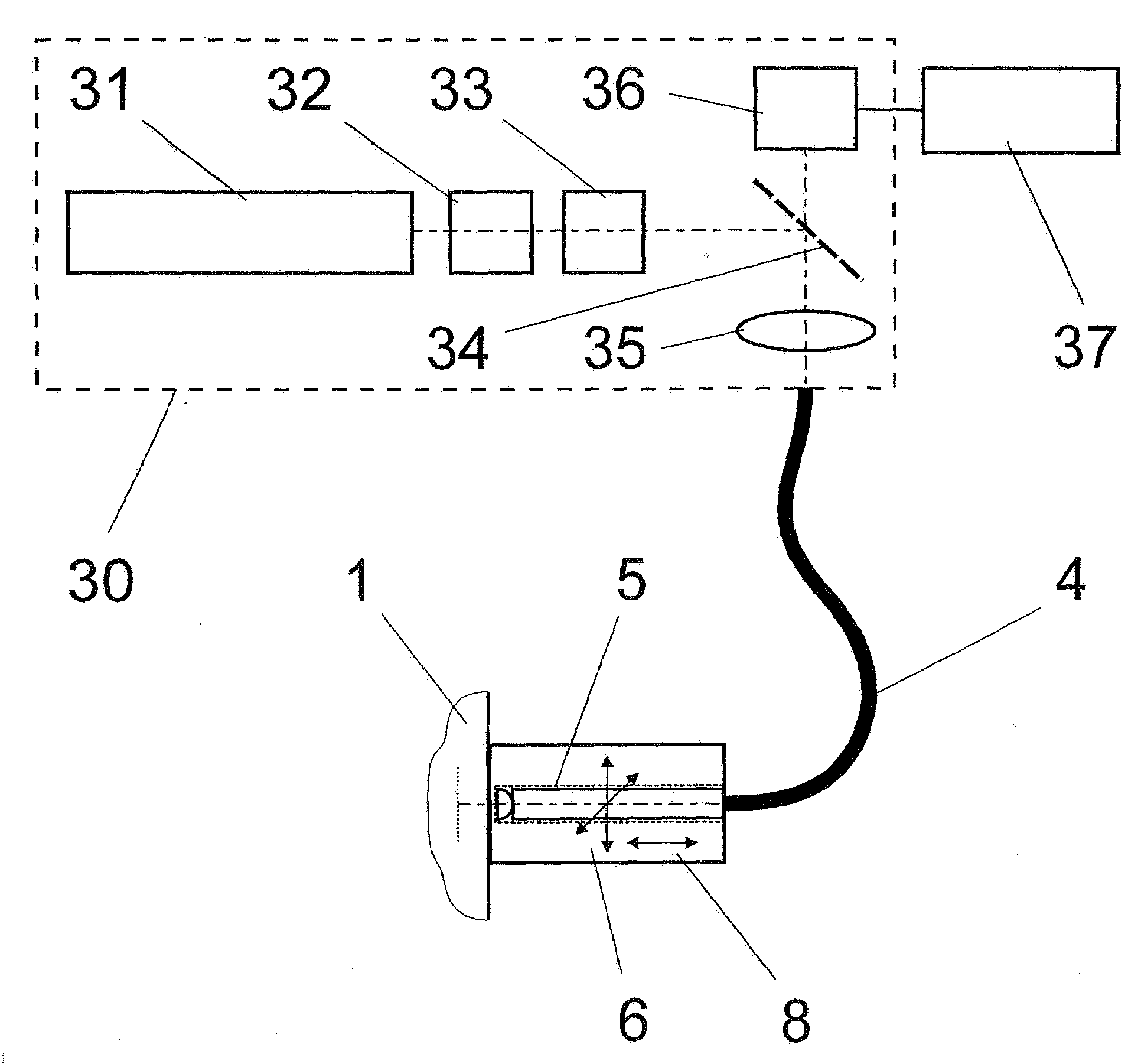
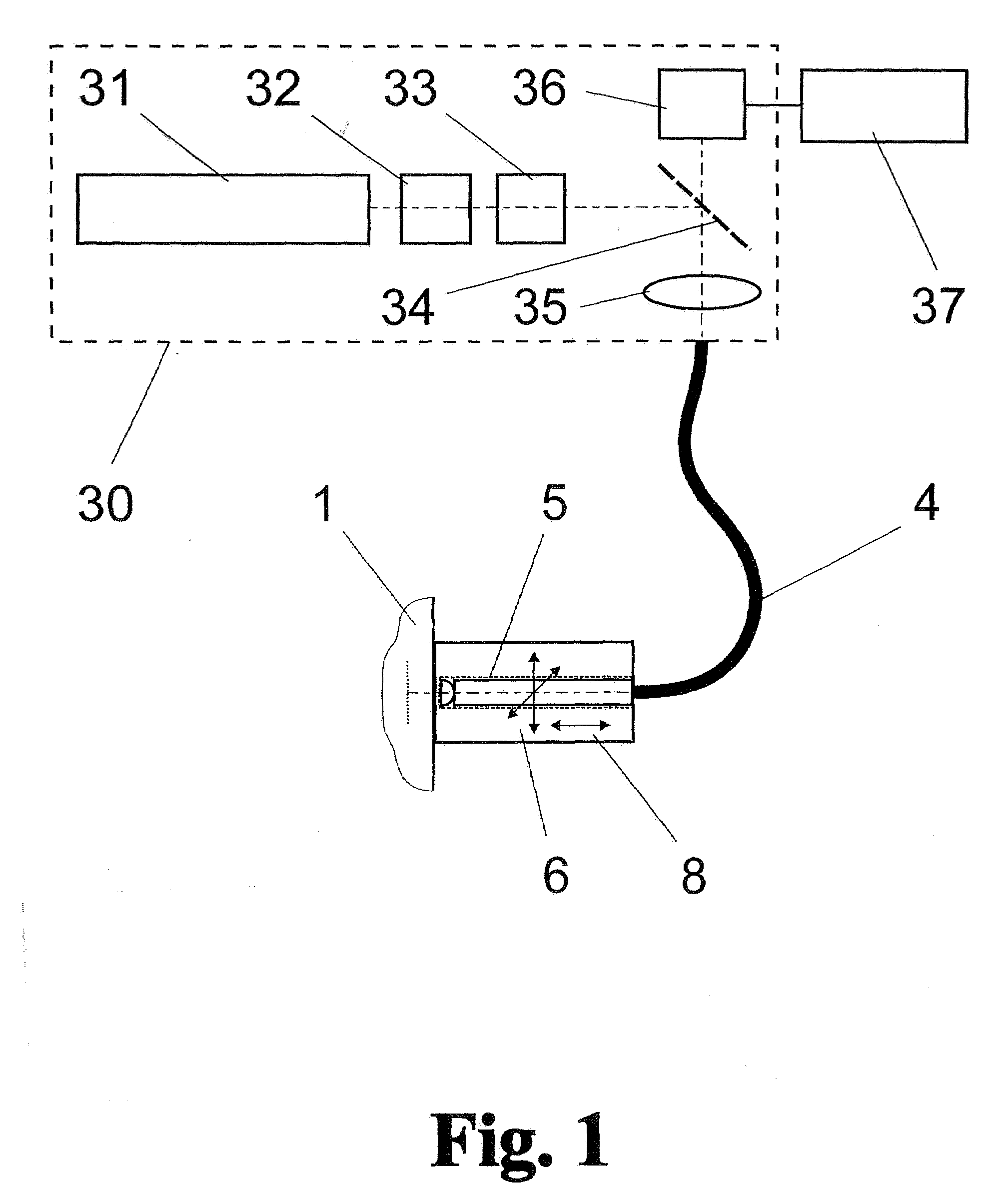
[0070]The method according to the invention will be described in the following—without limiting its universality for the application of other laser-assisted imaging methods—with reference to a femtosecond laser microscope such as is used for single-photon, two-photon and multiphoton microscopy.

[0071]As is shown in FIG. 1, the arrangement comprises an illumination and detection device 30 in which pulsed radiation with an emission wavelength of 780 nm generated by an 80-MHz titanium-sapphire femtosecond laser 31 is transmitted through a shutter 32, a beam attenuator 33 and a dichroic splitter mirror 34 and provided in a focused manner by microscope optics 35 and in which a secondary radiation (e.g., single-photon, two-photon or multiphoton fluorescence, SHG, THG, luminescence, etc.) returning from an object 1 through laser excitation is coupled out through the splitter mirror 34 to a photon detector 36.

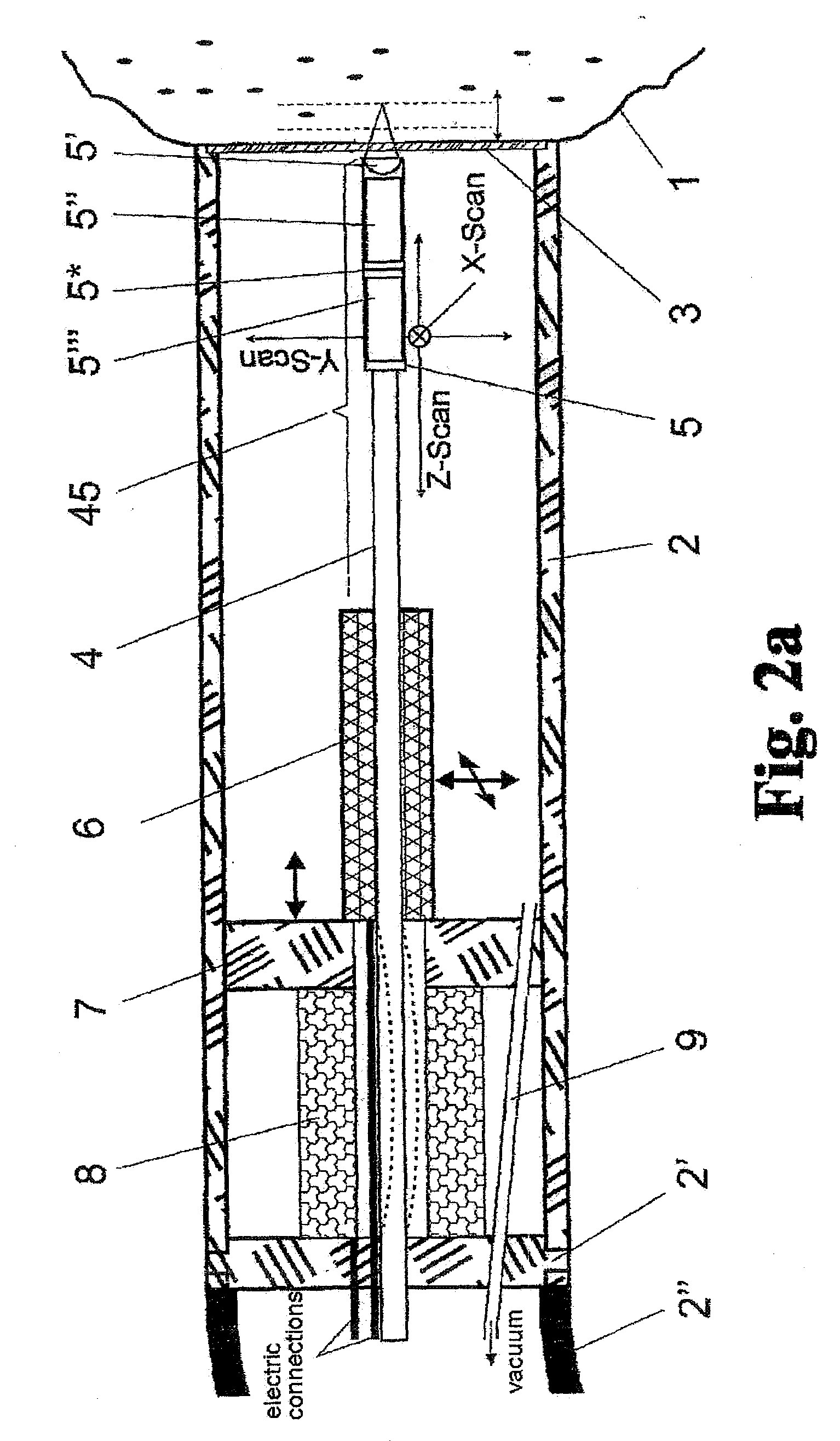
[0072]The microscope objective 35 of the illumination and detection device 30 coupl...


PUM


Abstract

The invention is directed to a method and an arrangement for high-resolution microscopic imaging in laser endoscopy based on laser-induced object reaction radiation and for performing microscopic cuts in biological tissue. In using multiphoton processes for endoscopic applications in biological materials with an accuracy of under one millimeter, radiation of a pulsed femtosecond laser is focused into an object by means of a transmission focusing optics unit comprising a transmission system and miniature focusing optics having a high numerical aperture greater than 0.55 to trigger a local object reaction radiation in the micrometer to nanometer range, and the distal end of the transmission focusing optics unit is moved in at least two dimensions for highly spatially resolved scanning of the object and for transmitting object reaction radiation which is scanned in a locally progressive manner to an image-generating system with a photon detector. In an other embodiment the femtosecond laser radiation is energy enhanced is applied to the same transmission focusing optics unit to perform microendoscopic surgery in biological tissue.

Description

CROSS-REFERENCE TO RELATED APPLICATIONS[0001]This application claims priority of German Application No. 10 2006 046 554.7, filed Sep. 28, 2006 and German Application No. 10 2006 046 925.9, filed Sep. 29, 2006, the complete disclosures of which are hereby incorporated by reference.BACKGROUND OF THE INVENTION[0002]a) Field of the Invention[0003]The invention is directed to a method and an arrangement for generating high-resolution microscopic imaging in endoscopy based on laser-induced object reaction radiation and for performing microscopic processing of biological material in endomicrosurgery.[0004]b) Description of the Related Art[0005]The invention is preferably applied in endoscopy based on multiphoton processes by means of femtosecond laser technology. In particular, it can be used in microimaging processes, laser scanning microscopy, optical coherence tomography, and single-photon and multiphoton imaging and is applied especially in endomicroscopy and endomicrosurgery due to th...

Claims


Application Information

Patent Timeline
Patent Type & Authority Applications(United States)
IPC IPC(8): A61B1/07A61B18/22
CPCA61B1/00172A61B5/0068A61B5/0084A61B5/415G02B26/101A61B18/20G02B23/2423G02B23/2469A61B5/418
Inventor KOENIG, KARSTENTCHERNOOK, ANDREI
Owner JENLAB
Why Eureka
Try Eureka