System and Method for Proteomics

a proteomics and system technology, applied in the field of systems and kits, can solve the problems of poor resolution, low throughput greatly limit the process, and most biomarker proteins are undetected by current methods, and achieve the effects of increasing resolution, low conductivity, and high field strength

Inactive Publication Date: 2008-11-06
PROTEIN FOREST
View PDF10 Cites 14 Cited by
  • Summary
  • Abstract
  • Description
  • Claims
  • Application Information

AI Technical Summary

Benefits of technology

[0012]Low conductivity buffers are preferred to increase resolution at high field strengths. It has been discovered that addition of a reducing agent such as dithiothreitol (“DTT”) and/or beta mercaptoethanol to the running buffer will enhance resolution a

Problems solved by technology

Human plasma is rich in biomarkers, however, most biomarker proteins are undetected by current methods.
The bottleneck in the process is at

Method used


Image

  • System and Method for Proteomics
  • System and Method for Proteomics
  • System and Method for Proteomics

Examples


example 1

Comparison of Recovery Using 0.1 Step pI Trap to No Trap.0

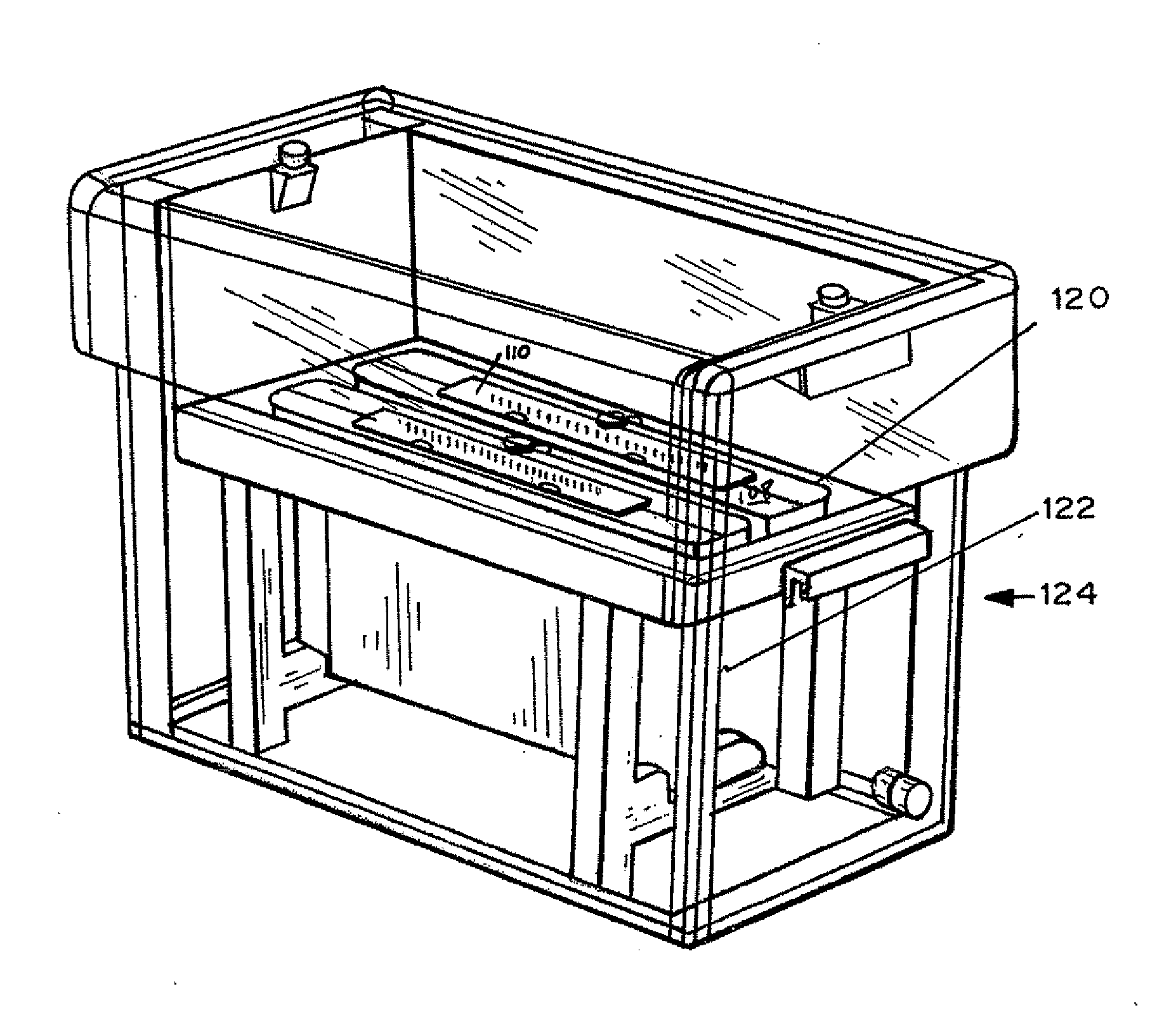
[0079]dPC™ manufacture. A glass blank with forty one 1 mm×2 mm holes was treated with silane to ensure glass to polymer adhesion. An array of pIs was created by mixing acrylamido buffers between a range of pH 4.2 and 6.2 at a 0.1 pH step. The range pH 4.2-6.2 is repeated twice in the chip. The final concentration of the immobiline buffers are between 12-30 mM, 6% C, 8% T. Using a robotic dispenser, 2.2 ul of the acrylamido buffer solution was dispensed into each hole followed by 8 minutes UV photopolymerization with a methylene blue / DPIC / STS system. 20 plugs ranging from 4.20, 4.30, 4.40 to 6.20 were into the first 20 plugs and then repeated for the next 20 plugs. To create a two pH layered chip, 0.2 ul of acrylamido buffer pk 3.6 was added to one side of the gel plug followed by an additional 22 minutes photopolymerization. No additional acrylamido buffer was added to the single layer plugs. After polymerization, the chip wa...

example 2

Comparison of Recovery Using Pharmalytes in the Running Buffers

[0083]dPC manufacture: dPC chips were manufactured as in Example 1 except no second pH layer was applied, the pH range was pH 4.2-6.2 and the step was 0.05 pH. The sample was pretreated and run as Example 1 with or without 0.25% Pharmalytes pH 2.5-5 in the anode and 0.25% Pharmalytes pH 5-8 in the cathode. dPC separated proteins were transferred to a second dimension gel as described earlier and Sypro Ruby stained.

[0084]FIGS. 5A and 5B are photographs comparing proteome coverage of an E. coli lysate separation between pH 4.2 and 6.2 by dPC™ isoelectric focusing and SDS PAGE 4-20% gels second dimension. FIG. 6A is the separation of E. coli lysate without pharmalytes and DTT in running buffers. FIG. 6B shows the separation of E. coli lysate with 0.25% pharmalytes and 40 mM DTT in running buffers. The comparison clearly shows higher protein yield and resolution through the addition of Pharmalytes to the running buffers.

example 3

Comparison of Recovery Using 0.1 step pI Traps to No Trap; with Ampholyte Added to Buffer

[0085]dPC manufactured and run as in Example 1. The sample was reduced and alkylated [125I]-ovalbumin spiked into 30 μg E. coli lysate. After isoelectric trapping, the individual gel plugs were extruded from the glass chip, placed in 100 μl SOLVABLE™ Packard Instruments and stored overnight at ambient temperature. 10 μl was added to 300 ul ULTIMAGOLD™ liquid scintillation cocktail prior to radioactive counting. [125I]-ovalbumin isoelectric.

[0086]As shown by FIGS. 6A and 6B, trapping collection increased by 70% with the pI 0.1 step traps.


PUM

PropertyMeasurementUnit
Fractionaaaaaaaaaa
Fractionaaaaaaaaaa
Molar densityaaaaaaaaaa

Abstract

Significantly higher yield and better resolution in pI gels are obtained by creating traps having two or more layers of gel containing closely stepped immobilized pH buffers. Proteins move from a pH at which they are negatively charged towards an anode at which they are positively charged. Discrete regions containing immobilized pH buffers trap the proteins when the immobilized buffer pH and the protein pI are approximately the same. The protein is trapped within the second layer and not on the surface of or interface of the second layer. Significantly higher yields with better resolution can be obtained through the use of layered sample application gels prior to isoelectric focusing. Layered plugs are prepared with a range of immobilized pH buffers ranging, for example, over 2 pH units, with steps of 0.05 or 0.1 pH units. An array of multilayered plugs wherein each plug has different pH increments is also provided. The array can be used to isolate and trap a variety of proteins having different isoelectric pHs during a single run. Another embodiment provides plugs having at least three layers; a gate layer, a trap layer, and an exit layer. Another embodiment includes adding a carrier ampholytes to running buffers and or adding thiol containing reducing agents to reduce current and improve resolution and collection efficiency.

Description

FIELD OF THE INVENTION[0001]This application is generally in the field of systems, kits and components thereof, for use in a method of separation of biomolecules in complex samples, especially those present in relatively low quantities, in a rapid and repeatable manner.BACKGROUND OF THE INVENTION[0002]Human plasma is rich in biomarkers, however, most biomarker proteins are undetected by current methods. This is because there are more than 3000 different plasma proteins and abundant proteins, such as albumin, mask low abundant disease biomarkers and thereby prevent detection. Successful biomarker discovery requires fractionation prior to mass spectroscopy analysis to “dive below the tip of the proteomics iceberg.”[0003]Many products are available for separation of mixtures of biomolecules, such as high performance liquid chromatography (HPLC) and gel electrophoresis, including gels that separate by molecular weight, charge, and pH. Isoelectric focusing is when molecules are placed in...

Claims


Application Information

Patent Timeline
IPC IPC(8): G01F1/64
CPCG01N27/44795
Inventor JOHANSEN, JACKGARLICK, RUSSELL K.SKEA, WILLIAMHARALAMPU, STEPHENKAGAN, OREN
Owner PROTEIN FOREST
Why Eureka
Try Eureka