Nanoparticles for protein drug delivery
- Summary
- Abstract
- Description
- Claims
- Application Information
AI Technical Summary
Benefits of technology
Problems solved by technology
Method used
Image
Examples
example no.1
Example No. 1
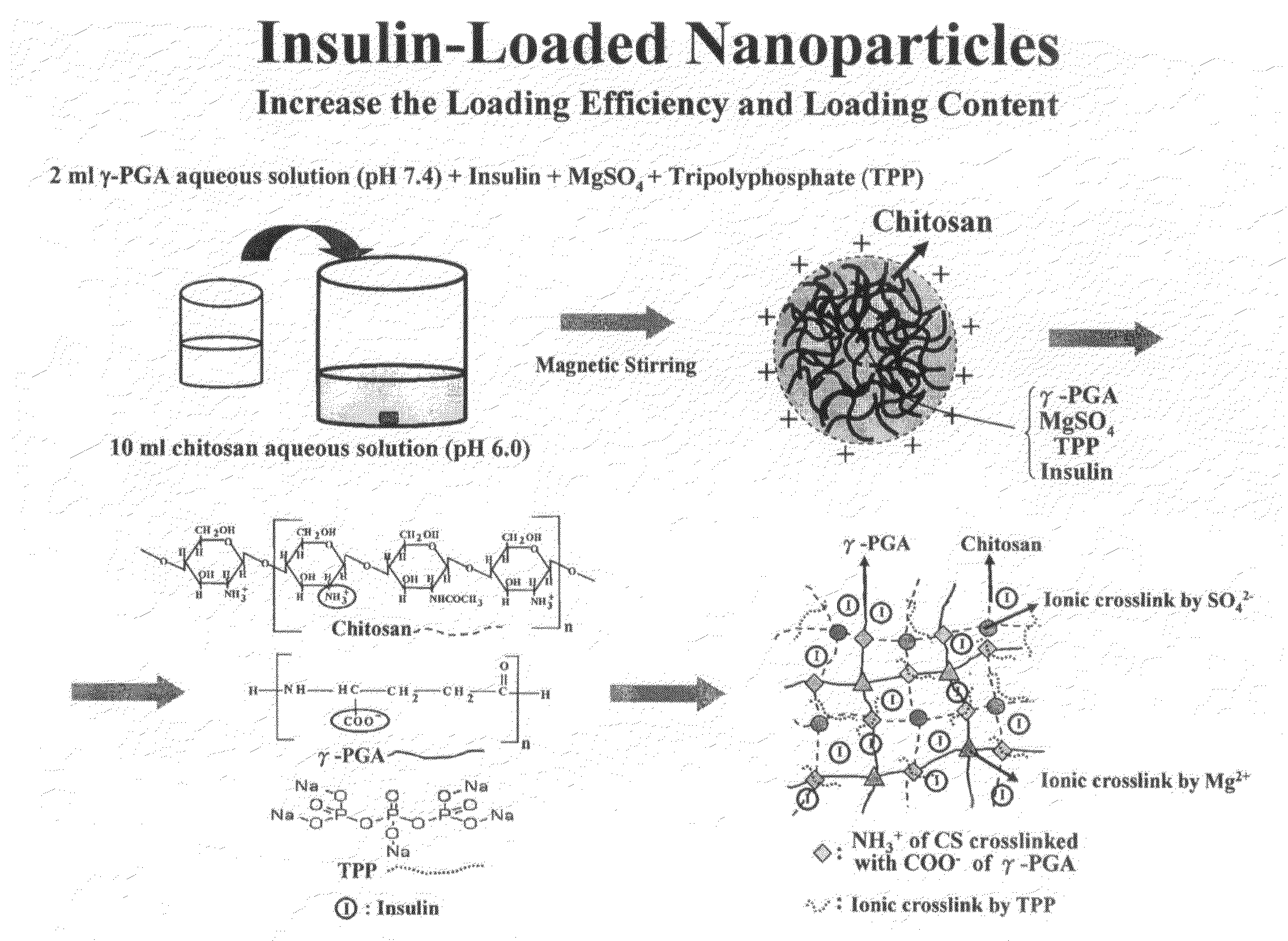
Materials and Methods of Nanoparticles Preparation
[0081]CS (MW˜2.8×10⁵) with a degree of deacetylation of approximately 85% was acquired from Challenge Bioproducts Co. (Taichung, Taiwan). Acetic acid, cellulase (1.92 units / mg), fluorescein isothiocyanate (FITC), phosphate buffered saline (PBS), periodic acid, sodium acetate, formaldehyde, bismuth subnitrate, and Hanks balanced salt solution (HBSS) were purchased from Sigma Chemical Co. (St. Louis, Mo.). Ethanol absolute anhydrous and potassium sodium tartrate were obtained from Merck (Darmstadt, Germany). Non-essential amino acid (NEAA) solution, fetal bovine serum (FBS), gentamicin and trypsin-EDTA were acquired from Gibco (Grand Island, N.Y.). Eagle's minimal essential medium (MEM) was purchased from Bio West (Nuaille, France). All other chemicals and reagents used were of analytical grade.
example no.2
Example No. 2
Depolymerization of CS by Enzymatic Hydrolysis
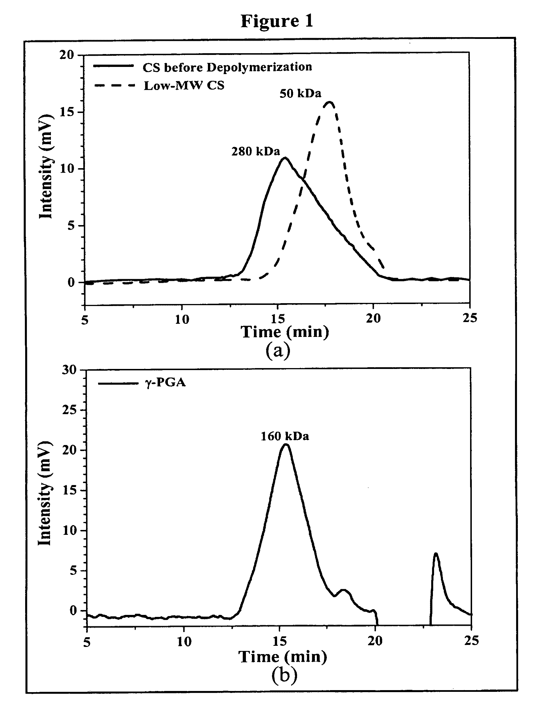
[0082]Regular CS was treated with enzyme (cellulase) to produce low-MW CS according to a method described by Qin et al. with some modifications (Food Chem. 2004; 84:107-115). A solution of CS (20 g / l) was prepared by dissolving CS in 2% acetic acid. Care was taken to ensure total solubility of CS. Then, the CS solution was introduced into a vessel and adjusted to the desired pH 5.0 with 2N aqueous NaOH. Subsequently, cellulase (0.1 g) was added into the CS solution (100 ml) and continuously stirred at 37° C. for 12 hours. Afterward, the depolymerized CS was precipitated with aqueous NaOH at pH 7.0-7.2 and the precipitated CS was washed three times with deionized water. The resulting low-MW CS was lyophilized in a freeze dryer (Eyela Co. Ltd, Tokyo, Japan).
[0083]The average molecular weight of the depolymerized CS was determined by a gel permeation chromatography (GPC) system equipped with a series of PL aquagel-OH columns (o...
example no.3
Example No. 3
Production and Purification of γ-PGA
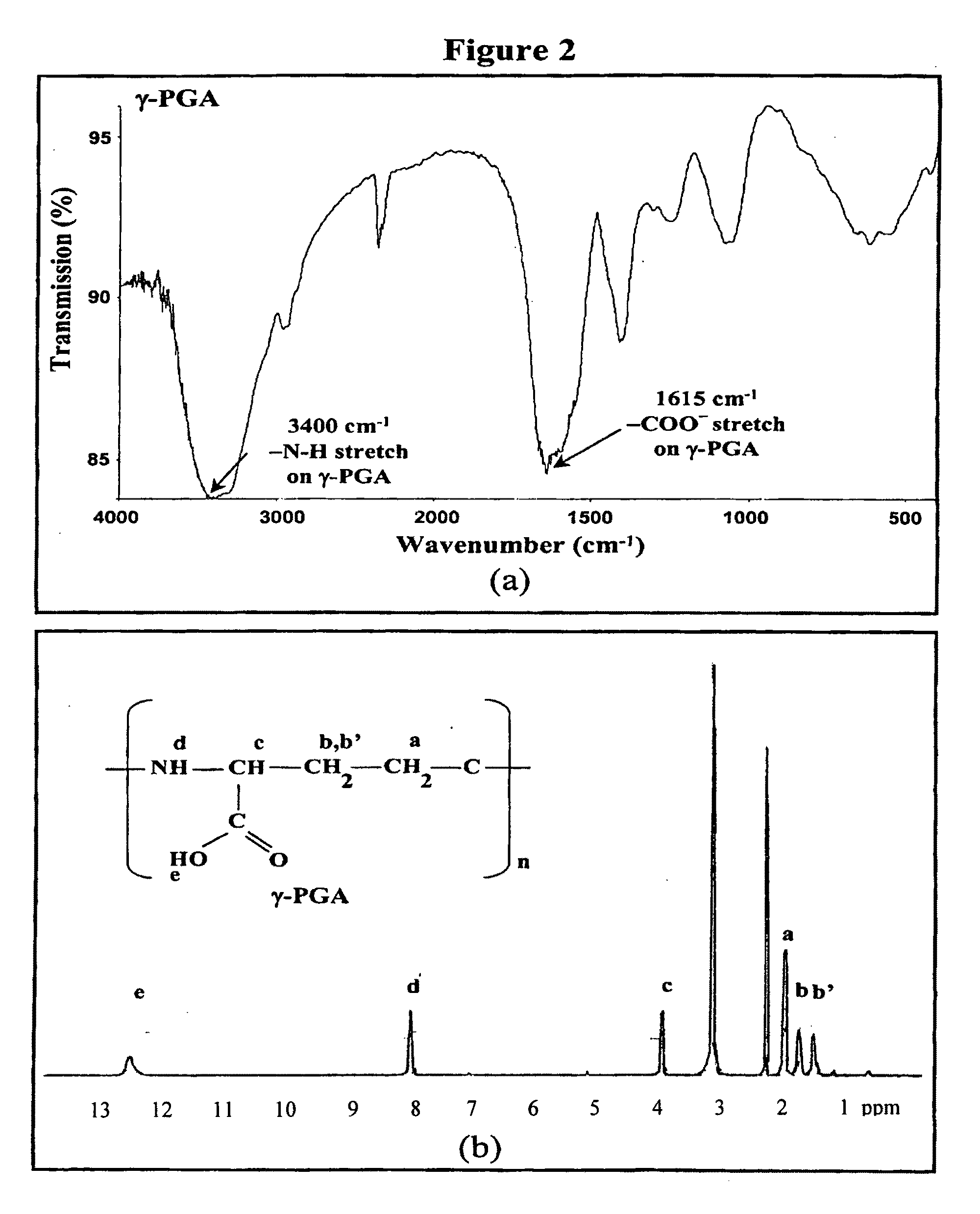
[0087]γ-PGA was produced by Bacillus licheniformis (ATCC 9945, Bioresources Collection and Research Center, Hsinchu, Taiwan) as per a method reported by Yoon et al. with slight modifications (Biotechnol. Lett. 2000; 22:585-588). Highly mucoid colonies (ATCC 9945a) were selected from Bacillus licheniformis (ATCC 9945) cultured on the E medium (ingredients comprising L-glutamic acid, 20.0 g / l; citric acid, 12.0 g / l; glycerol, 80.0 g / l; NH4Cl, 7.0 g / l; K2HPO4, 0.5 g / l; MgSO4.7H2O, 0.5 g / l; FeCl3.6H2O, 0.04 g / l; CaCl2.2H2O, 0.15 g / l; MnSO4.H2O, 0.104 g / l, pH 6.5) agar plates at 37° C. for several times. Subsequently, young mucoid colonies were transferred into 10 ml E medium and grown at 37° C. in a shaking incubator at 250 rpm for 24 hours. Afterward, 500 μl of culture broth was mixed with 50 ml E medium and was transferred into a 2.5-1 jar-fermentor (KMJ-2B, Mituwa Co., Osaka, Japan) containing 950 ml of E medium. Cells were cultured at...
PUM
| Property | Measurement | Unit |
|---|---|---|
| Particle size | aaaaa | aaaaa |
| Particle size | aaaaa | aaaaa |
| Atomic weight | aaaaa | aaaaa |
Abstract
Description
Claims
Application Information
 - R&D
- Intellectual Property
- Life Sciences
- Materials
- Tech Scout
- Unparalleled Data Quality
- Higher Quality Content
- 60% Fewer Hallucinations
Browse by: Latest US Patents, China's latest patents, Technical Efficacy Thesaurus, Application Domain, Technology Topic, Popular Technical Reports.
© 2025 PatSnap. All rights reserved.Legal|Privacy policy|Modern Slavery Act Transparency Statement|Sitemap|About US| Contact US: help@patsnap.com


