Method for preparing a cell-derived extracellular matrix membrane

a cell-derived extracellular matrix and membrane technology, applied in the field of cell-derived extracellular matrix membranes, can solve the problems of affecting the quality of patients' lives, bone marrow-derived blood clots, and it is difficult to expect successful healing using bone marrow stimulation

Inactive Publication Date: 2010-06-03
REGENPRIME
View PDF10 Cites 9 Cited by
  • Summary
  • Abstract
  • Description
  • Claims
  • Application Information

AI Technical Summary

Benefits of technology

[0022]Another features and embodiments of the present invention will be more clarified from the following detailed description and the appended claims.

Problems solved by technology

Once cartilage is damaged, its self-regeneration is extremely limited to ultimately cause osteoarthritis, which largely affects the quality of patients' lives.
Although the bone marrow stimulation is frequently used because of its minimal invasion procedure using an arthroscope in a short period of time, bone marrow-derived blood clots (including stem cells) are not maintained well during the operation so that the regenerated cartilage becomes fibrous cartilage rather than normal cartilage.
Therefore, it is difficult to expect successful healing using the bone marrow stimulation.
Although autologous chondrocyte implantation (ACI) is a clinically approved cell transplantation treatment (Brittberg, M. et al., New Eng. J. Med., 331:889, 1994), there is a problem in that the area of cartilage loss should be sutured after collecting the periosteum and the periosteum may be overgrown to cause pain at affected area after the surgery.
Moreover, it is troublesome to undergo a surgery process comprising two steps of isolating chondrocytes under arthroscopic operation to culture them for a long time in vitro, and then transplanting a cell suspension into a damaged area.
Also, because biodegradation of amniotic membrane cannot be controlled, it may remain in the body even long after transplantation.
Also, there is another difficulty of collecting tissues under a donor agreement.

Method used


Image

  • Method for preparing a cell-derived extracellular matrix membrane
  • Method for preparing a cell-derived extracellular matrix membrane
  • Method for preparing a cell-derived extracellular matrix membrane

Examples


example 1

Isolation of Pig Chondrocytes

[0059]2-week-old pigs without cholera, other viruses and contagious diseases were excessively anesthetized to kill according to animal ethics policy. In a sterile environment, cartilages were harvested from the stifle joint. The harvested cartilages were sliced into small pieces on a clean work table and treated with 0.1% collagenase for 12 hours. After filtering cells with a 0.4 μm filter, chondrocytes were isolated by centrifugation.

example 2

Preparing of a Chondrocyte-Derived ECM Membrane

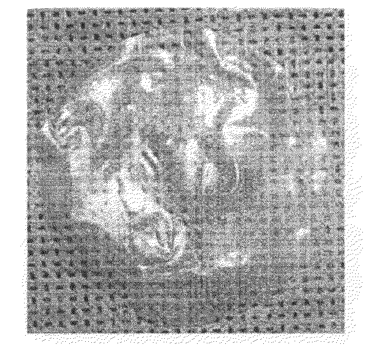
[0060]The chondrocytes isolated in Example 1 were seeded onto a 6-well culture dish at a concentration of 0.7×10⁵ cells / cm2 and then cultured in a culture medium (DMEM+20% FBS+1% penicillin-streptomycin+5 μg / mL ascorbic acid) for 3 weeks. The culture medium was replaced every three days. After 3 weeks, the ECM film was washed with PBS three times and separated from the 6-well culture dish. The prepared ECM membrane exhibited a semi-transparent thin membrane type (FIG. 1)

example 3

Analysis of Physicochemical Properties of an ECM Membrane

3-1: Microstructure Analysis of an ECM Membrane

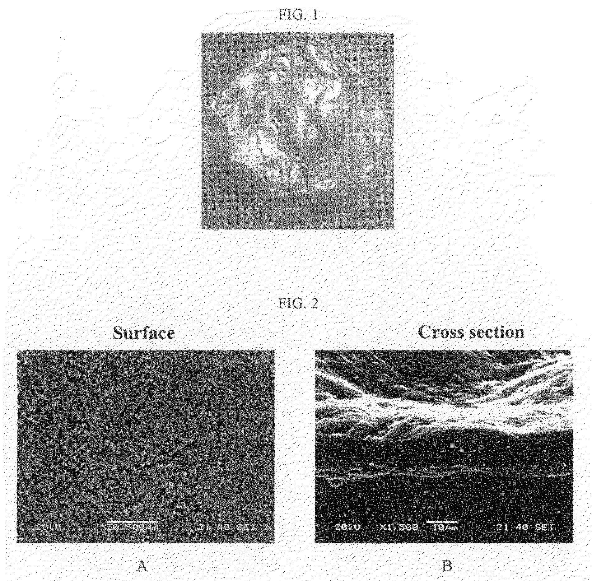
[0061]The microstructure of an ECM membrane was analyzed by scanning electron microscope (SEM). The ECM membrane prepared in Example 2 was fixed with 2.5% glutaraldehyde for 1 hour and washed with phosphate buffer solution. After dehydrating specimens with ethanol to dry, their surface and cross section were observed by an electronic microscope (JEOL, JSM-6380, 20KV, Japan). As a result, tough surface was observed due to structural bodies which seem to be cells and the cross section displayed about 10˜20 μm thickness.

3-2: Histological Observation of an ECM Membrane

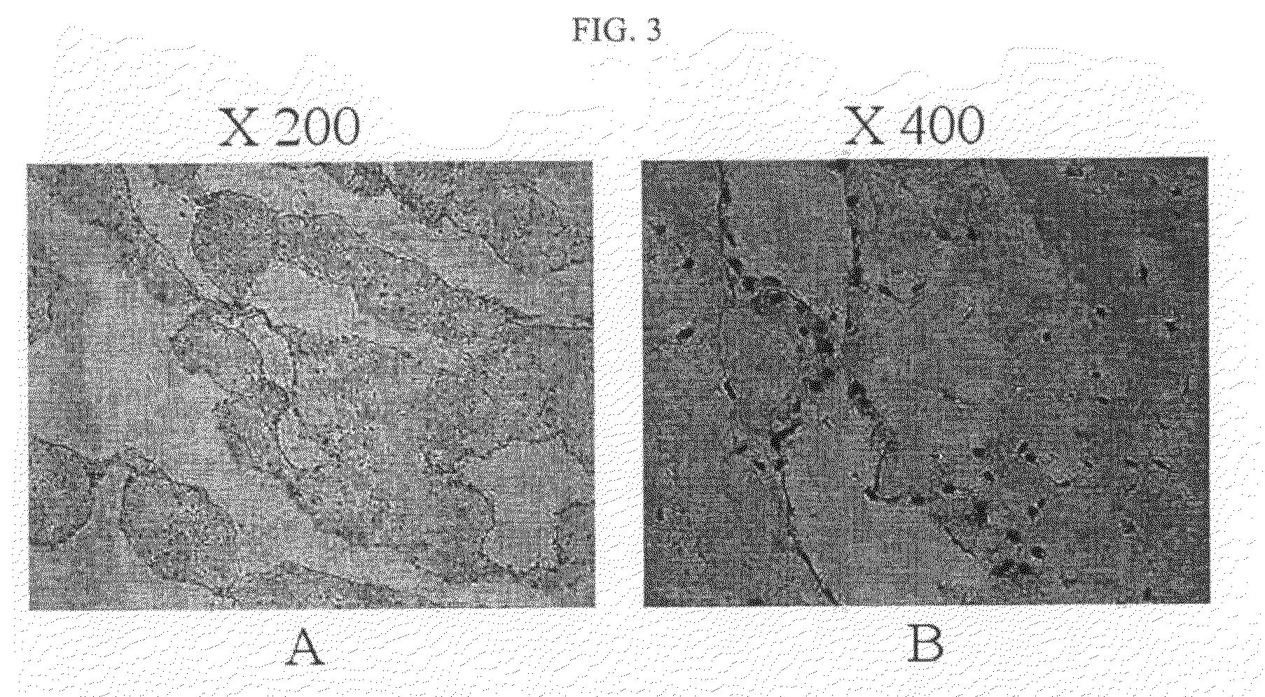
[0062]The ECM membrane prepared in Example 2 was fixed with 4% formalin solution for 24 hours, then embedded in paraffin and sectioned. The cross sections were stained with hematoxylin and eosin (H&E). The observation presented that cells having a nucleus were scattered inside of structural bodies which appear to be ECM ...


PUM

PropertyMeasurementUnit
compressive strengthaaaaaaaaaa
thicknessaaaaaaaaaa
temperatureaaaaaaaaaa

Abstract

The present invention relates to a method for preparing a cell-derived extracellular matrix membrane, more particularly, to a method for preparing a chondrocyte-derived ECM membrane, the method comprising the steps of forming a suitable thickness of ECM membrane by culturing chondrocytes derived from animal cartilage at a high concentration in vitro, and drying it after decellularization process. The cell-derived ECM membrane scaffold according to the present invention is composed of extracellular matrix secreted by chondrocytes so that the membrane has excellent biocompatibility as well as an immune-previlage effect specific to cartilage. Since the membrane also has a suitable compressive strength, it can be used to replace periosteum for cartilage regeneration or artificial collagen membrane and used as dura mater transplant material, a natural ECM membrane for treating skin loss, materials for cell transplantation and a growth factor delivery vehicle.

Description

TECHNICAL FIELD[0001]The present invention relates to a method for preparing a cell-derived extracellular matrix membrane (ECM membrane), more particularly, to a method for preparing a chondrocyte-derived ECM membrane, which comprises the steps of culturing chondrocytes derived from animal cartilage to form chondrocyte / ECM membrane, removing chondrocytes from the membrane, constructing a suitable thickness of ECM membrane and drying it for sterilization.BACKGROUND ART[0002]Articular chondrocytes are specialized mesoderm-derived cells found in only cartilage. Cartilage is an avascular tissue with characteristic physical properties depending on extracellular matrix produced by chondrocytes. Once cartilage is damaged, its self-regeneration is extremely limited to ultimately cause osteoarthritis, which largely affects the quality of patients' lives.[0003]Typical treatments for damaged cartilage include bone marrow stimulation to induce bone marrow-derived stem cells to the damaged site ...

Claims


Application Information

Patent Timeline
Patent Type & Authority Applications(United States)
IPC IPC(8): A61K38/16C12P19/26C12N5/00A61P43/00C12N5/077
CPCA61L27/3633A61L27/3683C12N2533/90A61L27/60C12N5/0655A61L27/3817A61P19/04A61P19/08A61P43/00C12N5/0652
Inventor MIN, BYOUNG-HYUNPARK, SO RACHOI, BYUNG HYUNE
Owner REGENPRIME
Why Eureka
Try Eureka