
Dinucleotide MRNA CAP Analogs

a technology of mrna cap and mrna cap, which is applied in the field of mrna cap analogs of mrna caps, can solve the problems of pseudosymmetric dinucleotides and the propensity to the 3 being almost impossible to d

Active Publication Date: 2010-10-14
APPL BIOSYSTEMS INC
View PDF4 Cites 83 Cited by
  • Summary
  • Abstract
  • Description
  • Claims
  • Application Information

AI Technical Summary

Benefits of technology

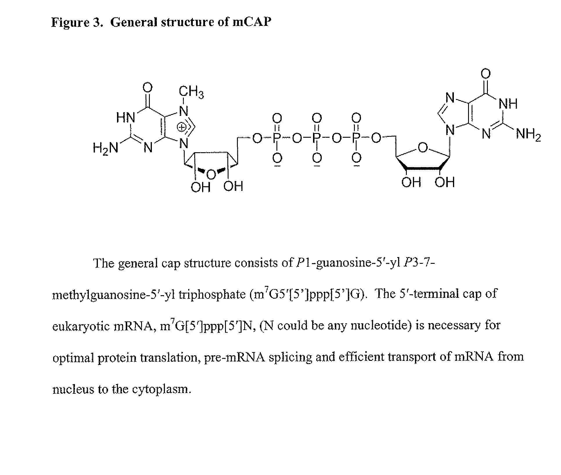
[0003]Eukaryotic mRNAs bear a “cap” structure at their 5′-termini that is well known to play an important role in translation. Naturally occurring cap structures consist of a 7-methyl guanosine that is linked via a triphosphate bridge to the 5′-end of the first transcribed nucleotide, resulting in m7G(5′)ppp(5′)N, where N is any nucleotide. The mRNA cap plays an important role in gene expression. It protects the mRNAs from degradation by exonucleases, enables transport of RNAs from the nucleus to the cytoplasm, and participates in assembly of the translation initiation complex. m7G(5′)ppp(5′)G (mCAP) has been used as the primer in transcription with T7 or SP6 RNA polymerase in vitro to obtain RNAs having a cap structure in their 5′-termini. In vivo, the cap is added enzymatically. However, over the past 20 years or so, numerous studies have required the synthesis of proteins in an in vitro translation extract supplemented with in vitro synthesized mRNA. The prevailing method for the in vitro synthesis of capped mRNA employs a pre-formed dinucleotide of the form m7G(5′)ppp(5′)G as an initiator of transcription. A disadvantage of using mCAP, a pseudosymmetrical dinucleotide, has always been the propensity of the 3′-OH of either the G or m7G (m7Guo) moiety to serve as the initiating nucleophile for transcriptional elongation. This leads to the synthesis of two isomeric RNAs of the form m7G(5′)pppG(pN)n and G(5′)ppp7G(pN)n, in approximately equal proportions, depending upon the ionic conditions of the transcription reaction. This may be problematic for various downstream processes, such as in vitro translation or crystallization studies.
[0006]Transcription of RNA usually starts with a nucleoside triphosphate (usually a purine, A or G). When transcription occurs in vitro, it typically includes a phage RNA polymerase such as T7, T3 or SP6, a DNA template containing a phage polymerase promoter, nucleotides (ATP, GTP, CTP and UTP) and a buffer containing magnesium salt. The synthesis of capped RNA includes the incorporation of a cap analog (e.g., N7 methyl GpppG or m7GpppG) in the transcription reaction. Excess m7GpppG to GTP (4:1) favors to increase the opportunity that each transcript will have a 5′ cap. The mMESSAGE mMACHINE© kit from Ambion (Ambion, Inc., Austin, Tex., a business of Applied Biosystems) recommends this ratio and will typically yield 80% capped RNA to 20% uncapped RNA, although total yields of total RNA are lower as GTP concentration becomes rate limiting as GTP is necessary for the elongation of the transcript.
[0009]Capped RNA encoding specific genes can be transfected into eukaryotic cells or microinjected into cells or embryos to study the effect of translated product in the cell or embryo. If uncapped RNA is used, the RNA in these experiments is rapidly degraded and the yield of translated protein is much reduced.

Problems solved by technology

A disadvantage of using mCAP, a pseudosymmetrical dinucleotide, has always been the propensity of the 3′-OH of either the G or m7G (m7Guo) moiety to serve as the initiating nucleophile for transcriptional elongation.
This may be problematic for various downstream processes, such as in vitro translation or crystallization studies.
The addition of the cap to the 5′ terminal end of RNA occurs after transcription but immediately after transcription initiation so that it is almost impossible to detect.

Method used


Image

  • Dinucleotide MRNA CAP Analogs
  • Dinucleotide MRNA CAP Analogs
  • Dinucleotide MRNA CAP Analogs

Examples


example 1

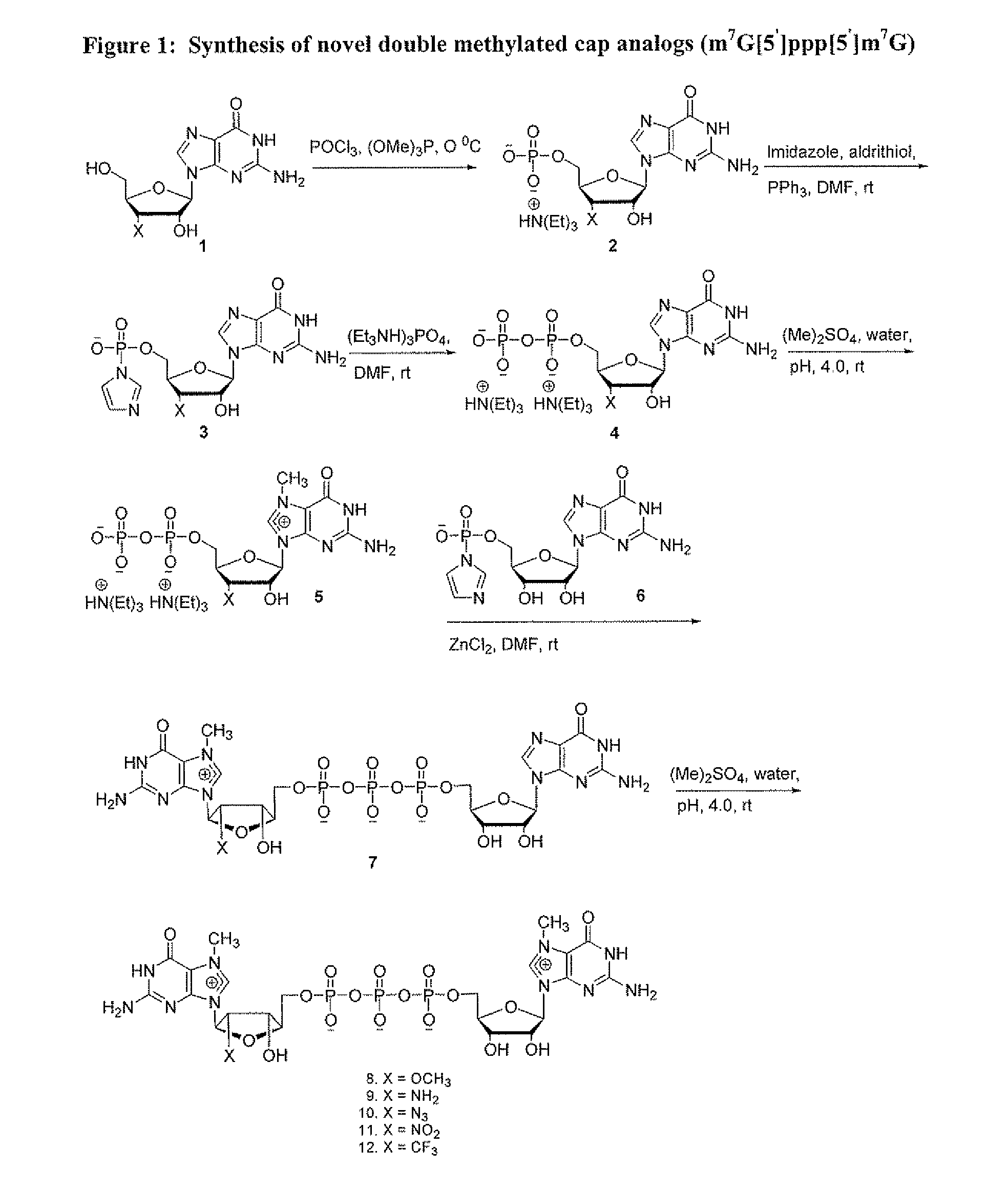
[0073]Synthesis of 7-Methyl Guanosine 5′-diphosphate In a clean, dry 2000 mL round bottom flask equipped with a stirring bar and under a stream of argon slowly dissolve dry and finely powdered guanosine 5′-diphosphate (1), (10.0 g, 20.5 mmol), either in free acid or sodium as a counter ion form, in 200 mL water, adjusting pH to 4.0 with glacial acetic acid. Dimethyl sulfate (20 mL, 119.04 mmol) was then added over a period of one hour with constant stirring at room temperature and the reaction continued for an addition hour during which time a decrease in pH was observed but pH was kept between pH 3.8 to 4.0 by drop-wise addition of 10 mM NaOH and methylations was monitored by analytical HPLC for progress. Methylation was determined to be 98% complete within 2 h. After 2 h, the reaction mixture was extracted with CHCl3(3×200 mL) to remove unreacted excess dimethyl sulfate.

[0074]The resulting aqueous layer was further evaporated on a rotary evaporator to remove any chloroform traces,...

example 2

Synthesis of Nucleoside-5′-diphosphates

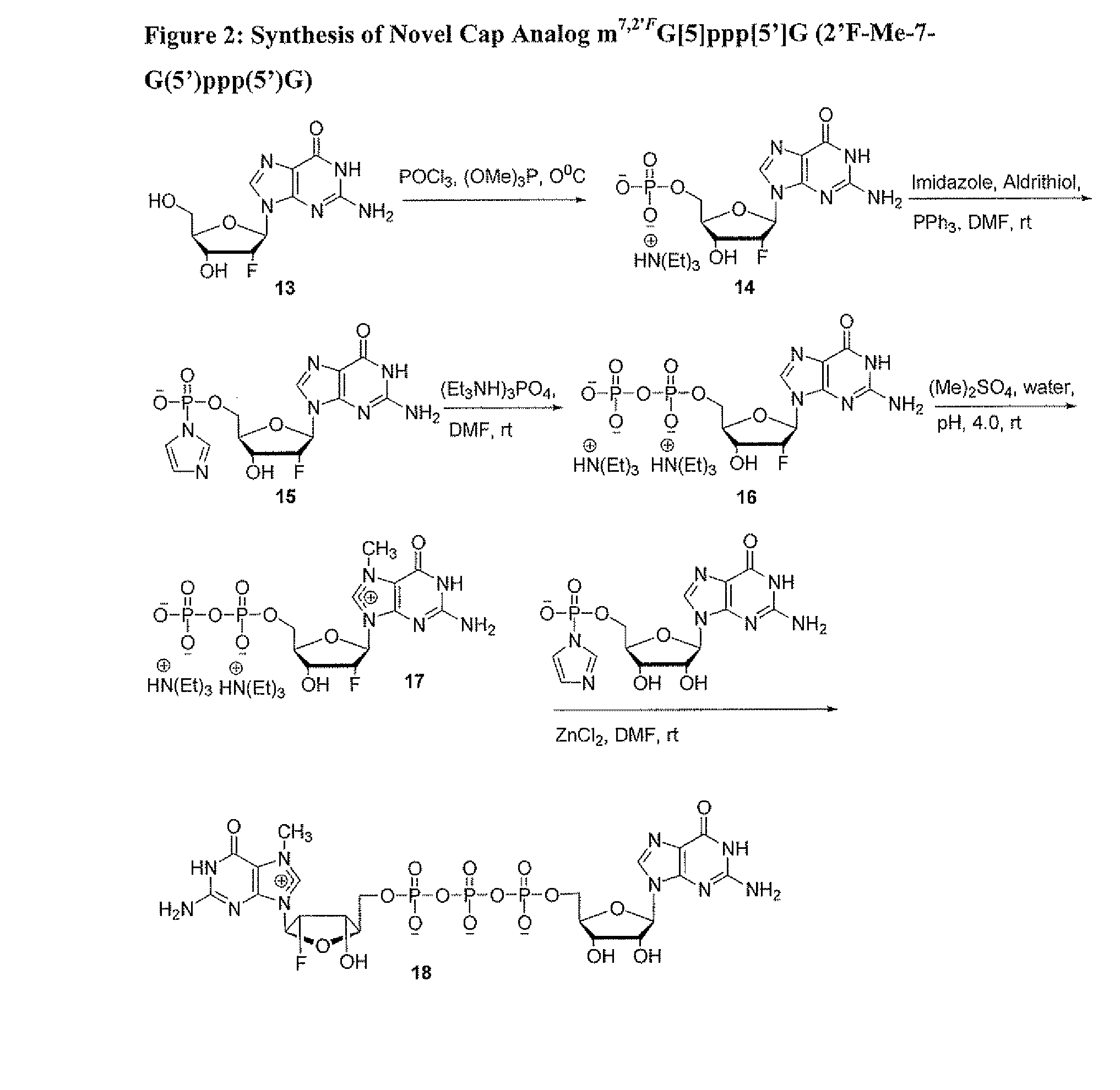
[0075]Although the following procedure illustrates synthesis of guanosine-5′-diphosphate, one of skill in the art would be able to use the procedure for the synthesis of adenosine-5′-diphosphate, uridine-5′-diphosphate, and cytidine-5′-diphosphate and analogs thereof. In a clean, dry 500 mL round bottom flask equipped with a stirring bar the triethylammonium salt of guanosine 5′-monophosphate (10.0 g, 21.5 mmol) in anhydrous dimethylformamide (200 mL) was stirred together, triethylamine was added (2.4 mL, 142.8 mmol) and allowed to stir for 5 min, followed by the addition of Imidazole (5.86 g, 86.1 mmol), 2,2′-dithiodipyridine (7.4 g, 33.58 mmol), and triphenylphosphine (8.9 g, 33.9 mmol). Stirring was continued for 2 h at room temperature. The reaction was allowed to go to completion as determined by HPLC and then poured slowly into a mixture of sodium perchlorate (7 g) in acetone (1500 mL), and then cooled for 30 min. at 4° C. The reaction mi...

example 3

Synthesis of 3′-O-Methyl Guanosine Monophosphate (3′-O-Me-GMP) TEA Salt (Compound 2)

[0077]In a clean, dry 500 mL round bottom flask equipped with a stirring bar and under a stream of argon slowly add dry and finely powdered 3′-O-Me-Guanosine (1), (6 g, 20 mmol) to a mixture of trimethylphosphate ((OMe)3P) (50 mL) and phosphorous oxychloride (POCl3) (6 mL, 60 mmol) at 0° C. in small portions with continuous stirring under argon. The mixture was kept at 0-4° C. and allowed to stir at least 19 hrs. Diethyl ether (200 mL) was added to extract the excess phosphorous oxychloride and to simultaneously precipitate the 3′-O-methylguanosine-5′-phosphodichloridate, which was then pelleted by centrifugation and dissolved in 100 mL ice-cold 5% NaHCO3 in water. The resulting aqueous solution was adjusted to pH ˜1.5 using 1 N NaOH. After stirring at 0-4° C. for an additional 20 h, the pH was adjusted to 7.0 and the resulting mixture was applied to a column of DEAE Sephadex A25. The column was wash...


PUM

PropertyMeasurementUnit
Lengthaaaaaaaaaa
Luminous fluxaaaaaaaaaa

Abstract

Novel cap analogs which are easily synthesized, resulting in high levels of capping efficiency and transcription and improved translation efficiencies are provided. Such caps are methylated at the N7 position of one or both guanosines of the dinucleotide cap as well as at the 3′ position on the ribose ring. Substituent groups on the ribose ring also result in the cap being incorporated in the forward orientation. Also provided are methods useful for preparing capped analogs and using mRNA species containing such analogs are also contemplated herein, as well as kits containing the novel cap analogs.

Description

CROSS-REFERENCE TO RELATED APPLICATIONS[0001]The present application is a National Stage of PCT / US2007 / 015896, filed Jul. 10, 2007, which claims priority benefit from earlier filed U.S, Provisional Application No. 60 / 820,771, filed Jul. 28, 2006, both of which are herein incorporated by reference in their entities.STATEMENT REGARDING FEDERALLY FUNDED SPONSORED RESEARCH OR DEVELOPMENT[0002]Work described herein was funded at least in part by an SBIR grant (SBIR Phase II, No. R44GM070156-02) awared by National Institutes of Health. The U.S. Government may therefore have certain rights therein.INTRODUCTION[0003]Eukaryotic mRNAs bear a “cap” structure at their 5′-termini that is well known to play an important role in translation. Naturally occurring cap structures consist of a 7-methyl guanosine that is linked via a triphosphate bridge to the 5′-end of the first transcribed nucleotide, resulting in m7G(5′)ppp(5′)N, where N is any nucleotide. The mRNA cap plays an important role in gene...

Claims


Application Information

Patent Timeline
IPC IPC(8): C12P19/34C07F9/02C07H21/02C12N15/11C12N15/113
CPCC07H21/02C07H21/04C12N2320/51C12N15/113C12N2310/317C12N15/111
Inventor KORE, ANILKUMAR R.MUTHIAN, SHANMUGASUNDARAM
Owner APPL BIOSYSTEMS INC
Learn More