
Method for stimulating mammalian cells and mammalian cell

a technology of applied in the field of stimulating mammalian cells and mammalian cells, can solve the problems of low therapeutic value, limited utility of these and other potential drugs, and no known therapy to remove harmful amyloids or cure damaged pancreatic tissues, etc., to achieve slow down major tissue pathology, reduce beta-amyloid levels, and efficient and specific

Inactive Publication Date: 2011-05-05
MEDEIA THERAPEUTICS
View PDF0 Cites 1 Cited by
  • Summary
  • Abstract
  • Description
  • Claims
  • Application Information

AI Technical Summary

Benefits of technology

[0018]An advantage of the invention is that a new efficient and specific method for preventing and treating amyloid accumulating disorders is provided compared to the current treatment protocols, which do not alter the levels of amyloid or trigger degradation of beta-amyloid to protect the brain against toxic amyloid protein. By reducing beta-amyloid levels in the brain, the present invention slows down major tissue pathology, which is the progression of amyloid accumulation. The treatment described in the present invention is cheap and relatively easily carried out.
[0019]A further advantage of the invention is that the migration of the cells of the present invention has high relative tissue selectivity, as the stimulatory treatment can be given inside the central nervous system by administration to the cerebrospinal fluid. This tissue selectivity prevents the potential side effects that would be caused by systemic stimulation of bone marrow cells or the like.
[0020]A further advantage of the invention is that the cells of the present invention can have either allogenic or autogenic origin, making it possible to use individual's own cells without the risk of rejection.
[0021]Another advantage is that the transplanted cells of the present invention have distinct properties, making them different from endogenous brain microglia, which do not respond by phagocytic activity as efficiently as the cells of the present invention, but may release, instead, neurotoxic molecules upon immunological stimulation.
[0022]Still another advantage is that the cell of the present invention may originate from non-embryonic tissue, such as bone marrow, umbilical cord or hematopoietic stem cells, and it is therefore accessible without ethical or technical difficulties.
[0023]Still another advantage compared to brain-derived cells, such as microglia, is that the cells of the present invention are available in high amounts sufficient to carry out transplantations.

Problems solved by technology

The use of a nerve growth blocking peptide or Congo Red, an amyloid stain, inhibits PrP-res formation and replication of scrapie agent, but has little therapeutic value when infection has reached the central nervous system.
Together with inherent toxicity, the utility of these and other potential drugs is very limited.
There is no known therapy to remove the harmful amyloid or cure the damaged pancreatic tissue.
When the interaction between these proteins is disturbed, the process leading to cell death in Parkinson's disease may occur.
Generally patients with Parkinson's disease are treated with drugs which either increase dopamine concentrations or reduce acetylcholine concentrations in the brain, but these drugs loose their effect with time, and have no impact on disease progression.
The disease is the leading cause of dementia, a condition that involves gradual memory loss, decline in the ability to perform routine tasks, disorientation, difficulty in learning, loss of language skills, impairment of judgment, and personality changes.
As the disease progresses, people with Alzheimer's disease become unable to care for themselves.
The loss of brain cells eventually leads to the failure of other systems in the body.
Additional genetic complexity is caused by the fact that the epsilon 4 allele of apolipoprotein E is the main risk factor for late-onset, sporadic form of AD.
Beta-amyloid is toxic to neurons both in the aggregated and soluble forms.
Some glutamate is needed for learning and memory, but too much can overstimulate and damage nerve cells.
However, neither cholinesterase inhibitors nor memantine are able to slow down or stop the progression of the disease and therefore they offer only temporary ease for some patients.
However, vitamin E has not been shown to have any significant effect on disease progression or symptoms.
However, this approach has been shown to have severe side effects, such as brain hemorrhages and encephalopathy.
Unable to function, the muscles gradually weaken, waste away (atrophy), and twitch (fasciculations).
Eventually, the ability of the brain to start and control voluntary movement is lost.
Eventually, all muscles under voluntary control are affected, and patients lose their strength and the ability to move their arms, legs and body.
When muscles in the diaphragm and chest wall fail, patients lose the ability to breathe without ventilatory support.
Free radicals are highly unstable molecules produced by cells during normal metabolism.
If not neutralized, free radicals can accumulate and cause random damage to the DNA and proteins within cells.
Although it is not yet clear how the SOD1 gene mutation leads to motor neuron degeneration, researchers have theorized that protein aggregates containing SOD1 accumulate free radicals, which may result from the faulty functioning of this gene.
No cure has yet been found for ALS.
Riluzole, which is believed to reduce damage to motor neurons by decreasing the release of glutamate, may prolong survival by months, mainly in those with difficulty swallowing.
There is no cure for Huntington's disease, and there is no known way to stop progression of the disorder.
However, no evidence has been provided that microglia would be responsible for beta-amyloid clearance after lipopolysaccharide stimulation in vivo (Mitrasinovic O. M. et al., Neurosci Lett.
However, the transplanted eGFP-positive BM-derived cells did not show increased migration through the blood brain barrier.
However, the use of human embryonic cells for development of therapy has not been legally or even ethically approved in the USA or numerous other Western countries, and utilization of cells from human newborns has similar ethical problems.
Also, brain tissue as a source of the cells to be utilized for the treatment is both ethically and technically very challenging and complicated.
Further, microglia needs to be purified from the brain which limits the cell number available for transplantation and includes a risk of impurities (such as material of other cell populations).

Method used


Image

  • Method for stimulating mammalian cells and mammalian cell
  • Method for stimulating mammalian cells and mammalian cell
  • Method for stimulating mammalian cells and mammalian cell

Examples


example 1

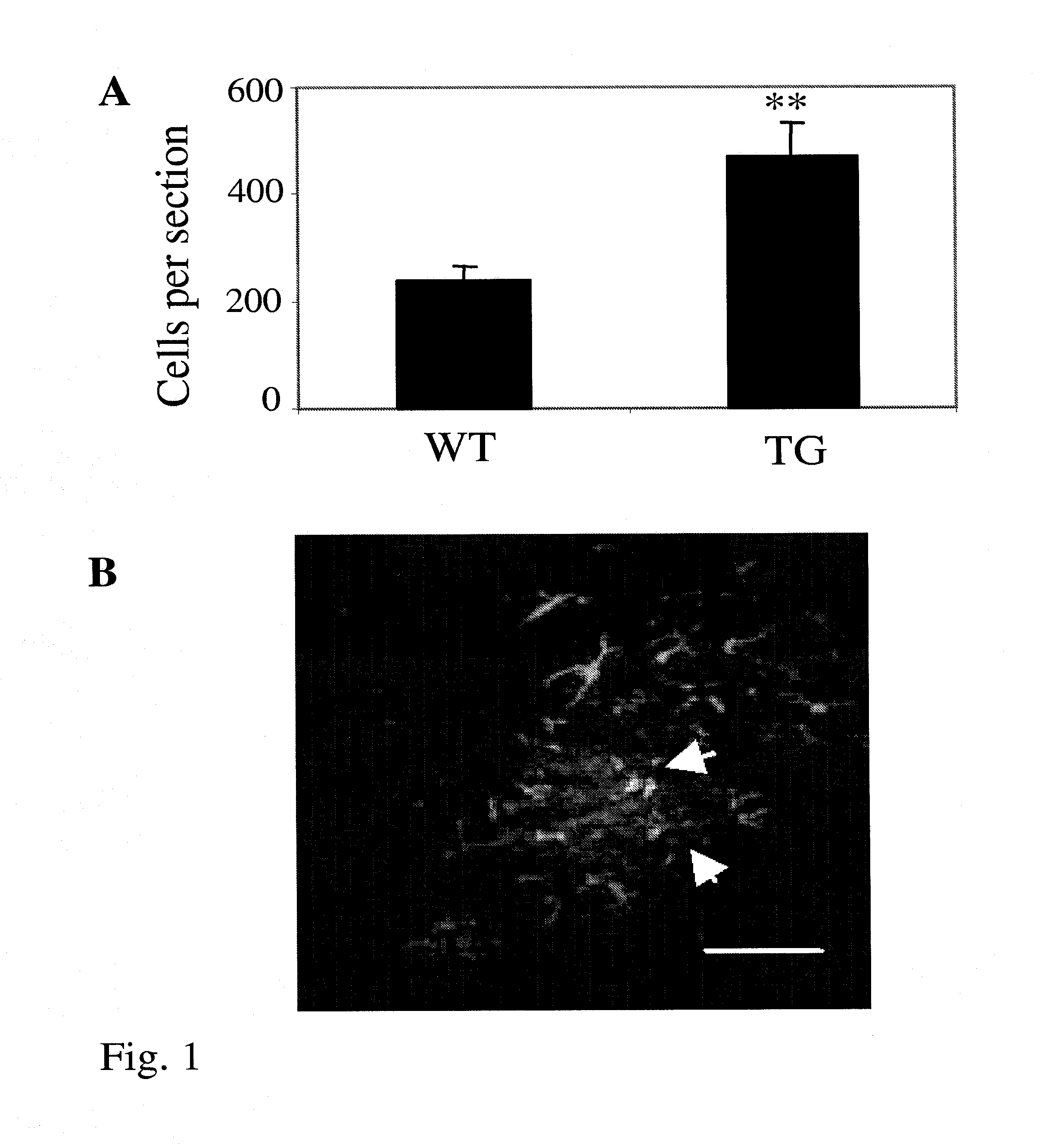
Transplantation of BM Cells into Transgenic Alzheimer Mouse and Increased Engraftment of Transplanted BM Cells into Alzheimer Mouse Brains

Transgenic Mice

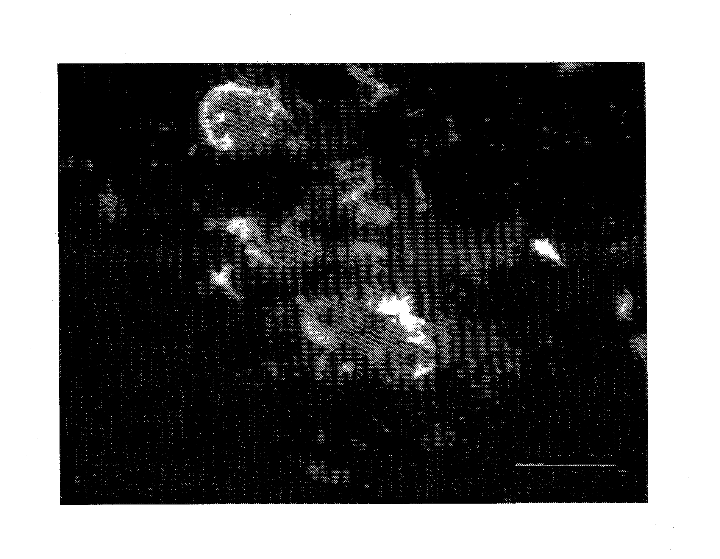
[0053]The mice used were generated by co-injection of chimeric mouse / human APPswe and human PS1-dE9 vectors, both controlled by their own mouse prion protein promoter element (Jankowsky et al., Hum Mol Genet. 13:159-170 (2004)). The double transgenic mice (APPdE9) were backcrossed to C57BL / 6J strain for six generations. Altogether 5 2.5-month-old female APPdE9 transgenic and 5 age-matched wild-type controls were used in this study. Enhanced GFP (eGFP) overexpressing mice (Okabe et al., FEBS Lett. 407:313-319 (1997)), were purchased from Jackson Laboratories (Maine, USA) and were maintained in C57BL / 6J strain in the Animal Facilities of the National Public Health Institute in Kuopio, Finland. All animal experiments were carried out according to the National Institute of Health (NIH) guidelines for the care and use of laboratory anima...

example 2

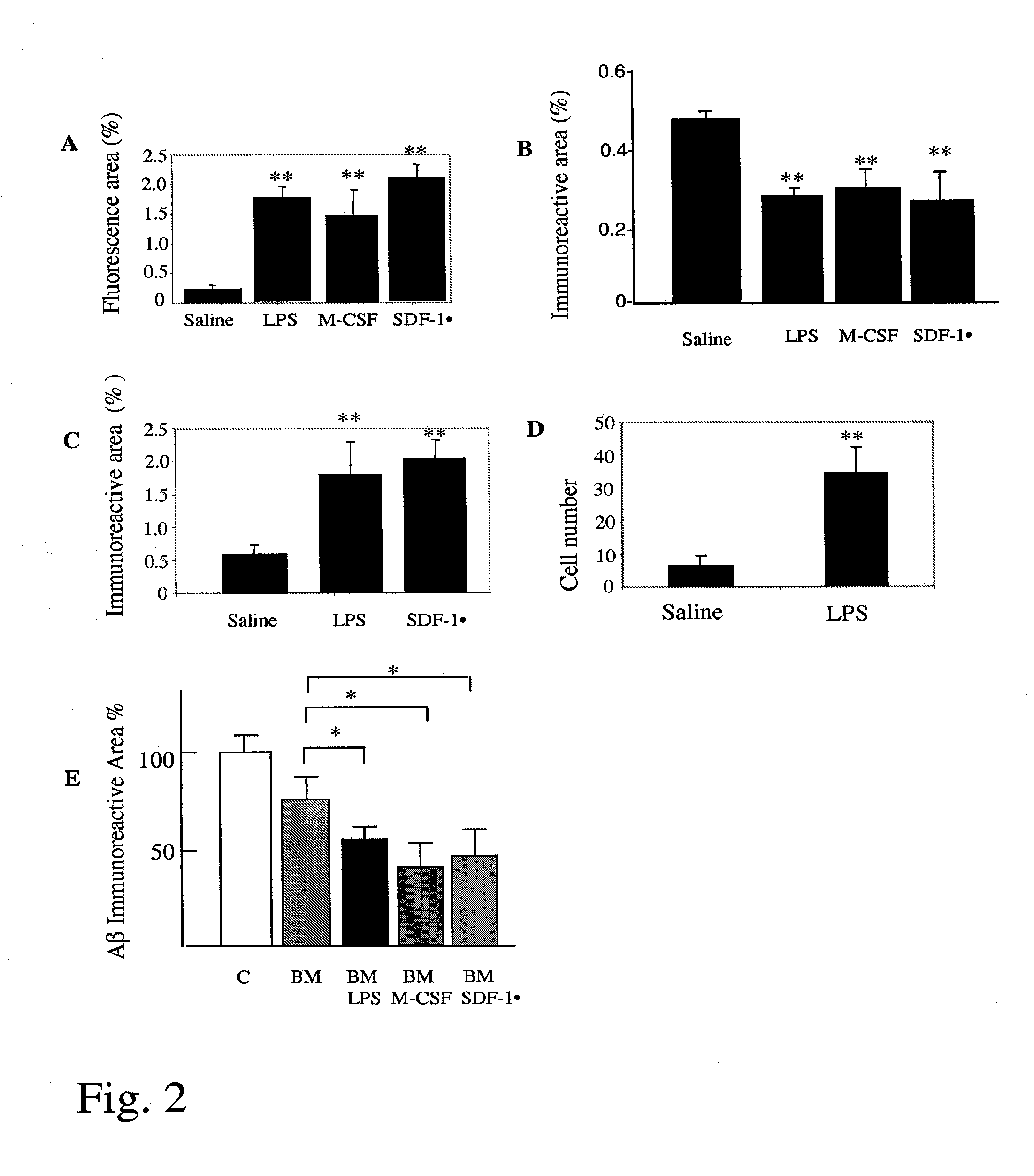
Increased Brain-Specific Engraftment of Transplanted BM Cells into the Brain and Activation of Transplanted BM Cells to Phagocytose and Clear Amyloid

[0059]Double transgenic female mice carrying chimeric mouse / human APP695 harboring the Swedish mutation (K595N / M596L) and human PS1 with familial AD-linked A246E mutation (Borchelt et al., Neuron. 19:939-945 (1997)) were used in this experiment. The parental APP695swe and PS1 (A246E) mice were backcrossed 13-14 generations to C57BL / 6 strain after which they were intercrossed to create double transgenic mouse line (APP+PS1 mice). Altogether 8 25-month-old APP+PS1 mice and 8 age-matched wild-type controls per age group were used in this study.

[0060]Flow cytometry analysis of eGFP expression in peripheral blood cells was done as described in Example 1 and showed that about 90% of the CD11b-immunoreactive monocytes were GFP positive, indicating successful engraftment of BM transplants.

Stimulation of BM-Derived Cells with LPS, M-CSF and SDF-...

example 3

LPS, M-CSF and SDF-1α Enhanced Beta-Amyloid Degradation (Clearance) by BM Cells In Vitro

[0065]BM cells from adult eGFP transgenic mice were cultured as described (Servet-Delprat et al., BMC Immunol. 3:15 (2002). Aged double transgenic APP+PS1 (see (2)) were perfused with saline and the brains containing human beta-amyloid deposits were frozen on dry ice. Sagittal sections (10 μm) were cut on a cryostat (Leica), mounted on poly-L-lysine-coated coverslips, transferred to two-well chamber slides and used immediately or stored at −80° C. until use. BM-derived eGFP expressing cells were seeded in the chamber at a density of 5×10⁶ cells in 1 ml of assay medium (DMEM / F12, G5 supplement, 0.2% bovine serum albumin (BSA), penicillin and streptomycin) and the cultures were maintained for 24 h or longer at 37° C. (Wyss-Coray et al., Nat Med. 9, 453-457 (2003)). The amyloid burden was analyzed using immunohistochemistry and image analysis. Beta-amyloid was detected using a pan-Aβ antibody. The p...


PUM

PropertyMeasurementUnit
timeaaaaaaaaaa
solubleaaaaaaaaaa
insolubleaaaaaaaaaa

Abstract

The current invention relates to methods for stimulating mammalian cells to enhance their ability to cross the blood-brain-barrier and to phagocytose and degrade beta-amyloid plaques in the brain. The current invention also relates to cells obtained by the method of the invention. The current invention also relates to methods for prevention and treatment of amyloid-accumulating disorders.

Description

FIELD OF THE INVENTION[0001]The present invention relates to methods for preparing stimulated mammalian cells originated from bone marrow, umbilical cord, any source of hematopoietic stem cells or any other monocytic lineage to enhance their ability to phagocytose and degrade beta-amyloid plaques and beta-amyloid peptides in the brain and to cross the blood-brain barrier. The current invention also relates to cells obtained by said method. The current invention also relates to methods and substances useful for prevention and treatment of disorders or diseases associated with amyloid accumulation, such as Alzheimer's disease or the like.BACKGROUND OF THE INVENTION[0002]Disorders associated with amyloid accumulation result when misfolded proteins amass to form amyloids, which are plaque-like deposits that crowd in different organs of the body. These diseases include for example Alzheimer's disease, Parkinson's disease, Huntington's disease, amyotrophic lateral sclerosis (ALS), transmi...

Claims


Application Information

Patent Timeline
Patent Type & Authority Applications(United States)
IPC IPC(8): A61K38/19A61K35/32A61P25/28A61P25/16A61P25/00A61P3/10A61K35/12C12NC12N5/077
CPCA61K2035/124C12N2501/22C12N2501/052C12N5/0647A61P3/08A61P3/10A61P21/04A61P25/00A61P25/14A61P25/16A61P25/28A61P31/12A61P43/00
Inventor KOISTINAHO, JERIKOISTINAHO, MILLA
Owner MEDEIA THERAPEUTICS
Learn More