Integrated Method for Enriching and Detecting Rare Cells from Biological Body Fluid Sample

a biological body fluid and enrichment method technology, applied in the field of integrated methods for enriching and detecting rare cells from biological body fluid samples, can solve the problems of difficult subsequent analysis and research thereto, unable to capture a lot of tumor cells, and proving time-consuming, and achieves good cell morphology and high recovery rate.

Inactive Publication Date: 2011-08-11
CYTTEL BIOSCI BEIJING
View PDF4 Cites 22 Cited by
  • Summary
  • Abstract
  • Description
  • Claims
  • Application Information

AI Technical Summary

Benefits of technology

[0005]The present invention provides a set of novel and unique integrated methods, comprising: removing plasma protein, lysing red cells, adding immunomicrospheres or immune materials to remove white blood cells, adding density centrifugation based on a special cell separation medium to separate the circulating rare cells in the biological body fluid sample. The present method consisting of concentrating and enriching can rapidly enrich the circulating rare cells in the biological body fluid specimens, e.g. in peripheral blood, and also has a high recovery rate. The enriched cells have a good cell morphology that can be used for image analysis. Moreover, most of the white blood cells in the patient specimen also can be effectively recovered for application in other researches and analysis, for instance, research of gene profile. In the present invention, no special equipment, e.g. cell separation tube or magnet, is required.
[0012]In the present method for enriching the rare cells in the biological body fluid, the step of lysing the red blood cells to remove the red blood cells is carried out prior to, after or while adding the immunomicrospheres or immunoadsorbent to incubate. If the present method does not comprise the step of adding a red cell lysis solution to carry out red cell lysis, the red blood cells can be separated and removed via a long-time centrifugation.
[0014]Up to now, technical means for detecting the circulating rare cells are all based on immunofluorescence. However, the main drawback of nonspecific combination with the target cells resulted from the negative charge of the fluorescence dye itself is inevitable. It seriously troubles people when distinguishing true and false positive staining signals. The present invention provides a whole set of optimized and novel multicolor staining method based on immunohistochemistry so as to avoid nonspecific staining brought by the immunofluorescence and allow the stained circulating rare cells to be detected by a ordinary optical microscope or a scanner based on microscope principle. This method has been proved to be a high specific, rapid and simple technical means and have low cost, and no longer needs any fluorescence dye or expensive fluorescence microscope.
[0025]The unique combination of the two methods for enriching and staining in the present invention can greatly improve popularization and application of detecting the rare cells such as circulating tumor cells in blood. The novel and unique methods for enriching and staining have been proved to have low cost and can rapidly, effectively and high specifically enrich and quantitatively detect the rare cells in blood.

Problems solved by technology

However, such method for directly capturing the circulating tumor cells by using the antibody-coupled magnetic beads has some well-known disadvantages (Mocellin et al, 2006 Trends in Molecular Medicine 12:130): due to the heterogeneity of the expression of markers on tumor cells surface, a lot of tumor cells cannot be captured with this method, which has been proved by a lot of clinical cases; in addition, as the tumor cells are “contacted” and stimulated by the magnetic beads coupled to the antibody, these tumor cells that are captured and enveloped by a great deal of immune granules are caused to be no longer cells in a natural state, thus, it is hard to make subsequent analysis and research thereto.
Although some single experimental methods for removing or separating specific cell population have been reported, such as density centrifugation method (U.S. Pat. No. 4,927,750), immune magnetic granule method (U.S. Pat. No. 4,177,145), red cell lysis method (hemolysis method), and a method of primary combining immune magnetic granule with density centrifugation which must utilize a special cell separation tube (U.S. Pat. No. 5,840,502), all these methods have been proved to be time-consuming, with a low removal rate of white blood cells and red blood cells and a low recovery rate of target cells, as well as inconvenience in operation brought by some special equipments required.
However, the inevitable high nonspecific staining signal, expensive fluorescence microscope and indispensable but inconvenient working environment (e.g. darkroom) greatly limit the development of detecting the circulating tumor cells and circulating endothelial cells based on immunofluorescence.

Method used


Image

  • Integrated Method for Enriching and Detecting Rare Cells from Biological Body Fluid Sample

Examples


example 1

Enriching the Circulating Tumor Cells in Peripheral Blood of a Breast Cancer Patient

[0063]5 ml of human peripheral blood is collected in a blood collection tube (BD, New Jersey, USA) containing ethylene diamine tetraacetic acid (EDTA) anticoagulant. The supernatant can be absorbed out with a pipette or automatic liquid-absorption device so as to remove plasma proteins after the blood samples are centrifuged (700×g, 10 minutes). The deposit obtained after centrifugation is resuspended in 30 ml of a red cell lysis solution (BD Pharmingen, California, USA) and incubated for 20 minutes. The specimen centrifugation is carried out (700×g, 10 minutes) so as to separate the lysed red blood cell chips in the supernatant. The deposit (i.e. deposited cells) is resuspended in 5 millilitre of phosphate buffer (pH 7.4) after the supernatant is removed. 0.5 millilitre of magnetic beads coated with a monoclonal antibody against white blood cell surface antigen such as CD45 (Invitrogen, California, ...

example 2

Tricolor Staining the Circulating Tumor Cells Enriched in Peripheral Blood of a Breast Cancer Patient

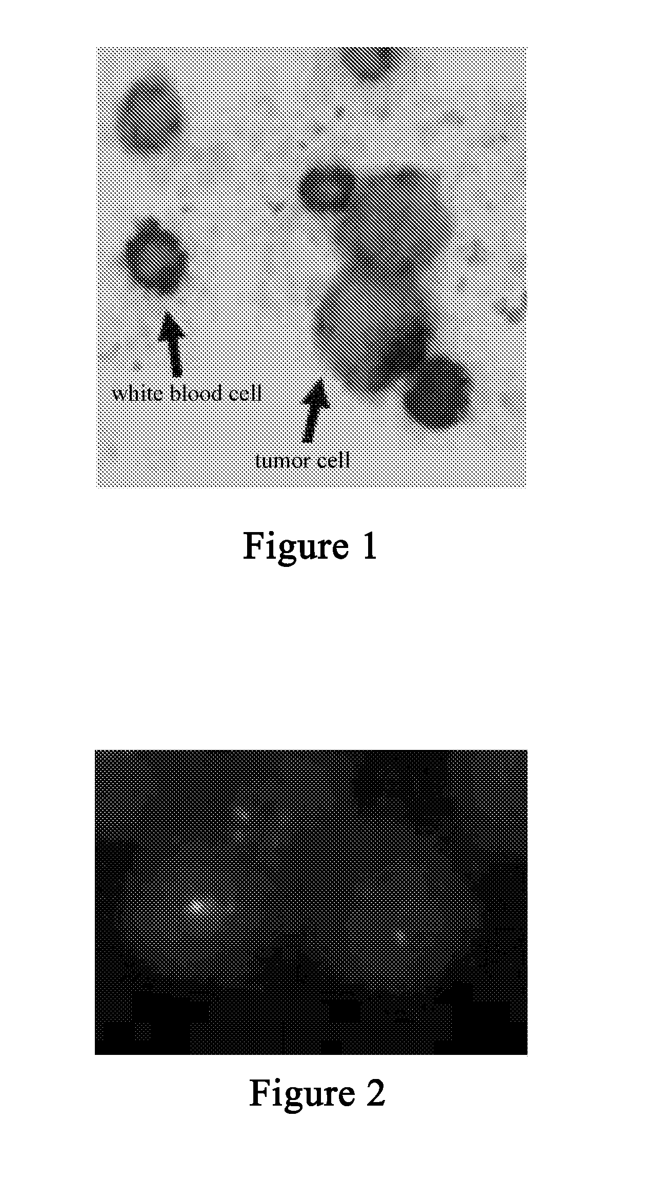
[0065]The enriched circulating tumor cells are put on the glass slide and fixed by 2% of paraformaldehyde prepared from phosphate buffer for 2 hours at room temperature, followed by washing thrice with phosphate buffer. The cells and a mixture (diluted by phosphate buffer) containing biotin (Pierce, Ill., USA) labelled monoclonal antibody (Abcam, UK, 1 μg / ml) against keratins 8+18+19 and rhodamine (Pierce, Ill., USA) labelled monoclonal antibody (Abcam, UK, 1 μg / ml) against CD45 are incubated for 30 minutes at a room temperature. After the glass slide is washed thrice with the phosphate buffer, it is incubated for 30 minutes at the room temperature with a mixture (diluted by phosphate buffer) containing alkaline phosphatase labelled monoclonal antibody (Sigma, Missouri, USA, 1 μg / ml) against biotin and peroxidase (Pierce, Ill., USA) labelled monoclonal antibody (Abcam, UK, 1 μg / ml) a...

example 3

Detecting the Circulating Tumor Cells by a Chromosomal Fluorescence in situ Hybridization

[0067]The enriched tumor cells are put on the glass slide as specimens. The glass is rinsed with SSC buffer after the stained specimens are treated with 20 milligramme / millilitre of RNA enzyme for 1 hour. The specimens are dehydrated with absolute ethyl alcohol for 10 minutes and then heated to 70° C., holding for 5 minutes for denaturation. The specimens are dehydrated with absolute ethyl alcohol for 10 minutes again, and hybridized and incubated with a probe at 45° C. overnight. The specimens are observed by a fluorescence microscope after being washed with the SSC buffer. The specimens can be the enriched tumor cells stained with the method in Example 2. The object of carrying out chromosomal fluorescence in situ hybridization is further confirming authenticity of detecting the tumor cells by tricolor staining based on immunohistochemistry. For the sake of rapid diagnosis, the specimens may n...


PUM

PropertyMeasurementUnit
diameteraaaaaaaaaa
diameteraaaaaaaaaa
pHaaaaaaaaaa

Abstract

The present invention relates to an integrated method for enriching and detecting rare cells in biological body fluid sample. The enriching method comprises: (a) removing plasma protein by centrifugation; (b) optionally adding a red cell lysis solution to carry out red cell lysis so as to remove the red blood cells; (c) adding immunomicrospheres or immunoadsorbent to incubate; and (d) carrying out density centrifugation based on a special cell separation medium for separating the circulating rare cells, residual red blood cells after removing red blood cells and the white blood cells combined on the immunomicrospheres. The method for detecting the enriched rare cells according to the present invention comprises combining immunohistochemistry based staining with immunofluorescence, or bicolor, tricolor or multicolor staining based on immunohistochemistry, and observing and identifying by fluorescence or ordinary optical microscope or a scanner based on microscope principle. The novel and unique method for enriching and staining has been proved to have low cost and can rapidly, effectively and high specifically enrich and quantitatively detect the rare cells in blood.

Description

FIELD OF THE INVENTION [0001]The present invention generally relates to an integrated method for enriching and detecting rare cells from biological body fluid sample.BACKGROUND OF THE INVENTION[0002]Since the technology of directly capturing the circulating tumor cells in the human peripheral blood of the U.S. Immunicon / Veridex (Philadelphia, USA) was examined and approved by U.S. Food and Drug Administration in 2004, important scientific researches and clinical significance on obtaining and detecting circulating tumor cells, circulating endothelial cells, tumor stem cells and some immune cells have been continuously and widely reported (Cristofanilli et al, 2004 New Eng J. Med. 351:781; Braun and Marth, 2004 New Eng. J. Med. 351:824).[0003]However, such method for directly capturing the circulating tumor cells by using the antibody-coupled magnetic beads has some well-known disadvantages (Mocellin et al, 2006 Trends in Molecular Medicine 12:130): due to the heterogeneity of the exp...

Claims


Application Information

Patent Timeline
Patent Type & Authority Applications(United States)
IPC IPC(8): C12Q1/68C12M1/00C12N5/00
CPCC12N5/0693G01N33/5005G01N33/6893G01N33/574G01N33/6803G01N33/5011
Inventor LIN, PING
Owner CYTTEL BIOSCI BEIJING
Why Eureka
Try Eureka