Fluorescent silica nanoparticle with radioactive tag and the detecting method of pet and fluorescent dual imaging using thereof

a fluorescent silica nanoparticle and radioactive tag technology, applied in the direction of organic chemistry, drug compositions, therapy, etc., can solve the problems of not observing the extent of a tumor during diagnosis or operation, difficult to find the transfered lymph node, and difficult to let the gamma probe approach along the route of transfer

Inactive Publication Date: 2011-10-27
SEOUL NAT UNIV R&DB FOUND +1
View PDF7 Cites 11 Cited by
  • Summary
  • Abstract
  • Description
  • Claims
  • Application Information

AI Technical Summary

Benefits of technology

[0034]Functionalized silica nanoparticles of this invention have promising potential as a role for sentinel lymphatic tracer through PET and fluorescent dual imaging in surgical guidance.

Problems solved by technology

Although medical doctors can observe the extent of a tumor beforehand through said imaging, they can not observe the extent of a tumor during diagnosis or operation such as endoscopy, laparoscopy etc.
So, they are hard to find transferred lymph node or happen not to remove the tumor completely.
But it is hard to let the gamma probe approach along the route of transfer in abdominal cavity, because transfer direction in the case of abdominal cavity is too widespread in contrast to breast cancer.
But the practical use is restricted because quantum dot use the Cadmium (Cd) which is forbidden to use to human body.
However, the low molecular weights of fluorophore dyes mean that their residence times at sentinel nodes are limited, and thus, researchers have been trying to develop new materials for this purpose.
However, despite their potential benefits, the practical applications of quantum dots are limited by poor bio-compatibility and potential toxicity (Hardman R., Environ Health Perspect 2006; 114:165-72; Zhang T, Stilwell J L, Gerion D, Ding L, Elboudwarej O, Cooke P A, et al., Nano Lett 2006; 6:800-8).
However, although several studies have concluded that functionalized silica nanoparticles can be applied in various biological and medical areas, functionalized silica nanoparticles was not applied to in vivo animal study using optical imaging.
But these functionalized silica nanoparticles have not been applied to the in vivo animal research in the nuclear medicine and optical imaging.
Also, in conventional examination of sentinel lymph node, we can not confirm radioisotpe nanoparticle during operation, and dying material was so small that it pass the sentinel lymph node.
Also, conventional nano fluorescent quantum dots use the Cd, so, it is hard to apply those to human body.

Method used


Image

  • Fluorescent silica nanoparticle with radioactive tag and the detecting method of pet and fluorescent dual imaging using thereof
  • Fluorescent silica nanoparticle with radioactive tag and the detecting method of pet and fluorescent dual imaging using thereof
  • Fluorescent silica nanoparticle with radioactive tag and the detecting method of pet and fluorescent dual imaging using thereof

Examples


embodiment 1

Animals and Chemicals

[0046] Animals

[0047]Specific pathogen-free six-week-old female BALB / c nude mice were obtained from SLC Inc. (Japan). All animal experiments were performed after receiving approval from the Institutional Animal Care and Use Committee (IACUC) of the Clinical Research Institute at Seoul National University Hospital. In addition, the National Research Council (NRC) guidelines for the care and use of laboratory animals (revised 1996) were observed throughout.

[0048] Chemicals

[0049]Rhodamine β isothiocyanate (RITC), 3-(aminopropyl)triethoxysilane (APTS), and phosphate buffered saline (PBS, pH 7.4) were obtained from Sigma (St. Louis, Mo.). Tetraethyl orthosilicate (TEOS), and 29 wt % aqueous ammonia solution were from Aldrich (Milwaukee, Wis.). 2-[Methoxy(polyethylenoxy)propyl] trimethoxysilane (PEG-silane, 90%) were from Gelest (Morrisville, Pa.).

embodiment 2

The Preparation of Radioisotope Labeled Fluorescent Silica Nanoparticles

[0050] The Preparation of Silica Nanoparticles Doped with Fluorescent Dyes

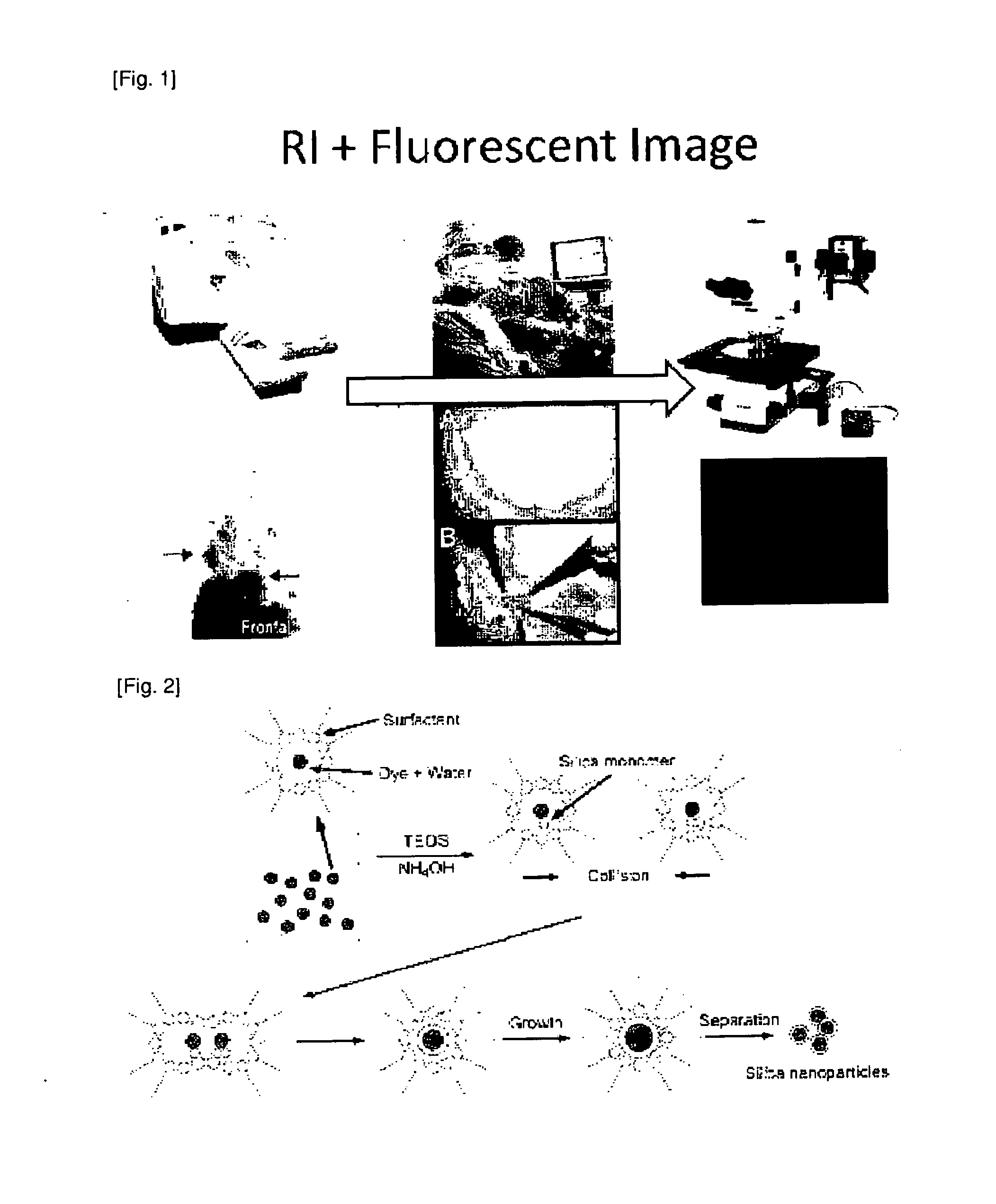
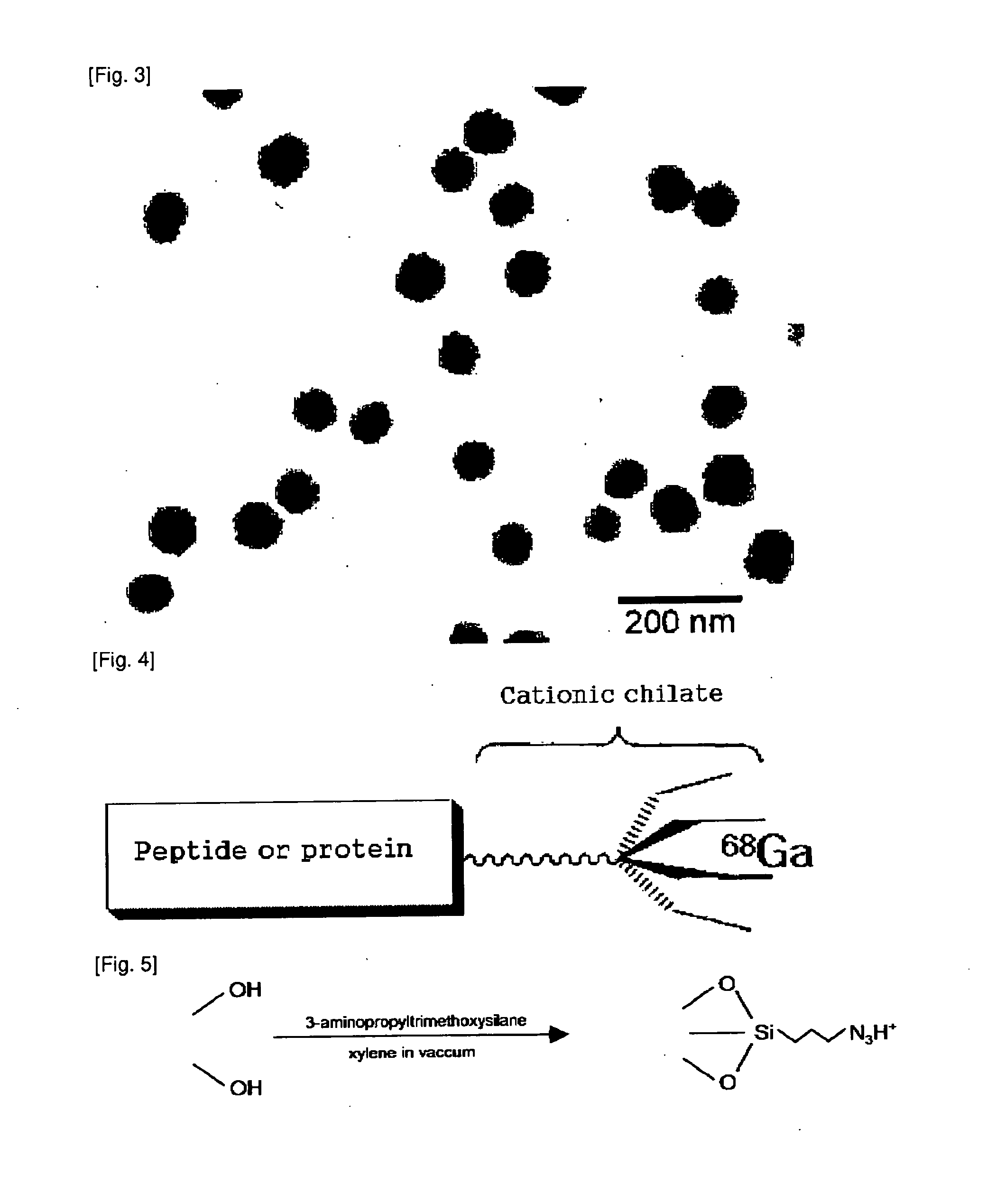
[0051]Silica nanoparticles were made by reverse micro emulsion method (FIG. 2). FIG. 3 is the TEM image of it. Silica nanoparticles doped with fluorescent dyes is manufactured by introducing RITC to said silica nanoparticles.

[0052] The Preparation of Ga-68 Labeled NOTA-Silica Nanoparticles for PET

[0053]Labeled 68Ga-NOTA is much stable and was stable in 6M HNO3 for over 6 hours. So, 68Ga labeled silica nano particles doped with fluorescent dyes are stable. We introduce —NH2 group at the surface of silica nanoparticle before the introduction of 68Ga labeling, and then coupled NCS-NOTA (2-(4′-isocyanatobenzyl)-1,4,7-triazacyclononanetriacetic acid). The NOTA-silica nanoparticles react with 68GaCl3 solution eluted from 68Ge / 68Ga generator and then 68 Ga-NOTA-silica nanoparticles are synthesized (FIG. 4 to FIG. 6).

[0054]In the concrete, To the ...

embodiment 3

PET / fluorescence dual imaging of sentinel lymph node

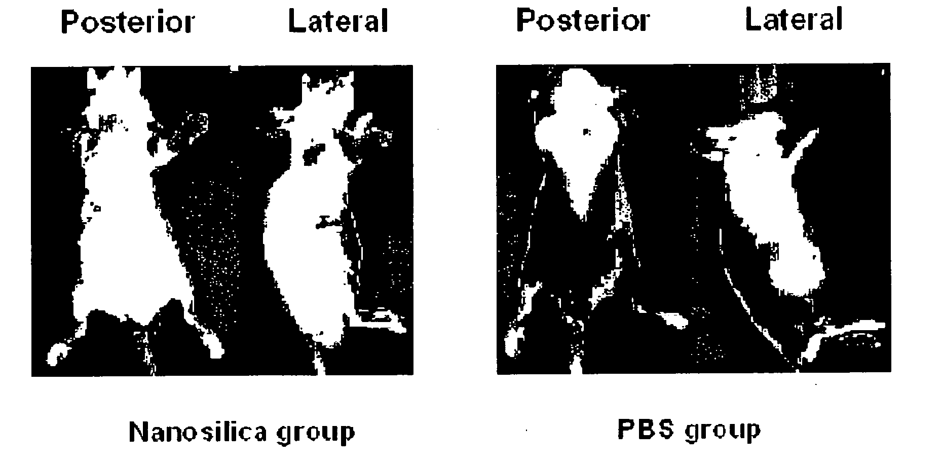
[0055] Fluorescence Imaging

[0056]We use nude mouse without fur in order to get the full fluorescent imaging. In order to confirm the optimized dosages, kinds (Tetramethylrhodamine-5, Indocyanine green etc.) and size (10-100 nm) of fluorescent materials, we get the full imaging without radioisotope labeling according to the time using Xenogen IVIS 100 under 2% isoflurane gas anesthesia after injecting to hypodermis of nude mouse. We confirm lymph node after cutting the portion showing fluorescence, we take a picture using Xenogen IVIS 100 and observe through fluorescent microscope, and confirm the lymph node with H&E dyeing. We measure the fluorescence remained in body through full photography of mouse after cutting the lymph node.

[0057]Fluorescence images were obtained using a Maestro In Vivo Imaging System (CRi Inc., Woburn, Mass.) for data acquisition and analysis. Before imaging, mice were anesthetized i.p. with a solution conta...


PUM

PropertyMeasurementUnit
half lifeaaaaaaaaaa
half lifeaaaaaaaaaa
pHaaaaaaaaaa

Abstract

The present invention relates to nuclear medicine using fluorescent silica nanoparticle and detecting method of optical dual imaging, and more particularly to radioisotope labeled fluorescent silica nanoparticles which are used for PET (positron emission tomography) and fluorescence detecting, and detecting method of PET and fluorescent dual imaging using thereof. Functionalized silica nanoparticles of this invention have promising potential as a role for organic lymphatic tracer in biomedical imaging such as pre- and intra-operative surgical guidance.

Description

TECHNICAL FIELD[0001]The present invention relates to nuclear medicine using fluorescent silica nanoparticle and detecting method of optical dual imaging, and more particularly to radioisotope labeled fluorescent silica nanoparticles which are used for PET (positron emission tomography) and fluorescence detecting, and detecting method of PET and fluorescent dual imaging using thereof.BACKGROUND ART[0002]Imaging technique capable of investigating location, extent, and transition of a tumor such as PET, MRI etc has been widely used. Although medical doctors can observe the extent of a tumor beforehand through said imaging, they can not observe the extent of a tumor during diagnosis or operation such as endoscopy, laparoscopy etc. So, they are hard to find transferred lymph node or happen not to remove the tumor completely.[0003]Sentinel lymph node detection using gamma probe after injecting radioisotope is used clinically in breast cancer surgery. But it is hard to let the gamma probe...

Claims


Application Information

Patent Timeline
Patent Type & Authority Applications(United States)
IPC IPC(8): A61K51/04A61P43/00C07F7/18
CPCA61B5/0071A61K49/0002A61K49/0021A61K51/1244A61K49/0041A61K49/0093A61K49/0034A61P35/00A61P43/00
Inventor CHUNG, DOOSOOKANG, KEONWOOKJEON, YONGHYUNKIM, YOUNGHWAALOTHMAN, ZEID A.AHMED, AHMED YACINE HADJCHOI, KIHWANAIMAJID, ABDUULLAH M.PIAO, JINGYUALOTHMAN, ASMA A.QUAN, BO
Owner SEOUL NAT UNIV R&DB FOUND
Why Eureka
Try Eureka