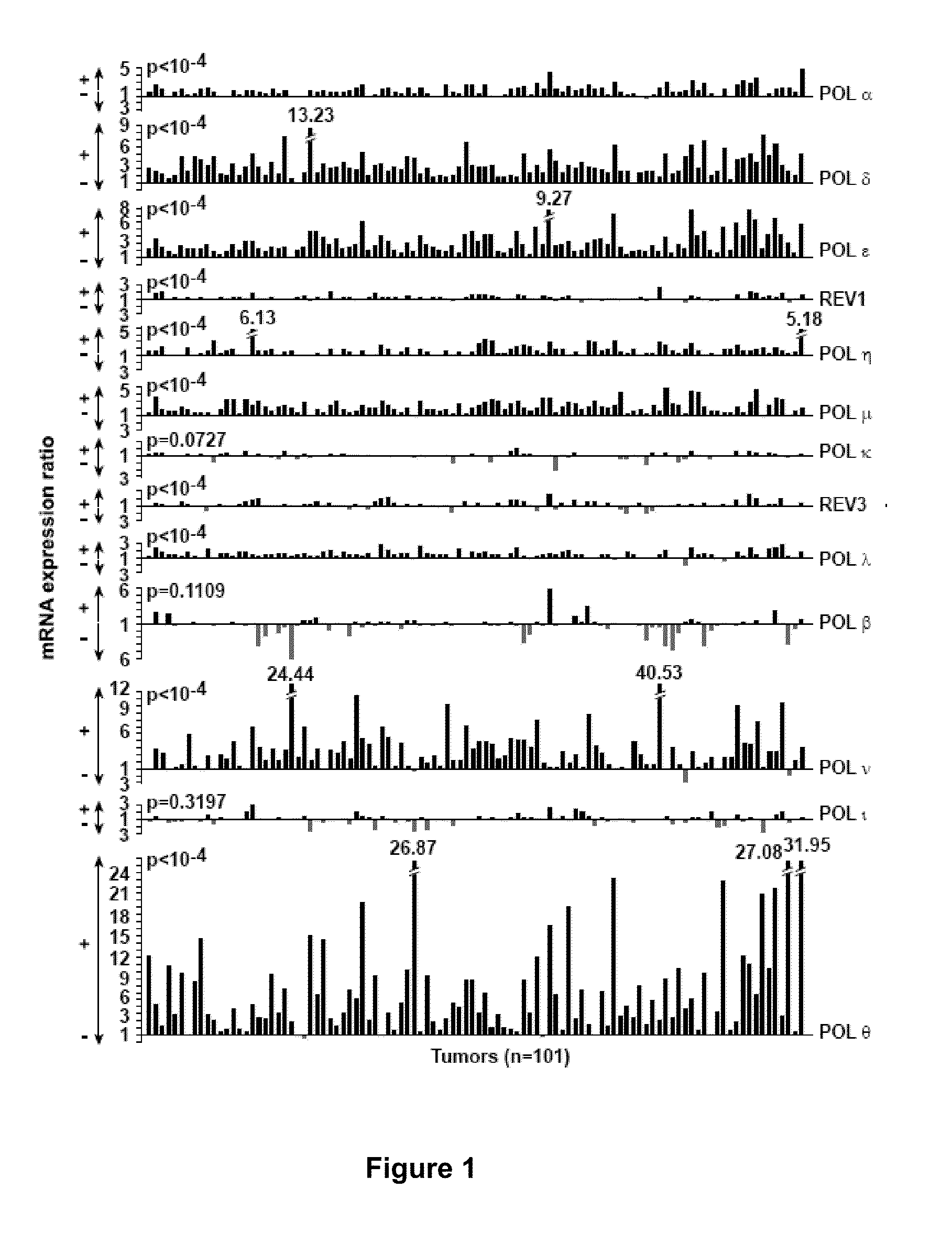
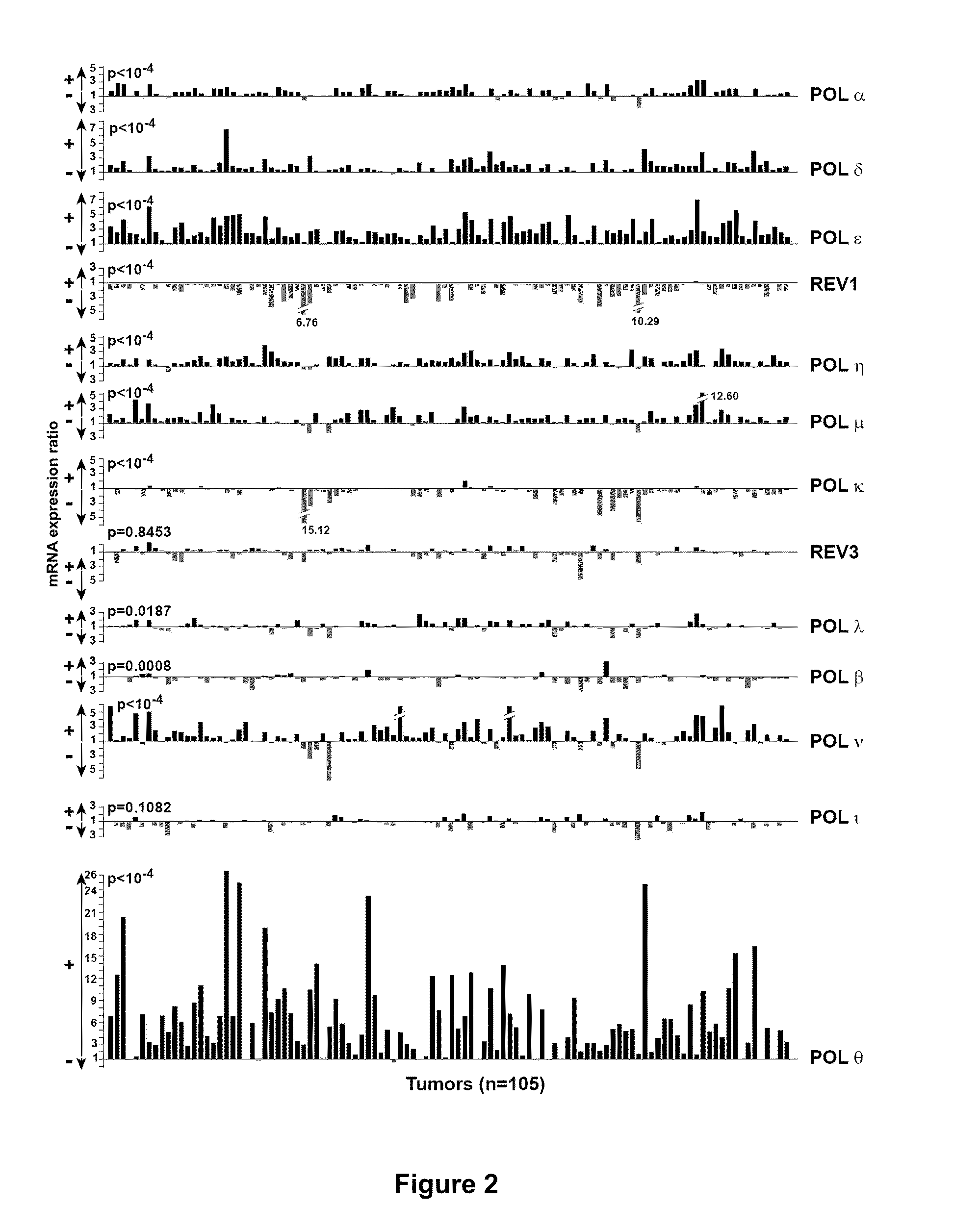
[0021]In another embodiment, the method of the invention may be used for prognosing the survival of a patient which has a tumor expressing a p53wt cDNA (the sequence does not contain any mutations by reference to sequence NC 000017-9 from
GenBank). In this embodiment, a high level of POLQ expression in a patient with a tumor expressing a p53wt cDNA indicates aggressiveness of the tumor, and results in poor
survival prognosis, whereas low level of POLQ expression in a patient with a tumor expressing a p53wt cDNA is associated to a much better diagnosis. Indeed, the inventors have shown that a low level of POLQ expression in a patient with a tumor expressing a p53wt cDNA is associated with a probability of survival of 98%. In another aspect, the method of the invention allows for the detection of cancer cells displaying genetic instability, i.e.
DNA damage or “replicative stress” caused by a perturbation of origin firing. Deregulated expression of POLQ leads to increased mitotic abnormalities, such as chromatid breaks, chromosomal end-to-end fusions, dicentric chromosomes, and other abnormalities, thus triggering constitutive activation of the γH2AX-ATL-CHK2
DNA damage checkpoint. Thus POLQ overexpression leads to
DNA damage and
chromosome instability.
[0027]It is understood that both methods can be combined. Therefore, in a further aspect, the invention relates to a method for diagnosing the aggressiveness of a cancer in a patient and for the detection of cancer cells displaying genetic instability. The method of the invention thus presents the
advantage of allowing the detection of a genetic event associated with both a bad
survival prognosis and genetic instability. As used herein, the terms “cancer” and “cancerous” refer to or describe the
physiological condition in mammals that is typically characterized by unregulated
cell growth. The terms “cancer” and “cancerous” as used herein are meant to encompass all stages of the
disease. Thus, a “cancer” as used herein may include both benign and malignant tumors. Examples of cancer include but are not limited to,
carcinoma,
lymphoma,
blastoma,
sarcoma, and
leukemia or lymphoid malignancies. More particular examples of such cancers include, but not limited to, squamous
cell cancer (e.g., epithelial squamous
cell cancer),
lung cancer including small-cell
lung cancer, non-
small cell lung cancer,
adenocarcinoma of the lung and
squamous carcinoma of the lung, cancer of the
peritoneum,
hepatocellular cancer, gastric or
stomach cancer including
gastrointestinal cancer and gastrointestinal stromal cancer,
pancreatic cancer,
glioblastoma,
cervical cancer,
ovarian cancer,
liver cancer,
bladder cancer, cancer of the urinary tract, hepatoma,
breast cancer, colon cancer, rectal cancer,
colorectal cancer, endometrial or uterine
carcinoma, salivary gland
carcinoma,
kidney or renal cancer,
prostate cancer, vulval cancer,
thyroid cancer,
hepatic carcinoma, anal carcinoma, penile carcinoma,
melanoma, superficial spreading
melanoma,
lentigo maligna
melanoma, acral lentiginous melanomas, nodular melanomas,
multiple myeloma and B-
cell lymphoma (including low grade / follicular non-Hodgkin's
lymphoma (NHL); small lymphocytic (SL) NHL; intermediate grade / follicular NHL; intermediate grade diffuse NHL; high grade immunoblastic NHL; high grade lymphoblastic NHL; high grade small non-cleaved cell NHL; bulky
disease NHL; mantle
cell lymphoma; AIDS-related
lymphoma; and Waldenstrom's
Macroglobulinemia);
chronic lymphocytic leukemia (CLL); acute
lymphoblastic leukemia (ALL); hairy cell
leukemia; chronic
myeloblastic leukemia; and post-transplant lymphoproliferative disorder (PTLD), as well as abnormal
vascular proliferation associated with phakomatoses,
edema (such as that associated with brain tumors), Meigs' syndrome, brain, as well as
head and neck cancer, and associated metastases.
[0040]According to the present invention, a “threshold value” is intended to mean a value that permits to discriminate samples in which the expression level ratio of the gene of interest corresponds to an expression level of said gene of interest in the patient's cancer sample that is low or high. In particular, if a
gene expression level ratio is inferior or equal to the threshold value, then the expression level of this gene in the patient's cancer sample is considered low, whereas if a
gene expression level ratio is superior to the threshold value, then the expression level of this gene in the patient's cancer sample is considered high. For each gene, and depending on the method used for measuring the expression level of the genes, the optimal threshold value may vary. However, it may be easily determined by a skilled artisan based on the analysis of several control cancer samples in which the expression level (low or high) is known for this particular gene, and on the comparison thereof with the expression of a control gene, e.g. a
housekeeping gene. In particular, the inventors determined that a unique threshold value of 15.8 is particularly useful. The inventors have shown that the
patient survival is significantly decreased for expression level ratios above this value. This unique threshold value of 15.8 is specifically useful when the expression level of all genes is measured at the
mRNA level using quantitative PCR.
[0047]Such genetic instability, and notably the increased frequency of
DNA breaks, may have consequences concerning the selection of an
adjuvant therapy. In particular, while genetic instability may favor
tumor cells mutations and thus deregulation of proliferation and adhesion, the presence of DNA damage might also be used against
tumor cells. Indeed, for continued proliferation, those
DNA breaks have to be repaired, and cells with a high number of DNA damage are usually prone to cell death. As a result, while radiotherapy may not be efficient in all circumstances, its efficiency on tumor cells that already present an increased frequency of
DNA breaks may be improved. For example, radiotherapy may be highly efficient against the POLQ-overexpressing tumor cells identified by the method of the invention, because these cells contain high levels of
DNA breakage and chromosomal instability. In the same manner, the use of
DNA repair inhibitors might permit to freeze DNA breaks and lead to tumor cells death.


