Proteins and method for detection of lyme disease

a lyme disease and protein technology, applied in the field of lyme disease detection methods, can solve the problems of not being diagnosed, unable to detect, and unable to treat, and achieve the effect of accurate detection of ld, time-saving, and cost-effectiv

Inactive Publication Date: 2012-06-07
ROCKLAND IMMUNOCHEM
View PDF2 Cites 38 Cited by
  • Summary
  • Abstract
  • Description
  • Claims
  • Application Information

AI Technical Summary

Benefits of technology

[0023]There is a need for a test for LD that is cost-effective, time-efficient, can be performed on varying types of bodily fluids, and can accurately detect LD despite the mutations and other changes during the life cycle of B. burgdorferi.

Problems solved by technology

Late, delayed, or inadequate treatment can lead to late manifestations of LD which can be disabling and difficult to treat.
Serological testing is useful to survey for B. burgdorferi-specific antibodies present in the patient's blood, but is not diagnostic due to the high degree of false positive and false negative error rates for the assays commonly in use today.
A significant limitation of LD western blot testing is the associated variability from laboratory to laboratory and the subjectivity in the visual interpretation of the test strips, requiring a high level of expertise and discretion on the part of the person reviewing the test.
Furthermore, western blot testing is time consuming; taking more than 4 hours to perform in a clinical laboratory-based setting.
While both the MarBlot and ViraBlot assays have been approved by the U.S. Food and Drug Administration (FDA) they do not address the following three problems: (i) both use in vitro cell culture-derived B. burgdorferi-specific antigens and as mentioned above, certain B. burgdorferi proteins are only expressed throughout the course of naturally occurring infections.
Therefore, there is a potential false positive when whole cell lysates are used; and (iii) both assays rely on western blot technology, are time consuming and require qualified personnel and / or robotic automation.
The use of several different spirochete strains causing LD would considerably increase the overall costs of routine diagnosis, since every strain would need to be represented on a separate immunoblot strip.
Standardization of the whole-cell lysate blot is problematic due to differential expression of immunodominant proteins as well as the discrimination between specific and non-specific antigens.
An additional drawback of the LD western blot is that cultured spirochete cell lysates contain numerous highly conserved housekeeping proteins, which antibodies resultant of other bacterial infections may recognize.
The presence of these housekeeping proteins potentially could indicate a false positive signal.
However, the PCR-based assay has not been standardized for routine diagnosis of LD.
Also, B. burgdorferi localized in tissue cannot be detected by the use of blood specimen, which limits the utility of PCR testing methods.
While PCR, if performed correctly, is a highly sensitive assay for the detection of B. burgdorferi DNA; there are a number of associated technique drawbacks including: (i) the time required for sample preparation, PCR reaction and the required electrophoresis; (ii) the inability of the PCR-based assay to detect the bacterial antigen when such is localized in different body tissues; (iii) the requirement of qualified personnel and expensive equipment; (iv) the direct correlation between sample preparation and achievable results; (v) the non-standardization of the test resulting in result variations between independent laboratories; and (vi) the inability of inter-strain gene amplification using highly strain-specific PCR primers in the event of non-conserved or mutated primer binding regions.
The PCR method for LD detection also shows high false negative results.
The use of only one spirochete-specific antigen limits the overall detection capacity of the test and could result in false negative diagnoses.
Currently available methods have not achieved a sufficiently high accuracy (i.e., low false negative and low false positive rates relative to the findings of the clinical exam) for the detection of LD.
The PCR and immunoblot strip-based approaches remain labor intensive and require qualified personnel to perform the work in a clinical laboratory setting.
As mentioned above, the PCR-based approach remains subject to interpretation due to the fact that it is difficult to be standardized.
A set of specific primers may amplify the DNA of one specific bacterial strain; however, may not detect a different strain if mutations or rearrangements occur within the primer binding regions.
The immunoblot strips displaying immobilized LD antigen fractions (MarBlot and ViraStripe) do not account for the processing time, the qualified personnel and equipment required for the costly analysis of human biological specimen.
Furthermore, these tests rely on LD-specific antigens produced using in vitro cultures and as mentioned above do not reflect the overall protein expression pattern encountered during naturally occurring infections in vertebrate animals.
In summary, the currently applied methods for the detection of B. burgdorferi (LD) are not only labor intensive, time-consuming and expensive; they require qualified personnel working in a specialized laboratory setting.
These assay requirements are not suitable for the Point-of-Care (“POC”) application in areas of the world where qualified laboratory personnel might be in short supply, including rural areas where LD might be prevalent.
In addition, the requirements of qualified personnel, specialized laboratories and a time intensive process make the detection of LD by the existing methods an expensive proposition, and therefore, these tests are not economically feasible for the poor and uninsured.
Increases in health care costs result in a greater number of people without health insurance, which may limit the accessibility and affordability of regular tests for LD for many individuals.

Method used


Image

  • Proteins and method for detection of lyme disease
  • Proteins and method for detection of lyme disease
  • Proteins and method for detection of lyme disease

Examples


example 1

Selection, Design and Production of Antigens for Borrelia-Specific Antibody Detection

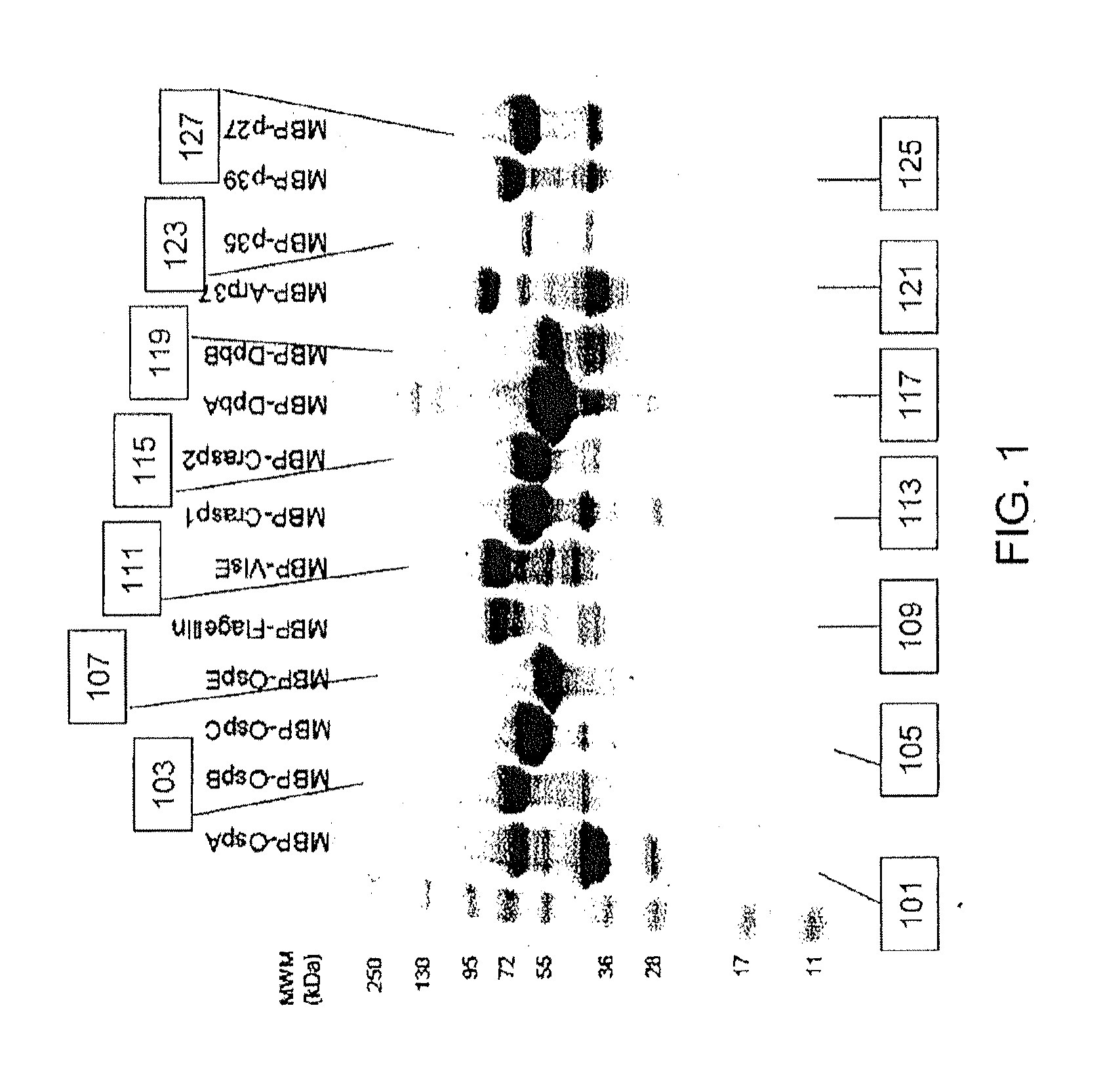
[0083]B. burgdorferi genomic DNA (Catalog #35210D-5) was obtained from the ATCC and fourteen (14) B. burgdorferi-specific proteins representative of the in vivo life cycle post infection: OspA, OspB, OspC, OspE, Flagellin, VlsE, Crasp-1, Crasp-2,DbpA, DbpB, Arp37, p35, p39 and p27 were selected to be included in the assay design. Gene-specific primers for the PCR-based amplification of individual bacterial genes were designed and synthesized. Amplified genes were inserted into a modified pMa1C2X vector (NEB #E8200S, New England Biolabs) by ligation independent cloning. Cloned inserts were verified by DNA sequencing and confirmed vectors were transformed into the E. coli expression strain BL21(DE3)pLysS. All fourteen MBP / B. burgdorferi fusion proteins were successfully expressed in E. coli upon IPTG induction (FIG. 9).

[0084]Cells from induced two liter cultures were harvested by centrifugation and ly...

example 2

Assay Optimization

[0086]Initial investigations to determine assay-specific parameters included the evaluation of human LD positive serum reactivity in a slot blot assay. Recombinant MBP / B. burgdorferi antigens as well as recombinant MBP at known concentrations ranging up to 1 μg were individually loaded into separate slots and adsorbed onto a nitrocellulose membrane overnight at 4° C. The membrane was rotated by 90° prior to application of 1:100 diluted serum from clinically characterized individual donors (n=12), obtained from SeraCare Diagnostics (Milford, Mass.), across all 14 B. burgdorferi antigens (OspA, OspB, OspC, OspE, VlsE, CRASP-1, CRASP-2, DbpA, DbpB, Flagellin, Arp37, P27, P35, and P39). Sera were incubated for 2 h at 4° C. prior to washing of the membrane and application of either HRP-conjugated anti-human IgG Fc or HRP-conjugated anti-human IgM Fc5μ antibodies. Results obtained from individual serum donors after substrate addition showed reactivity to specific B. burg...

example 3

Application of the Assay to Measure Borrelia-Specific Antibodies in Human Serum and Comparison with Current Assays Available on the Market

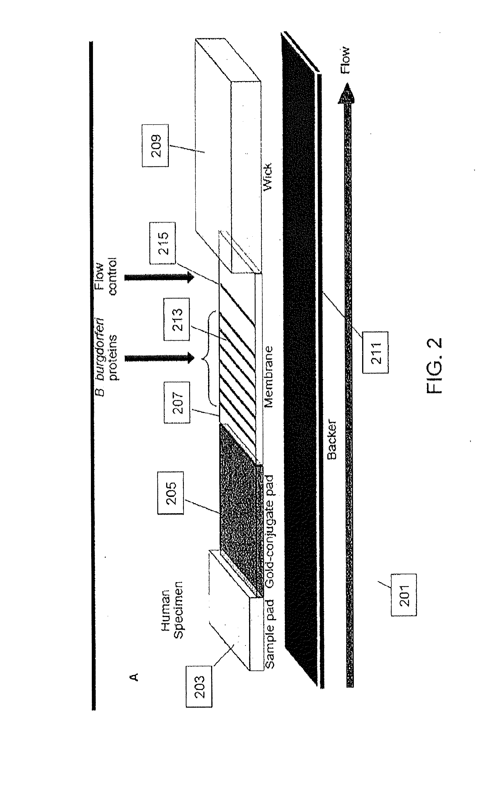
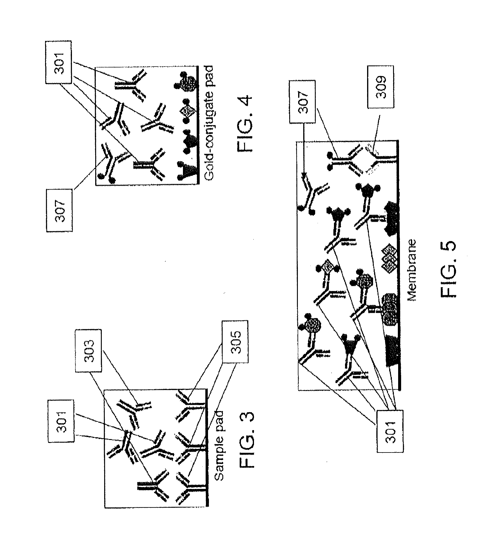
[0090]LD positive human sera were obtained from the CDC (n=35) and Tufts University (n=12). Samples from the CDC were accompanied by written results of a clinical exam as well as bioMérieux ELISA and MarBlot IgG and IgM WB results. LD positive human sera and negative control sera (n=5) were analyzed to demonstrate detection of strip immobilized B. burgdorferi antigens by antibodies present in sera of patients diagnosed with LD. IgG and IgM test strips were modified to receive sera premixed with liquid gold-labeled recombinant B. burgdorferi antigens and processed using all 52 sera. Antibody reactivity resulted in gold deposition for band visualization and was recorded after 20 min while wet and after 18 h when dried. No change in band reactivity between the two evaluation time points was noted. All LD positive sera showed significant reactivity to...


PUM

PropertyMeasurementUnit
molecular weightaaaaaaaaaa

Abstract

Recombinant B. burgdorferi proteins, representative of the life cycle, are membrane-immobilized to capture antibodies in biological samples. Lateral flow technology incorporating gold colloid deposition results in band visualization indicative of a positive test.

Description

[0001]This invention claims the priority of U.S. Provisional Patent Application No. 61 / 419,028, filed Dec. 2, 2010, which is incorporated herein by reference.[0002]This invention was made with government support under grant #1R43AI084296-01 awarded by the National Institute of Health. The government has certain rights in the invention.FIELD OF THE INVENTION[0003]The invention relates to assays used to test for Lyme disease. More specifically, the invention relates to a composition for testing for Borrelia burgdorferi sensu lato (the causal agent of Lyme disease, including antigens derived from B. burgdorferi, B. garinii and B. afzelii, and other sensu lato Borrelia species, hereinafter B. burgdorferi), an assay for testing for B. burgdorferi using the composition, and a method of testing for Lyme disease. The assay is amenable to use in a lateral flow device and can be used for on-site testing at the point of care. The assay relates to Lyme disease detection in vertebrate animals, i...

Claims


Application Information

Patent Timeline
Patent Type & Authority Applications(United States)
IPC IPC(8): G01N33/569G01N33/82G01N21/64
CPCG01N2800/26G01N33/56911Y02A50/30
Inventor ASCOLI, CARL A.CHIMENTO, DAVID P.TRAN, HIEP T.KOHL, THOMAS O.
Owner ROCKLAND IMMUNOCHEM
Why Eureka
Try Eureka