
Systems and methods for localization of a puncture site relative to a mammalian tissue of interest

a technology of puncture site and mammalian tissue, which is applied in the field of system and method for localizing a puncture site relative to a mammalian tissue of interest, can solve the problems of inconsistent delivery, significant decrease in the ability of the heart to pump blood, and chronic heart failure, so as to reduce the supply, shorten the operation time, and save costs

Inactive Publication Date: 2012-07-26
CVDEVICES
View PDF23 Cites 28 Cited by
  • Summary
  • Abstract
  • Description
  • Claims
  • Application Information

AI Technical Summary

Benefits of technology

[0027]In at least one embodiment of a method for facilitating mammalian organ treatment using a system for facilitating mammalian organ treatment of the present disclosure, the method comprises the steps of inserting the engagement catheter and puncture device into a mammalian body and into a right atrium of a heart, stabilizing an atrial septum at a first location using suction through the first lumen or a second lumen of the engagement catheter so that the open distal end reversibly engages the atrial septum at the first location, operating the scanner to obtain data relative to the distance between the second mammalian tissue and the at least a portion of the bodily access system positioned within the mammalian luminal organ, advancing at least part of the puncture device through the atrial septum at a fossa ovalis and into a left atrium to facilitate a therapy within the left atrium if / when the data relative to the distance between the second mammalian tissue and the at least a portion of the bodily access system is satisfactory, and performing the therapy within the left atrium, the therapy selected from the group consisting of delivering a mitral valve into the heart, placing a mitral valve within the heart, delivering a mitral valve prosthesis into the heart, placing a mitral valve prosthesis within the heart, delivering a mitral clip into the heart, placing a mitral clip within the heart, delivering a mitral ring into the heart, placing a mitral ring within the heart, treating mitral regurgitation, treating mitral stenosis, delivering a left atrial appendage occluder device into the heart, placing a left atrial appendage occluder device within the heart, treating stroke, reducing a risk of stroke, preventing a blood clot from a left atrial appendage from entering into a bloodstream, and delivering a drug therapy into the heart.
[0030]In at least one method for facilitating mammalian organ treatment of the present disclosure, the data relative to the distance between the second mammalian tissue and the at least a portion of the bodily access system is selected from the group consisting of data relative to a distance between a mitral valve annulus and the at least a portion of the bodily access system, data relative to a distance between a mitral valve portion and the at least a portion of the bodily access system, data relative to a distance between a left atrial appendage opening and the at least a portion of the bodily access system, and data relative to a distance between a left atrial appendage portion and the at least a portion of the bodily access system. In an additional embodiment, the method further comprises the step of repeating one or more of the stabilizing step and the operating step, prior to the advancing step, until the data relative to the distance between the second mammalian tissue and the at least a portion of the bodily access system is satisfactory. In yet an additional embodiment, the method further comprises the step of performing the therapy within the left atrium. In another embodiment, the therapy is selected from the group consisting of delivering a mitral valve into the heart, placing a mitral valve within the heart, delivering a mitral valve prosthesis into the heart, placing a mitral valve prosthesis within the heart, delivering a mitral clip into the heart, placing a mitral clip within the heart, delivering a mitral ring into the heart, placing a mitral ring within the heart, treating mitral regurgitation, treating mitral stenosis, delivering a left atrial appendage occluder device into the heart, placing a left atrial appendage occluder device within the heart, treating stroke, reducing a risk of stroke, preventing a blood clot from a left atrial appendage from entering into a bloodstream, and delivering a drug therapy into the heart.

Problems solved by technology

Indeed, the disease has had a profound impact worldwide, If left untreated, ischemic heart disease can lead to chronic heart failure, which can be defined as a significant decrease in the heart's ability to pump blood.
However, endoluminal drug delivery has several shortcomings, such as: (1) inconsistent delivery, (2) low efficiency of localization, and (3) relatively rapid washout into the circulation.
Such patients generally have an abnormality in conduction that causes the right and left ventricles to beat (i.e., begin systole) at slightly different times, which further decreases the heart's already-limited function.
Like the local delivery of drugs to the heart, the placement of CRT leads on the heart can be challenging, particularly when the target placement site is the left ventricle.
Problems with these methods of lead placement can include placement at an improper location (including inadvertent placement at or near scar tissue, which does not respond to the electrical signals), dissection or perforation of the coronary sinus or cardiac vein during placement, extended fluoroscopic exposure (and the associated radiation risks) during placement, dislodgement of the lead after placement, and long and unpredictable times required for placement (ranging from about 30 minutes to several hours).
The additional clearance tends to decrease the likelihood that the myocardium will be inadvertently punctured when the pericardial sac is pierced.
Although this technique works well in the normal heart, there are major limitations in diseased or dilated hearts—the very hearts for which drug delivery and CRT lead placement are most needed.
When the heart is enlarged, the pericardial space is significantly smaller and the risk of puncturing the right ventricle or other cardiac structures is increased.
Additionally, because the pericardium is a very stiff membrane, the suction on the pericardium provides little deformation of the pericardium and, therefore, very little clearance of the pericardium from the heart.
Therefore, it is not surprising that cardiac perforation can quickly result in tamponade, which can be lethal.
Ultimately, compression of the heart by a pressurized pericardial effusion results in markedly elevated venous pressures and impaired cardiac output producing shock which, if untreated, it can be rapidly fatal.
End-stage renal disease is a major public health problem.
Renal failure is a common cause of pericardial disease, producing large pericardial effusions in up to 20% of patients.
Purulent (bacterial) pericarditis in adults is rare, but always fatal if untreated.
Management of pericardial effusions continues to be a challenge.
There is no uniform consensus regarding the best way to treat this difficult clinical entity.
This procedure portends significant operative and anesthetic risks because these patients often have multiple comorbidities.
Less invasive techniques such as blind needle pericardiocentesis have high complication and recurrence rates.
However, practices in community hospitals have rarely been studied in detail, Buchanan C. L., Sullivan V. V., Lampman R., Kulkarni M. G. (2003).
Medical management is usually ineffective and should be used only while arrangements are made for pericardial drainage.
Surgical drainage (or pericardiectomy) is excessive for many patients.
“[Diagnosis and management of acute pericardial syndromes].” Rev Esp Cardiol 58(7): 830-41, This less-invasive technique resulted in a short operative time and decreased supply, surgeon, and anesthetic costs.

Method used


Image

  • Systems and methods for localization of a puncture site relative to a mammalian tissue of interest
  • Systems and methods for localization of a puncture site relative to a mammalian tissue of interest
  • Systems and methods for localization of a puncture site relative to a mammalian tissue of interest

Examples


Embodiment Construction

[0097]For the purposes of promoting an understanding of the principles of the present disclosure, reference will now be made to the embodiments illustrated in the drawings, and specific language will be used to describe the same. It will nevertheless be understood that no limitation of the scope of this disclosure is thereby intended.

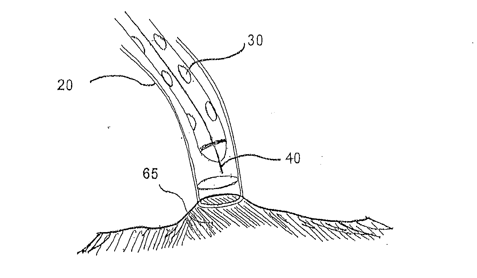
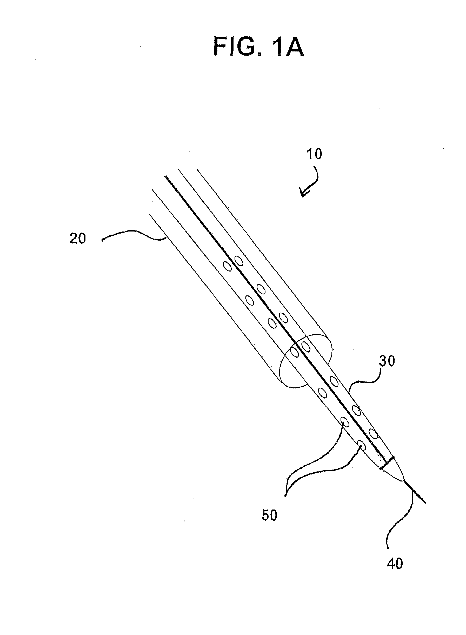
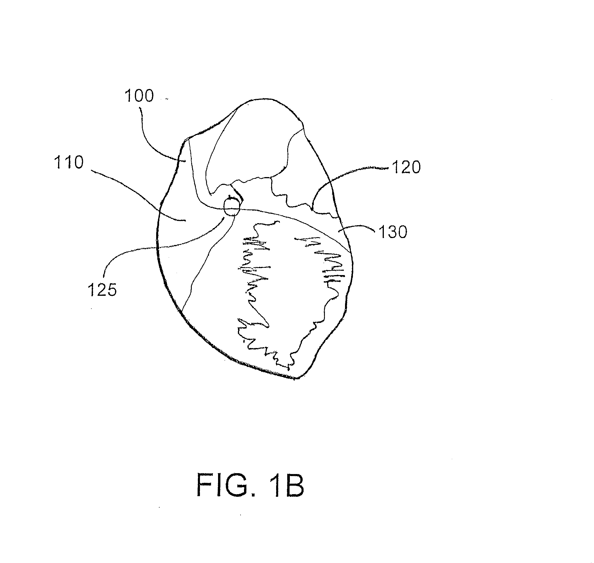
[0098]The disclosed embodiments include devices, systems, and methods useful for accessing various tissues of the heart from inside the heart. For example, various embodiments provide for percutaneous, intravascular access into the pericardial space through an atrial wall or the wall of an atrial appendage. In at least some embodiments, the heart wall is aspirated and retracted from the pericardial sac to increase the pericardial space between the heart and the sac and thereby facilitate access into the space.

[0099]Unlike the relatively stiff pericardial sac, the atrial wall and atrial appendage are rather soft and deformable. Hence, suction of the atri...


PUM


Abstract

Systems and methods for localization of a puncture site on an atrial wall relative to a mammalian tissue of interest. In at least one embodiment, such a system includes a bodily access system, having an engagement catheter with an open distal end, a first lumen therethrough, and configured to reversibly attach to a first tissue using suction, a puncture device, configured to fit at least partially within the first lumen, and a scanner configured to identify at least a portion of the bodily access system when positioned within the luminal organ and further configured to identify a distance between a second tissue and a portion of the bodily access system. When such a system is used in connection with a therapeutic procedure, the distance between the second tissue and the portion of the bodily access system is used to facilitate a puncture of the first tissue at a desired location.

Description

PRIORITY[0001]This U.S. continuation-in-part patent application is related to, and claims the priority benefit of, U.S. Nonprovisional patent application Ser. No. 13 / 084,102, filed Apr. 11, 2011, which is related to, claims the priority benefit of, and is a continuation-in-part of, U.S. Nonprovisional patent application Ser. No. 12 / 881,953, filed Sep. 14, 2010, which is related to, claims the priority benefit of, and is a continuation-in-part of, U.S. Nonprovisional patent application Ser. No. 12 / 596,968, filed Oct. 21, 2009, which is related to, claims the priority benefit of, and is a U.S. national stage application of, International Patent Application No. PCT / US2008 / 056666, filed Mar. 12, 2008, which (i) claims priority to International Patent Application No. PCT / US2007 / 015207, filed Jun. 29, 2007, and U.S. Provisional Patent Application Ser. No. 60 / 914,452, filed Apr. 27, 2007. The contents of each of these applications are hereby incorporated by reference in their entirety into...

Claims


Application Information

Patent Timeline
IPC IPC(8): A61B17/34A61F2/24A61M25/04
CPCA61B17/0057A61M2025/0089A61B17/3478A61B2017/00247A61B2017/00592A61B2017/00601A61B2017/00606A61B2017/00876A61B2017/306A61B2017/308A61B2017/3419A61B2017/3454A61B2017/3488A61B2018/00392A61M25/003A61M25/007A61M25/04A61M2025/0004A61M2025/0039A61M2025/004A61B17/3417
Inventor KASSAB, GHASSAN S.NAVIA, SR., JOSE A.
Owner CVDEVICES
Learn More