Biological tissue inspection method and system

a tissue inspection and biochemical technology, applied in the field of biomedical imaging, can solve the problems of damage in industrial laser-ultrasonics, limited probing depth, and limited imaging methods relying on ballistic photons, so as to achieve less absorption, reduce pulse duration, and effectively provide concentrated power

Inactive Publication Date: 2012-08-09
NAT RES COUNCIL OF CANADA
View PDF15 Cites 33 Cited by
  • Summary
  • Abstract
  • Description
  • Claims
  • Application Information

AI Technical Summary

Benefits of technology

[0022]Specific operating conditions involve permissible use of high detection laser power, with a large étendue demodulator, wherein the wavelength is chosen to provide little absorption, and the pulse duration is minimized, to effectively provide concentrated power.
[0023]The less absorbing the laser wavelength, the higher a dose is permitted, effectively ensuring a higher MPE. For example, choosing detection at 1.06 μm (fundamental of Nd-YAG laser) instead of 532 nm (frequency-doubled Nd-YAG) for which absorption is much stronger, allows a 5 fold increase in MPE. Using a wavelength longer than 1.5 μm and below 1.8 μm gives an even higher MPE: 1 J / cm2 compared to 0.5 J / cm2 calculated above.

Problems solved by technology

Damage in industrial laser-ultrasonics is usually caused by ablation from the generation laser and not the detection laser.
Those imaging methods rely on ballistic photons and are essentially limited by the light diffraction limit.
This technique still provides a high transverse resolution (typically of the order of 1 μm), a good depth resolution (typically 10 μm) but is limited in terms of the probing depth, typically 1 to 2 millimeters in biological tissue.
Diffuse optical tomography (DOT) relies instead on the detection of diffuse photons but has a poor spatial resolution (1 cm is typical).
There are obviously major drawbacks to applying PAT in some applications, such as small animal imaging where the water bath require immersing the animal with an air carrier (aerophore) as shown in FIG. 2.
It is also cumbersome in the case of human breast imaging for the detection of cancer which is another application for which PAT has been developed.
This coupling requirement also limits the application of PAT in the case of surgery or endoscopic examination.
Non-contact detection by optical means in PAT, as done in industrial laser-ultrasonics, would obviously be desirable but is not feasible with prior art knowledge, essentially because of two opposing requirements: the requirement of using a high power detection laser to get sufficient detection sensitivity; and the requirement of using a low power detection laser to avoid damaging the tissue.
Some efforts to apply non-contact PAT to biological phantoms and tissues have been reported in the past, but the reported approaches have failed to be practical or could not produce adequate sensitivity in the case of a tissue that has not been provided a high reflectivity coating.
These previous works do not teach or suggest that it was possible to apply non-contact PAT in such conditions.
Existing non-contact PAT techniques do not teach how to apply non-contact PAT on biological tissue in vivo or ex vivo, and does not even suggest that this would be possible.
The conflicting requirements for high power signaling without damaging tissues have so far led to the conclusion that non-contact optical detection of ultrasonic waves on a living organism is not possible, and no such technique or system has been reported.
Therefore the sensitivity calculated based only on the reflection of the surface of the tissue, assuming maximum permissible exposure for 1 mm spot size and other design parameters but neglecting all transmission losses through the various optical elements, is barely sufficient.
It would therefore appear inevitable that the sensitivity of optical detection of ultrasonic waves in tissues under non-damaging conditions is generally insufficient, and this confirms the belief that non-contact detection and therefore non-contact imaging of biological tissues using PAT-like excitation is expected to be infeasible, regardless of how desirable it would be.

Method used


Image

  • Biological tissue inspection method and system
  • Biological tissue inspection method and system
  • Biological tissue inspection method and system

Examples


example

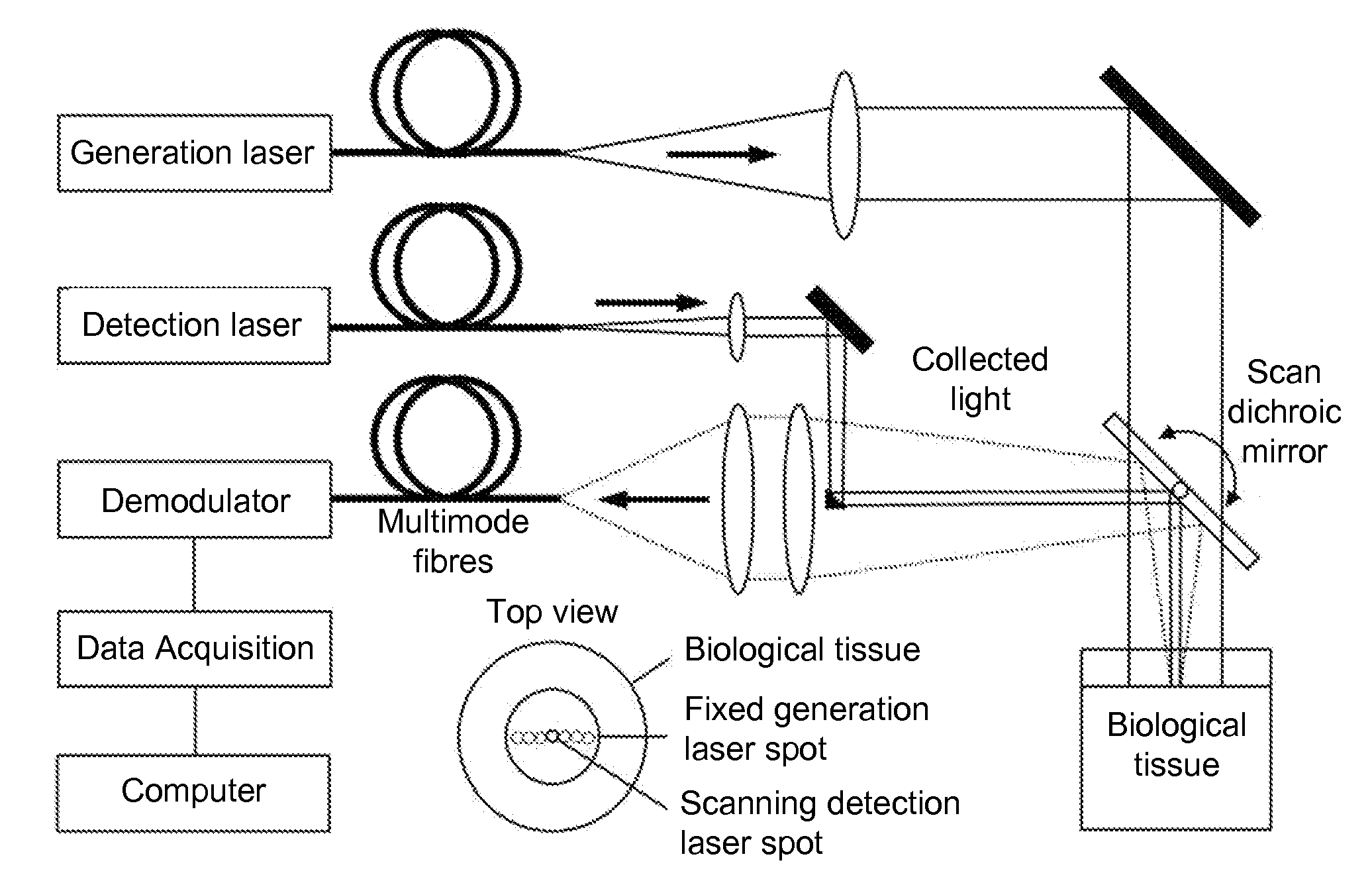
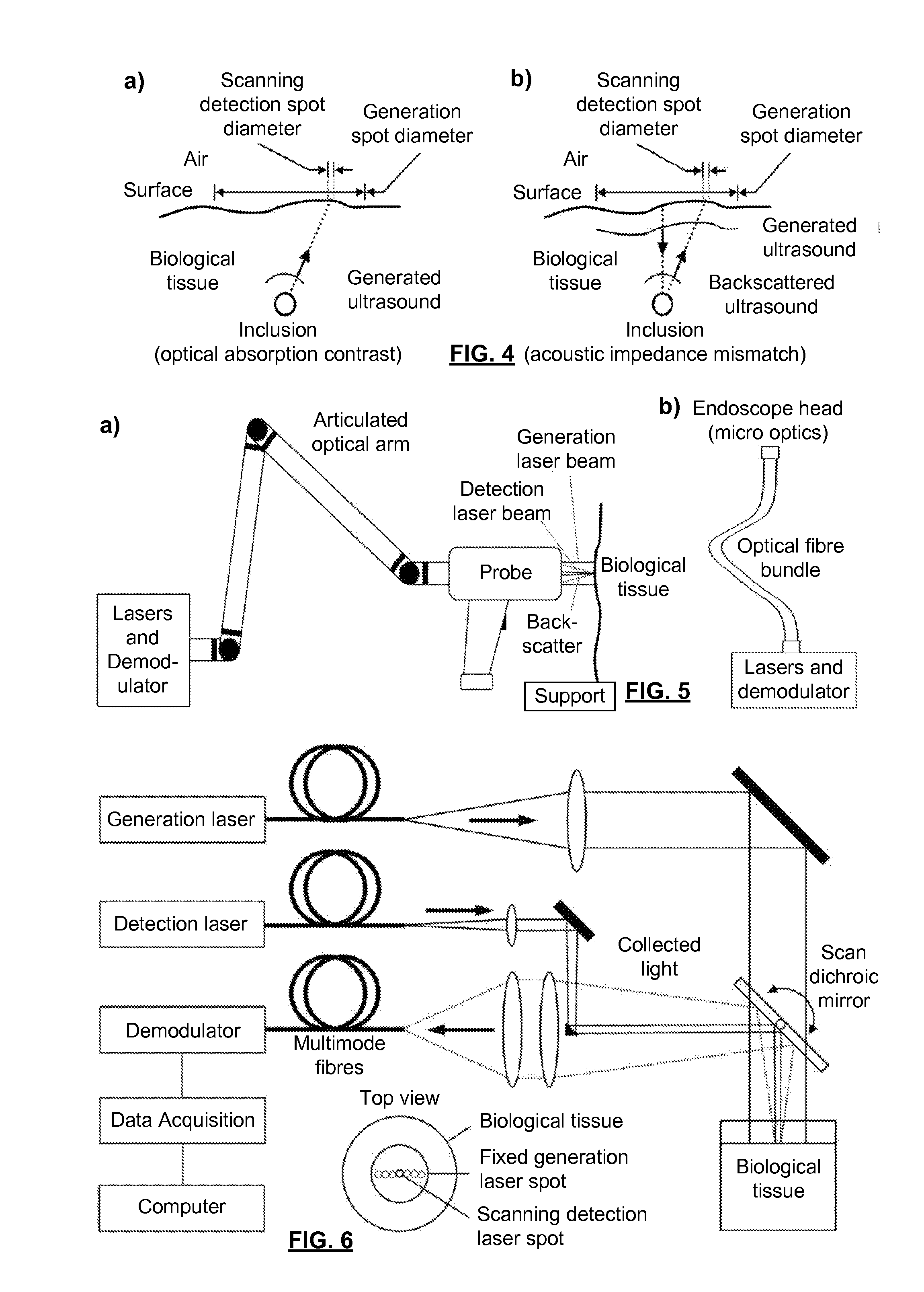
[0057]FIG. 6 is a schematic illustration of a system used to verify the present invention. The generation laser was a frequency doubled actively Q-switched laser emitting 5 ns pulses at 532 nm. This wavelength efficiently excites blood, making it an effective ultrasonic transducer by thermoelastic generation. The repetition rate was equal to 10 Hz and the pulse energy impinging on the surface was about 100 mJ. The GLB was oriented toward a BT onto a large spot size (about 25 mm in diameter giving an area of 5 cm2) which satisfies the maximum permissible exposure (MPE) at generation wavelength (20 mJ / cm2 at 532 nm). With a 10 Hz repetition rate, the average power density of the GLB was equal to 200 mW / cm2, which is also in accordance with the safety limit in the case of repetitive exposure at 532 nm. The detection laser beam was transmitted by a second optical fiber and focused onto the surface of the tissue after reflection by a computer controlled scanning mirror. The DLB had a wav...


PUM

PropertyMeasurementUnit
wavelengthaaaaaaaaaa
peak poweraaaaaaaaaa
depth resolutionaaaaaaaaaa

Abstract

A method and system for inspecting biological tissue that has no applied coatings or treatments to improve reflectivity comprises an optical detection system with an exposed surface for inspection by an optical detection system; and a laser for exciting an ultrasonic wave within the tissue, which wave propagates within the tissue at least near the surface. The optical detection system includes: a laser to emit a pulsed detection laser beam onto the surface at a detection spot, the pulsed laser beam having a wavelength at which there is large scattering and little absorption by the tissue, and a pulse duration is chosen to correspond with ultrasonic propagation times associated with a range of depths of the scan, whereby the fluence of the detection laser is not applied unless ultrasonic information regarding a given depth is being obtained; and large étendue collection optics for collecting reflected and backscattered light from the detection spot; and a demodulator to extract information from the ultrasonic wave from the collected light.

Description

FIELD OF THE INVENTION[0001]The present invention relates to the field of biomedical imaging and, in particular, to a laser and ultrasound-based method and system for in vivo or ex vivo, non-contact imaging of biological tissue.BACKGROUND OF THE INVENTION[0002]Laser-ultrasonics is a well established technique first developed for the non-destructive testing of industrial materials like metals, plastics and polymer-matrix composite materials. A typical laser-ultrasonic system is composed of two lasers and a phase demodulator, as sketched in FIG. 1. In laser-ultrasonics, ultrasound is generated and detected at a distance. The ultrasound is usually generated on the surface of the material and the detection of ultrasound is performed on the same surface with a detection laser and a phase demodulator collecting the reflected and / or scattered light. Laser-ultrasonics can be applied to parts having complex shapes without any surface preparation and at high temperature. This technique has be...

Claims


Application Information

Patent Timeline
Patent Type & Authority Applications(United States)
IPC IPC(8): G01N21/55G01B9/02
CPCG01N21/1702G01N2291/02475G01N29/2418G01N21/171A61B5/0059A61B5/0093A61B5/0095A61B5/0097
Inventor ROUSSEAU, GUYBLOUIN, ALAINMONCHALIN, JEAN-PIERRE
Owner NAT RES COUNCIL OF CANADA
Why Eureka
Try Eureka