Gut flora-derived extracellular vesicles, and method for searching for a disease model, vaccine, and candidate drug and for diagnosis using the same

Active Publication Date: 2012-08-30
AEON MEDIX
View PDF1 Cites 9 Cited by
  • Summary
  • Abstract
  • Description
  • Claims
  • Application Information

AI Technical Summary

Benefits of technology

[0066]Based on the finding that extracellular vesicles derived from intestinal bacteria that colonize the digestive tract cause local diseases characterized by mucosal inflammation and systemic disease, such as sepsis, characterized by systemic inflammatory responses upon introduction into blood, the present invention utilizes gut flora-derived extracellular vesicles in establishing a disease model, a method for screening candidate drugs preventive or therapeutic of diseases, and a vaccine for the prophylaxis or therapy of diseases.
[0067]In the present invention, it was found that gut flora-derived extracellular vesicles induced the secretion of an inflammatory factor from cells when applied to the cells, and caused mucosal inflammation when administered topically, and systemic diseases including sepsis, intravascular blood coagulation, emphysema, hypertension and osteoporosis when intraper

Problems solved by technology

However, no therapeutics targeting LPS have been successfully developed thus far (J. Hellman, Bacterial peptidoglycan-associated lipoprotein is released into the bloodstream in gram-negative sepsis and causes inflammation and death in mice.
However, these sepsis animal models suffer from the drawback of being low in reproducibility and great in error rate between operators and lacking the ability to properly express the phenotype of sepsis.
However, little is known about any method for ex vivo screening candidate drugs capable of regulating inflammat

Method used


Image

  • Gut flora-derived extracellular vesicles, and method for searching for a disease model, vaccine, and candidate drug and for diagnosis using the same
  • Gut flora-derived extracellular vesicles, and method for searching for a disease model, vaccine, and candidate drug and for diagnosis using the same
  • Gut flora-derived extracellular vesicles, and method for searching for a disease model, vaccine, and candidate drug and for diagnosis using the same

Examples


example 1

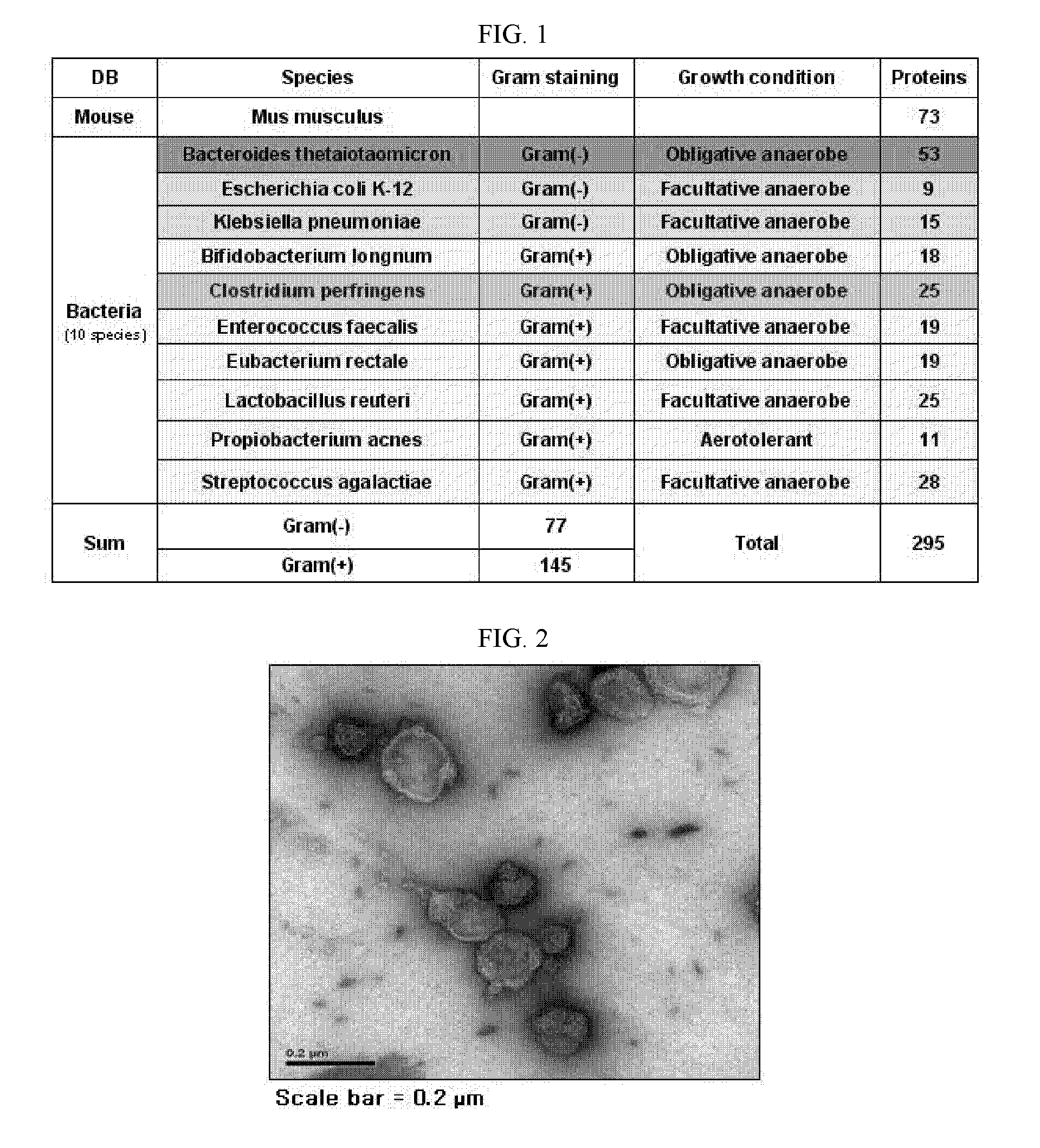
Proteomic Assay of Extracellular Vesicles Separated from Normal Mice

[0164]Extracellular vesicles were separated from mouse feces. In this regard, first, 25 g of feces excreted from 5-week-old, male C57BL / 6 mice was resuspended in 2 L of PBS (phosphate buffered saline) at 4° C. for 16 hours. After the centrifugation of the suspension at 4° C. and 10,000×g for 20 min, the supernatant was passed through a filter with a pore size of 0.45 nm. The filtrate thus free of bacteria was about 30-fold concentrated to 70 mL using QuixStand Benchtop System equipped with a membrane with 100 kDa cutoff. The concentrate was again centrifuged at 4° C. and 10,000×g for 20 min. This supernatant was added to 0.5 ml of 2.5 M sucrose solution (2.5 M sucrose / 20 mM HEPES / 150 mM NaCl, pH7.4) and 1 ml of 0.8 M sucrose solution (0.8 M sucrose / 20 mM HEPES / 150 mM NaCl, pH7.4) in an ultracentrifuge tube, followed by ultracentrifugation at 4° C. and 100,000×g for 4 hours. The layer containing extracellular vesicle...

example 2

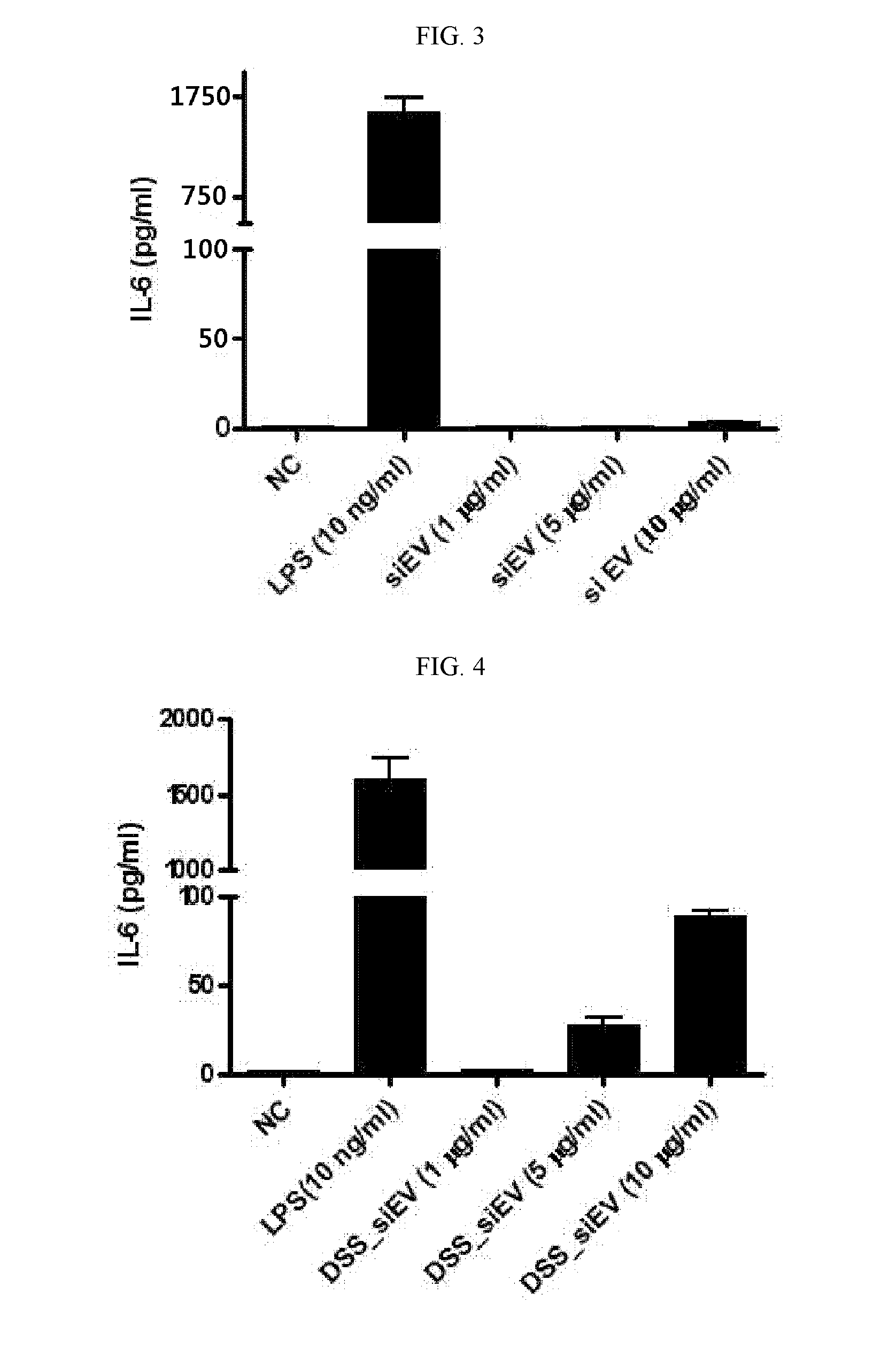
Secretion of Inflammatory Mediators by Extracellular Vesicles Separated from Small Intestinal Fluid of Normal and Diseased Mice

[0170]Extracellular vesicles were separated from small intestinal fluids of normal mice and DSS-induced mouse model of irritable bowel disease. To this end, mice were operated on to excise the small intestine. The peyer's patch and liquid were removed from the excised small intestine which was then cut at regular lengths of about 5 cm. The intestinal pieces were transversely cut and washed with physiological saline to remove impurities therefrom. They were again cut into pieces with dimensions of 1 cm×1 cm and vortexed five times for five sec per time in 30 mL of physiological saline. After the removal of intestinal tissues and impurities by centrifugation, the supernatant was ultra-centrifuged to separate extracellular vesicles.

[0171]FIG. 2 shows sizes and morphologies of the separated extracellular vesicles as observed by transmission electron microscopy (...

example 3

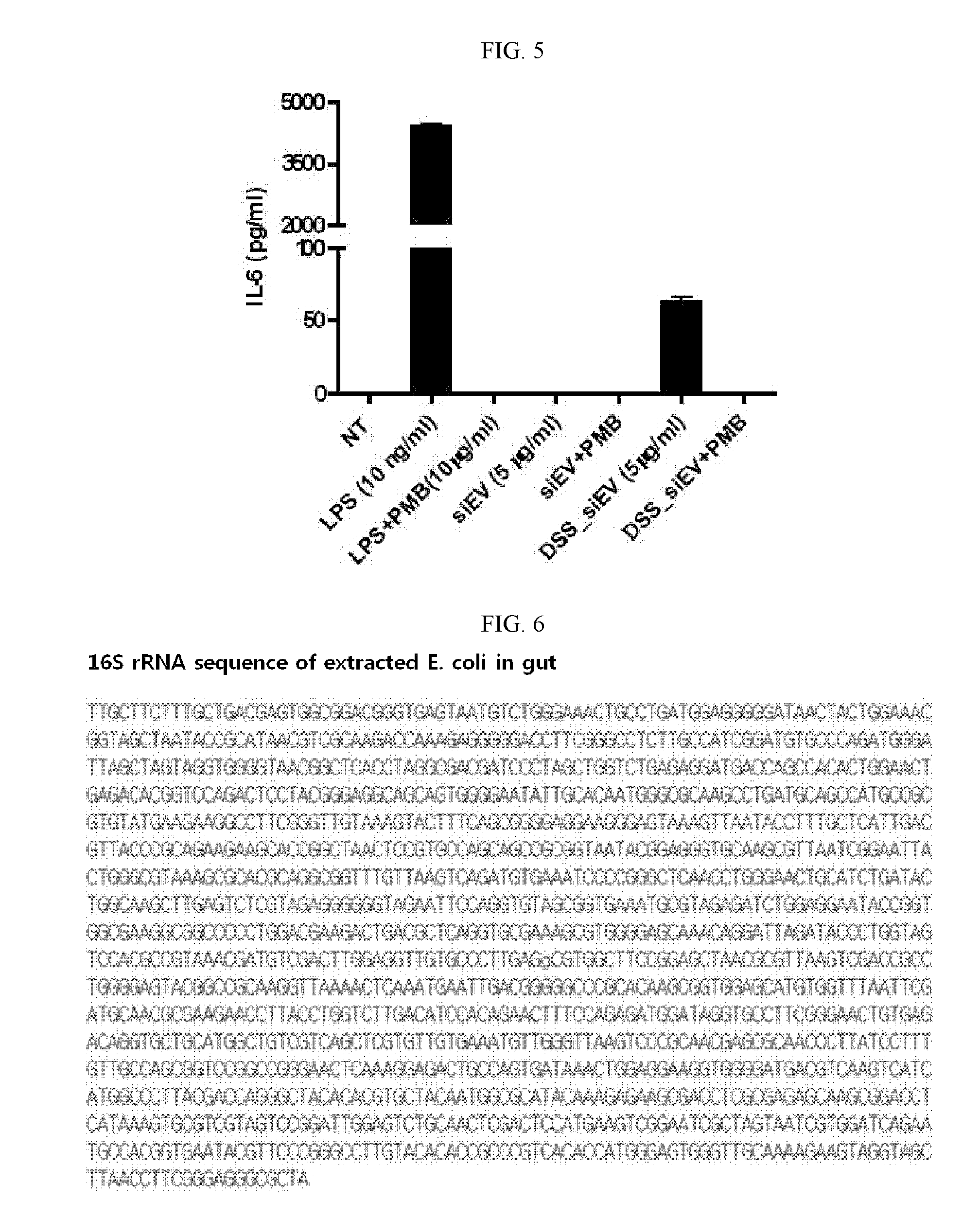
Characterization of Extracellular Vesicles Isolated from E. coli of Gut Flora in Peritoneal Fluid of CLP-Induced Animal Model of Sepsis

[0175]CLP (cecal ligation and puncture) was performed as previously reported. In brief, one C57BL / 6 (male, 6 weeks old) mouse was anesthetized and a midline incision was made through the linea alba. The cecum was located, ligated with a sterile thread and perforated twice with 18-gauge needle. Forty hours later, 3 mL of PBS was intraperitoneally injected into the mouse using a 5 mL syringe and well-mixed therein, after which 1 mL of peritoneal fluid was recovered. A mixture of 10 μl of the recovered fluid and 90 μl of LB (Luria Bertani) was 10,000-fold diluted, spread over LB agar plates and incubated at 37° C. for 8 hours in an incubator. One of the colonies thus formed was inoculated into 5 mL LB in a test tube and incubated for 8 hours in a 37° C. incubator. A mixture of 10 μl of this culture and 90 μl of LB was 10,000-fold diluted, spread over LB...


PUM

PropertyMeasurementUnit
Compositionaaaaaaaaaa
Immunogenicityaaaaaaaaaa

Abstract

The present application relates to a composition including gut flora-derived extracellular vesicles, and to an animal disease model using same. In addition, the present application relates to a method for using the gut flora-derived extracellular vesicles to efficiently search for a candidate drug which may prevent or treat diseases that occur due to gut flora-derived extracellular vesicles, and to a vaccine which can efficiently prevent or treat infections caused by gut flora or diseases that occur due to gut flora-derived extracellular vesicles. Further, the development of diagnostic technology to discover, using the gut flora-derived extracellular vesicles of the present application, the etiology of diseases that occur due to gut flora-derived extracellular vesicles, can be achieved.

Description

TECHNICAL FIELD[0001]The present invention relates to a composition comprising gut microbiota- or gut flora-derived extracellular vesicles from extracellular vesicles, a disease animal model established with the same, and a vaccine for the prevention and / or therapy of diseases caused by gut microbiota- or gut flora-derived vesicles.BACKGROUND ART[0002]Gut microbiota or flora consists of microorganisms that live in the digestive tracts of animals. The 10 trillion or so microbial cells living in the gut, exceed the number of human cells by 10 to 1.[0003]In the 1960s, electron microscopy revealed that Gram-negative cells release extracellular vesicles (EV) or outer membrane vesicles (OMV). Extracellular vesicles are spherical with a size of 20-200 nm and consist of phospholipid bilayers. Gram-negative bacterial extracellular vesicles have various outer membrane proteins as well as LPS (E. Y. Lee et al., Proteomics in gram-negative bacterial outer membrane vesicles. Mass. Spectrom. Rev....

Claims


Application Information

Patent Timeline
IPC IPC(8): A61K39/108C07K14/195G01N33/53A61K39/00A61K39/02A61K39/104A61K31/496A61K31/5377A61K31/404A61K31/551A61K31/55A61K31/4184A61P35/00A61P3/04A61P3/10A61P11/00A61P29/00A61P1/00A61P9/10A61P7/00A01K67/027
CPCC12Q1/025A01K67/027A01K2207/20A01K2217/075G01N33/56916A01K2267/0337A01K2267/035C12Q1/37A01K2227/105A61P1/00A61P1/02A61P1/04A61P1/16A61P1/18A61P11/00A61P19/02A61P19/10A61P25/00A61P29/00A61P3/04A61P31/00A61P31/04A61P35/00A61P43/00A61P7/00A61P9/10A61P9/12A61P3/10
Inventor KIM, YOON KEUNGHO, YONG SONGPARK, KYOUNG SUHONG, BOK SILKIM, JI HYUNKIM, YOU SUNLEE, WON HEEKIM, JUNG WOOKKIM, DAE-KYUM
Owner AEON MEDIX
Why Eureka
Try Eureka