Reagent kit for quantitatively detecting the mutations of epidermal growth factor receptor(EGFR)

a technology of epidermal growth factor receptor and quantitative detection, which is applied in the direction of microbiological testing/measurement, fluorescence/phosphorescence, biochemistry apparatus and processes, etc., can solve the problems of not producing quantitative, requiring a lot of human labor, and the restriction fragment length polymorphism method, so as to accurately and quantitatively determine the ratio of egfr mutations

Inactive Publication Date: 2012-09-20
BEIJING ACCB BIOTECH
View PDF3 Cites 1 Cited by
  • Summary
  • Abstract
  • Description
  • Claims
  • Application Information

AI Technical Summary

Benefits of technology

[0006]The detecting method of the present invention has the following advantages: easy manipulation, and easy standardization. Other methods, such as allele specific oligonucleotide probe hybridization method, are very much dependent on hybridization conditions, thus they need strict controlling of the experimental conditions. The restriction fragment length polymorphism method, on the other hand, needs a lot of human labor, and can not generate quantitative results. The method of the present invention has short experimental cycle, and can be completed with 2 hours. It doesn't need to verify the result by sequencing, whereas the direct sequencing and high resolution melting analysis need 4 days to 2 weeks. Sensitivity of the method of the present invention is high, which, after optimizing experimental conditions, can reach 1% for detecting mutations, whereas sensitivity of direct sequencing is 20-50%. Specificity of the method of the present invention is also high. Immunohistochemistry (IHC) method can easily get pseudo-positive and pseudo-negative results, and can not determine the position and types of point mutations. The unique advantage of the present invention is accurate quantification. By using absolute quantification method to analyze data, draw standard curve, and accurately determine the content of wild-type gene and mutant gene in the samples, one can obtain ratio of the mutant gene in the samples, which will be of help to clinical diagnosis and therapeutic selection. Further, the present invention is safe and non-toxic, other methods such as chemical breaking method of mismatch base need isotope and toxic chemical agents.
[0007]The question that the present invention addresses is to provide a quantitative detection kit for EGFR gene mutations, which can quantitatively detect the following mutations: EGFR Exon 18 (SEQ ID NO:1; SEQ ID NO:2) position 2155 G substituted with A; Exon 19 (SEQ ID NO:1; SEQ ID NO:3) position 2235-2249 deletion; Exon 19 position 2236-2250 deletion; Exon 19 2254-2277 deletion; and Exon 21 (SEQ ID NO:1; SEQ ID NO:4) position 2573 T substituted with G.
[0008]To address the above question, the present invention provides quantitative detection kit containing a mixture comprising Taq enzyme, 10×Taq buffer, MgCl2, dNTP mixture, PCR primers which can specifically amplify the sequences at EGFR gene mutation positions, and probes which can specifically identify wild-type sequences and mutant sequences, together with method of the detection.
[0009](1) Separately design upstream and downstream primers around the mutation positions of Exon 18, 19 and 21 of EGFR gene; and design specific probes according to each mutant site. Said probes can specifically bind wild-type sequences or the mutant sequences to be detected at specific EGFR sites, so as to determine whether the tested mutations occur at said sites.
[0010](2) To accurately and quantitatively determine the ratio of the EGFR mutations, standards were designed in the present invention.
[0011](3) Use fluorescent quantitative PCR to detect the samples and standards.

Problems solved by technology

The restriction fragment length polymorphism method, on the other hand, needs a lot of human labor, and can not generate quantitative results.
Immunohistochemistry (IHC) method can easily get pseudo-positive and pseudo-negative results, and can not determine the position and types of point mutations.

Method used


Image

  • Reagent kit for quantitatively detecting the mutations of epidermal growth factor receptor(EGFR)
  • Reagent kit for quantitatively detecting the mutations of epidermal growth factor receptor(EGFR)
  • Reagent kit for quantitatively detecting the mutations of epidermal growth factor receptor(EGFR)

Examples


example 1

[0029]Extracting genome DNA from fresh human tumor tissues, paraffin embedded tissues, peripheral blood, pleural effusion, and human cell lines

[0030]The tumor cell lines we tested included cell lines of: non-small-cell carcinoma (NSCLC; A549, H460, H838 and H1703), breast cancer (MCF-7, BT474 and HuL100), malignant mesothelioma (H513, H2052, H290, MS-1 and H28), colon cancer (SW480), head and neck cancer (U87), cervical carcinoma (Hela), sarcoma (Mes-SA, Saos-2 and A204).

[0031]The fresh human tumor tissues, peripheral blood, paraffin embedded tissues we tested included: NSCLC, mesothelioma, colon cancer, malignant melanoma, renal carcinoma, esophagus cancer, thyroid carcinoma, malignant cancer and ovarian cancer.

[0032]Extraction of Sample DNA

[0033]DNA extracting kit from Qiagen Inc., Promega Inc., or Roche Inc. can be used to extract genome DNA from the samples. Content and purity of the extracted DNA can be determined by using Nanodrop ND 1000 (Gene Inc.) (ODD260 / OD280 is about 1.8...

example 2

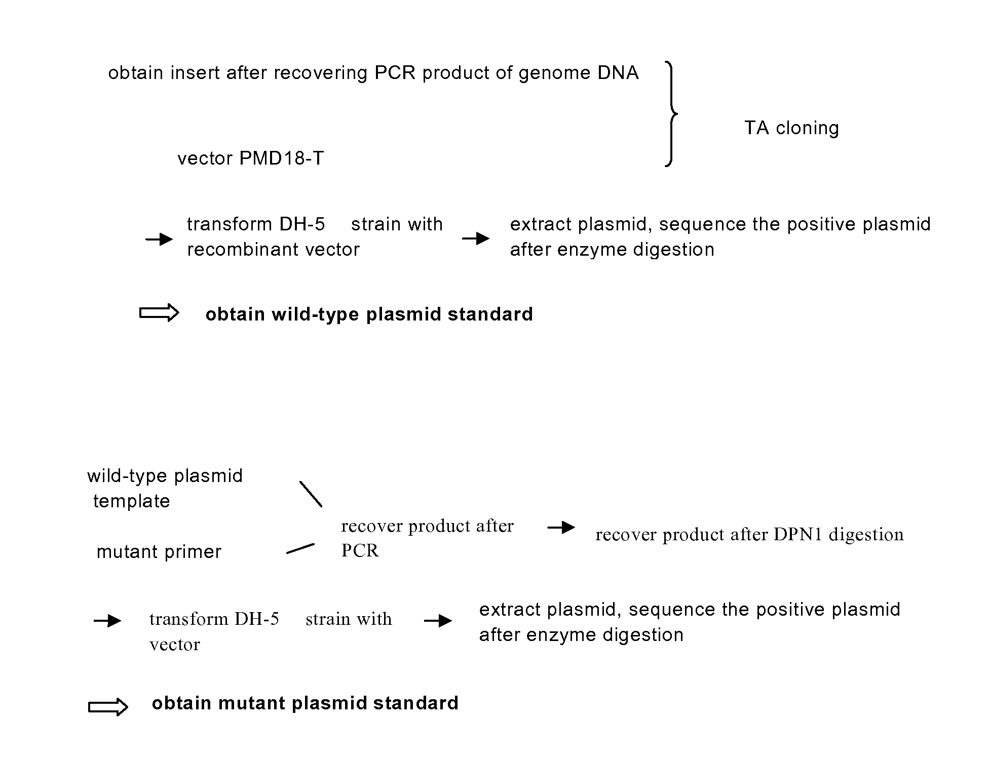
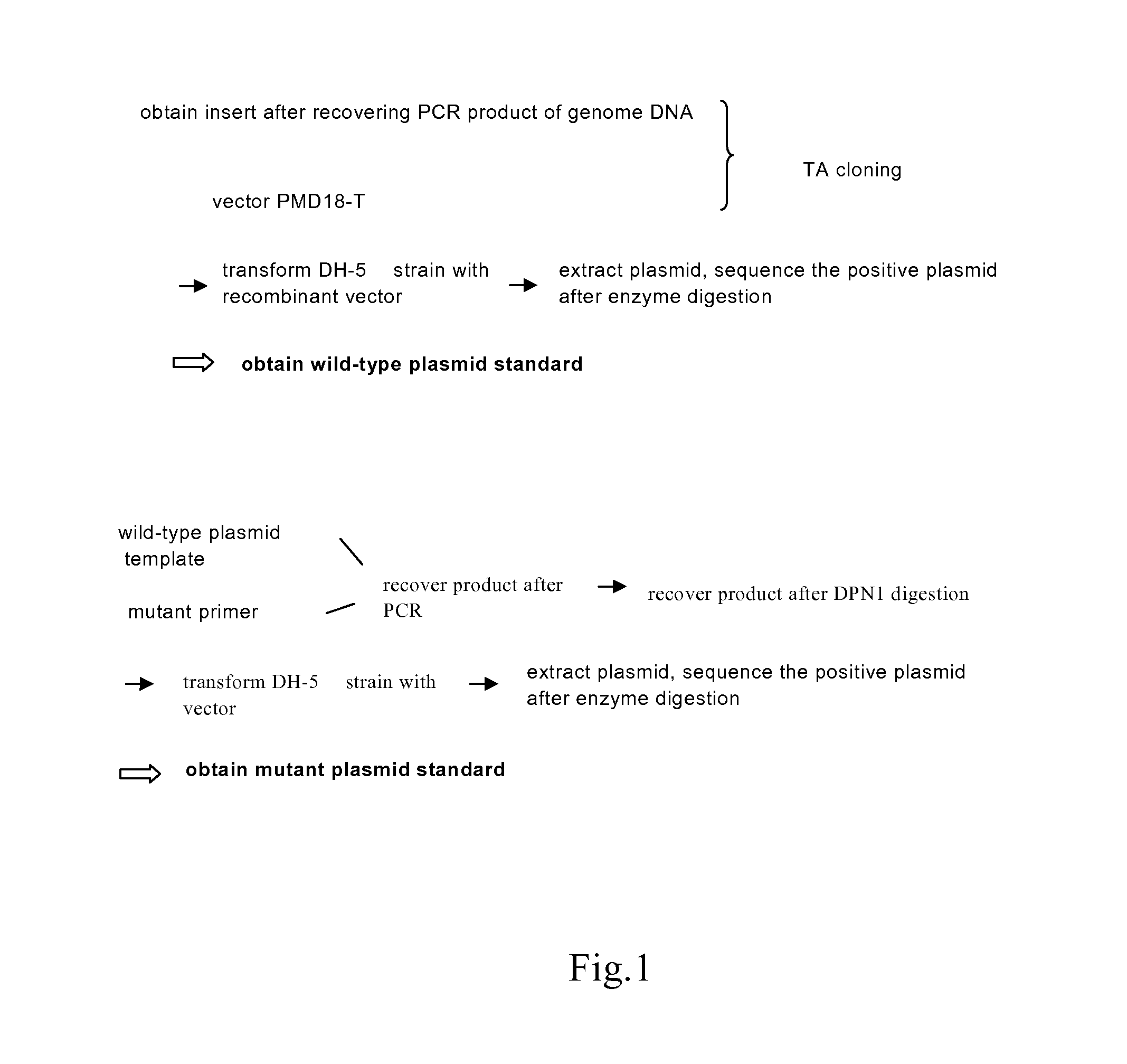
[0087]Preparation of the plasmid standards containing mutant and wild-type sequences

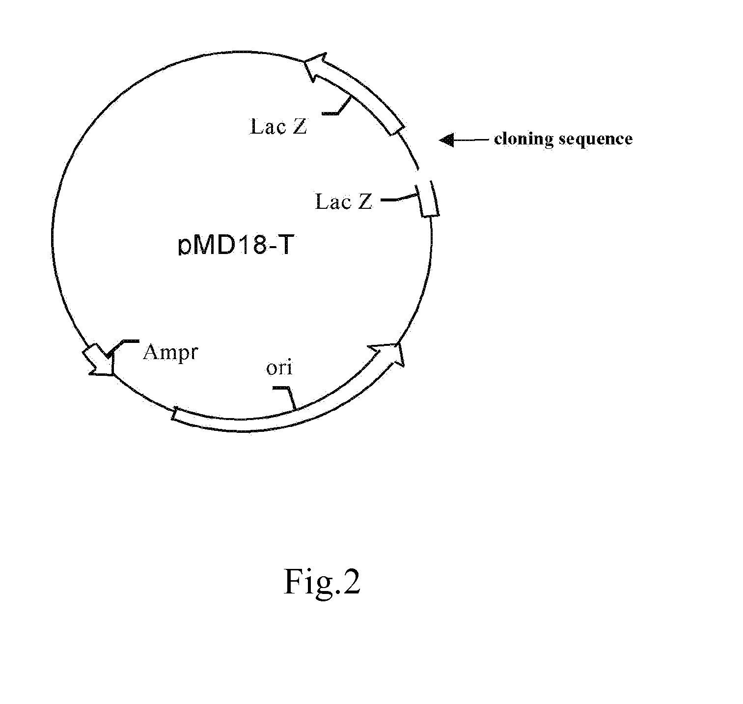
[0088]1. Construction of Wild-Type Plasmids (FIG. 1, FIG. 2)

[0089]1.1 Preparation of the Carrier

[0090]TA cloning carrier pMD18-T was purchased from TAKARA Inc.

[0091]1.2 Preparation of the Insert

[0092]The insert is prepared using PCR. The template of PCR is the sample genome DNA extracted in Step 1. The reaction system and amplification condition are shown in the following tables (Table 1, Table 2 and Table 3):

TABLE 1PCR reaction system (50 μl)reagentsamount(μl / tube)double-distilled water29.7510 × buffer (free of Mg2+)5MgCl2 (25 mM)7.5dNTP (10 mM)1.25upstream primer (25 μM)1.25downstream primer (25 μM)1.25Taq enzyme1DNA template3total volume50

[0093]For preparing the plasmid containing wild-type sequence at EGFR gene Exon 18 position 2155, it needs to add the primer sequences of E18-F-1 (SEQ ID NO:5) or E18-F-2 (SEQ ID NO:6) and E18-R-1 (SEQ ID NO:7) or E18-R-2 (SEQ ID NO:8) into the amplification syst...

example 3

[0105]Detection of EGFR mutations from genome DNA of human cell lines, human fresh tumor tissues, peripheral blood, and paraffin embedded tissues, using lung cancer and cervical carcinoma as examples.

[0106]1. The templates for fluorescent quantitative PCR were the genome DNA of lung cancer and cervical carcinoma samples extracted in Example 1, and the standards prepared in Example 2. Double-distilled water was served as negative control. For drawing the standard curves, the standards were diluted as 1 ng / μl. 0.5 ng / μl, 0.25 ng / μl, 0.125 ng / μl, 0.0625 ng / μl, 0.03125 ng / μl.

[0107]2. The reaction system and condition are shown in Table 2, Table 5, Table 6 and Table 7, wherein the fluorescent emission group bound to the probe is selected from FAM, TET, HEX or ROX, the quench group is selected from BHQ or TAMARA.

TABLE 5Reaction system for fluorescent quantitative PCR (20 μl / tube)reagentamount (μl / tube)double-distilled water9.910 × buffer (free of Mg2+)2MgCl2 (25 mM)3dNTP (10 mM)0.5upstrea...


PUM

PropertyMeasurementUnit
fluorescent quantitative PCRaaaaaaaaaa
fluorescentaaaaaaaaaa
fluorescent emittingaaaaaaaaaa

Abstract

The present invention relates to a detection method and a detection kit for EGFR gene mutations, which relates to the therapeutic efficacy of molecular-targeted anti-cancer drugs. Particularly, the present invention relates to a fluorescent quantitative PCR method and kit for detecting mutations at hotspots of EGFR gene, together with the use thereof. The present invention detects the mutations at specific sites of EGFR gene, and can predict the therapeutic efficacy of EGFR tyrosine kinase inhibitors. Therefore, it can provide a guidance to individualize treatments for cancer patients.

Description

CROSS REFERENCE TO RELATED APPLICATIONS[0001]This application claims the priority of Chinese Patent Application No. 200910176852.5, filed on Sep. 22, 2009, the disclosure of which is incorporated herein by reference.BACKGROUND OF THE INVENTION[0002]Epidermal Growth Factor Receptor (EGFR), a multifunctional glycoprotein widely spread on cell membranes of human tissues, is a member of HER / ErbB family, and is related with tumor propagation, vascular-genesis, tumor metastasis and resistance to apoptosis.[0003]Most of the tumor cells express EGFR and its natural ligand, which, after binding to each other, can cause self phosphorylation, and transfer signal into nucleus via series of reactions, thus influence tumor development and evolution by influencing the growth and apoptosis of tumor cells, and tumor vascular-genesis.[0004]It has been reported that EGFR plays an important role in cancer initiation and development. Targeting drugs directed to EGFR, e.g. EGFR tyrosine kinase inhibitors...

Claims


Application Information

Patent Timeline
Patent Type & Authority Applications(United States)
IPC IPC(8): G01N21/64C12N15/63
CPCC12Q1/6851C12Q2600/156C12Q1/6886
Inventor XU, JUNPUCHEN, ZHAOLI, JUN
Owner BEIJING ACCB BIOTECH
Why Eureka
Try Eureka