Specimen for detecting infiltrative large intestine tumors

a technology for colorectal tumors and specimens, applied in the field of specimens for diagnosing invasive colorectal tumors, can solve the problems of increasing false-negative rates, reducing the effectiveness of occult blood tests, and inability to provide an index to determine the degree of invasion of cancer or tumors, etc., to eliminate the need for new equipment investment, accurate diagnosis of the degree of invasion, and easy collection of specimens

Inactive Publication Date: 2012-10-18
SAPPORO MEDICAL UNIVERSITY
View PDF1 Cites 2 Cited by
  • Summary
  • Abstract
  • Description
  • Claims
  • Application Information

AI Technical Summary

Benefits of technology

[0037]The specimen, kit and method of the present invention use a colonic-mucous-layer detachment fluid obtained by washing the colonic mucous layer that adheres to the mucosa of the large intestine, in particular the mucosa of the lesion of a subject, and therefore the present invention is fundamentally different from conventional methods using biopsy samples of the colonic mucosa.
[0038]Surprisingly, the inventors have found that a larger amount of tumor cells can be collected from the colonic-mucous-layer detachment fluid, as the degree of invasion of cancer or tumor increases. Therefore, according to the specimen, kit, and method of the present invention, as the degree of invasion of cancer or tumor increases, a larger amount of tumor cells can be collected from the colonic mucous layer, so that more accurate diagnosis of degree of invasion can be made by cytological diagnosis using the tumor cells and by examination of disease-related markers including DNA testing. Accordingly, the specimen, kit, and method of the present invention are useful as a specimen, kit and method for diagnosing the degree of invasion of cancers or tumors, which can provide various beneficial effects that had been impossible to achieve by conventional methods.
[0039]Namely, because the specimen, kit, and method of the present invention can be performed by simple equipment, and a washing fluid that had been disposed of to date is used as a material, various risks that may be caused by other test methods (for example, risk of bleeding (biopsy), allergy, extended test time (magnifying endoscopy), expensive equipment(magnifying endoscopy, NBI) and others) do not occur, and since the collection of a specimen is easy, almost no burden is placed on physicians performing endoscopy and clinical nurses assisting them; furthermore, a commercially available endoscopic apparatus can be used as is, eliminating the necessity of new equipment investment.
[0040]In addition, in the biopsy wherein a part of a tumor is collected and examined, evaluation of only a “point” where the specimen has been collected is possible; whereas with the specimen, kit and method of the present invention, it becomes possi

Problems solved by technology

However, these test methods aim at detection of early cancer, measurement of therapeutic effects, provision of materials for determining recurrence and metastasis, or definite diagnosis, and any of these methods cannot provide an index to determine the degree of invasion of cancer or tumor.
However, effectiveness of occult blood test is decreased by the fact that bleeding from a colorectal tumor is intermittent, which increases false-negative rates.
In addition, since the amount of bleeding from small colorectal tumors with a diameter of less than 20 mm is as small as 1-2 ml per day, blood is not always detected by occult blood test.
Thus, fecal occult blood test is not a specific screening test for tumors such as colorectal tumors, and it is not necessarily sufficient as a preliminary diagnostic method of colorectal tumors.
Moreover, it is impossible to determine the degree of invasion of colorectal tumors by fecal occult blood test.
Furthermore, diagnosis of the degree of invasion of colorectal tumors by digital rectal examination is not possible.
However, tumor markers in the blood have the following drawbacks: they may be positive even in the absence of colorectal cancer, they may not be positive until colorectal cancer grows to a certain extent, and they may not be positive even for advanced colorectal cancer.
In addition, degrees of invasion of colorectal tumors cannot be determined by concentrations of tumor markers in the blood.
However, enema examination has problems that it is costly and subjects must bear a large burden, with a risk of complications.
Moreover, since enema examination only checks irregular morphology of the intestinal lumen, the degree of invasion of colorectal tumors cannot be determined by the enema examination.
PET usually requires a cyclotron and needs an expensive equipment that costs more than 1,000,000,000 yen.
In addition, radiation exposure cannot be avoided upon execution of PET.
Moreover, although approximate size of a tumor can be measured by PET, the degree of invasion of a colorectal tumor cannot be determined.
However, in endoscopy, since surface of the intestine is observed from the lumen side, it is impossible to determine the degree of invasion of colorectal cancer.
In tissue biopsy by endoscopy, only the tissue at a “point” is evaluated, and there is a limitation in extending the points to an “area.” Endoscopic tissue biopsy evaluates only a part of the lesion, and therefore, definite diagnosis of colorectal cancer is impossible depending on the biopsy site, or depending on the removal site of specimens even for lesions of endoscopic mucosal ablation.
Moreover, it is impossible to accurately determine the i

Method used


Image

  • Specimen for detecting infiltrative large intestine tumors
  • Specimen for detecting infiltrative large intestine tumors
  • Specimen for detecting infiltrative large intestine tumors

Examples


example 1

Investigation of Collection Method of Specimens

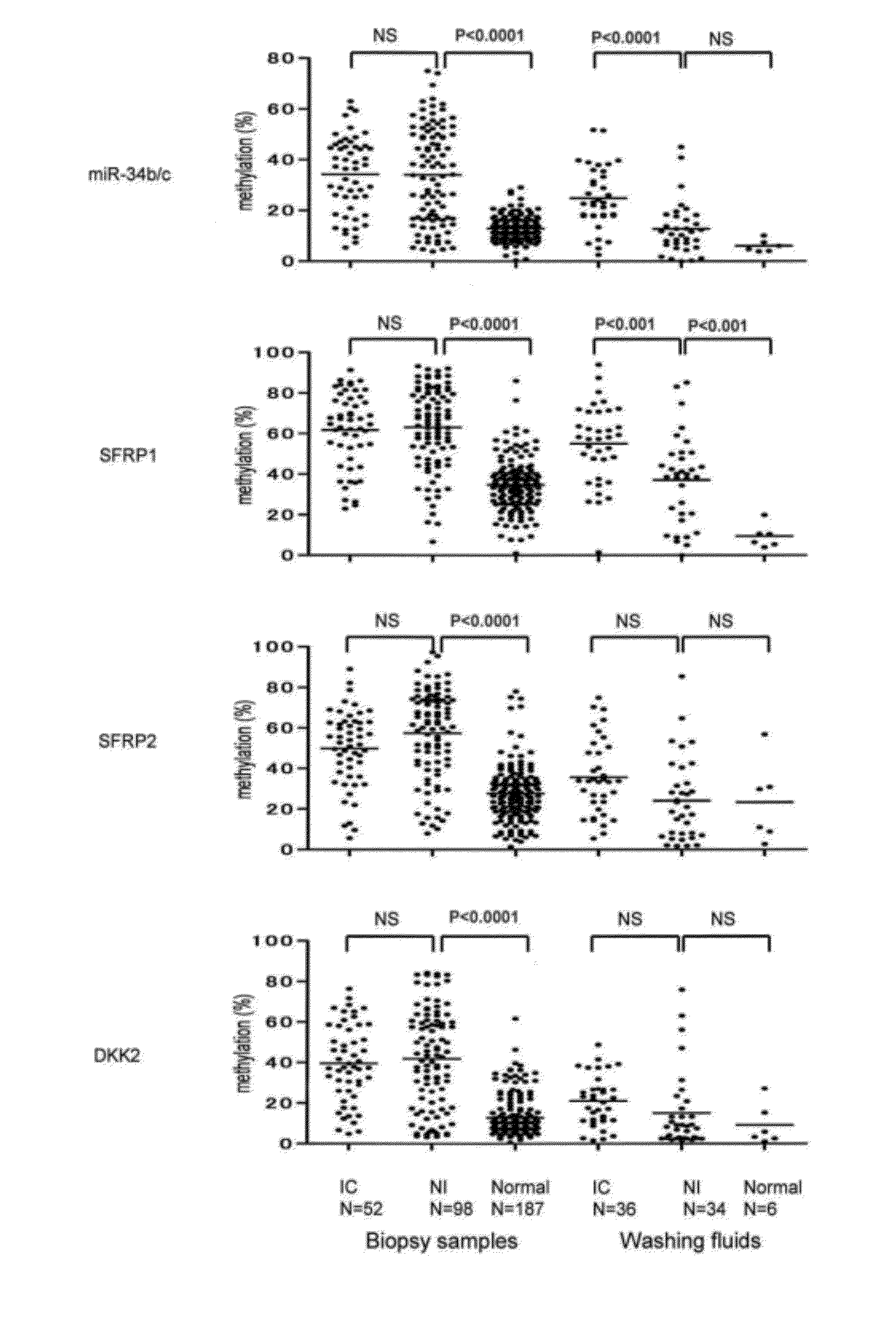
[0137]The specimens were collected during November 2008 and June 2009 in the Digestive Organs and Endoscopy Center of the Akita Red Cross Hospital. The subjects from which the specimens were collected include patients who have had colonic adenomas for a long time and who took colorectal endoscopy for follow-up purposes, or patients who took the same for the purpose of pre-operation of colorectal cancer surgery, or those who took the same for detailed examination by reason of positive results of fecal occult blood tests in check-ups. Biopsy was carried out for 52 specimens of invasive tumor, 98 specimens of non-invasive tumor, and 187 specimens of normal mucosa. Mucous-layer detachment fluid was collected for 34 specimens of invasive tumor, 36 specimens of non-invasive tumor, and 6 specimens of normal mucosa. Table 1 shows characteristics of subjects from which specimens were collected.

[0138]“Hyperplastic polyp,”“adenoma,”“early cancer,”...

example 2

Measurement of DNA Methylation Level and Measurement of K-RAS Mutation

[0148]Next, DNA methylation level was measured for all cases. First, colonoscopy was performed and when a tumor was observed by colonoscopy of the entire large bowel, a washing fluid was sprayed by a Pyoktanin-spraying tube (NT tube, Olympus) from an adjacent position of the tumor to detach the mucus from the surface of the lumen of the large intestine. At this time, the washing fluid was sprayed onto the colonic mucous layer at a rate of approximately 5 ml / s.

[0149]Then, after observation by a magnified endoscope (CF-260AZI), background normal mucosae at the cancerous site of the tumor, adenoma site, and periphery of the tumor (within 1 cm) were collected by biopsy, and treated with EMR or marking, etc. The collected specimens were stored in endfresh, and after frozen storage, DNA extraction by phenol-chloroform method was performed.

[0150]The biopsy samples and colonic-mucous-layer detachment fluids collected were...

example 3

Endoscopic Classification

[0161]Tumor morphology of hyperplastic polyp, adenoma, and early cancer was classified into protruded type (0-I), flat type (IIa, LST), or depressed type (IIc, IIa+IIc) by colorectal endoscopy. Early cancer was defined as those wherein cancer invasion reaches the submucosa, regardless of venous invasion or lymphatic invasion. Borrmann classification was applied to advanced cancer. All the colorectal tumor surgery and EMR specimens underwent clinical diagnosis in accordance with WHO classification, at the Pathological Department of the Akita Red Cross Hospital.


PUM

PropertyMeasurementUnit
Flow rateaaaaaaaaaa
Contentaaaaaaaaaa
Levelaaaaaaaaaa

Abstract

An object of the present invention is to provide a method for non-invasively diagnosing invasiveness or degree of invasion of colorectal tumors.
The present invention is characterized in that it enables to obtain a specimen that can be used to detect invasive colorectal tumors by spraying a washing fluid onto the colonic mucous layer of a subject to detach the mucus from the mucous layer, and collecting the detached mucus together with the washing fluid.

Description

TECHNICAL FIELD [0001]This application is a continuation-in-part of, and under 35 U.S.C. §120 claims the benefit of priority to, international application Ser. No. PCT / JP2010 / 064715, filed on Aug. 30, 2010, which claims the benefit of priority to US provisional application Ser. No. 61 / 238,106, filed Aug. 28, 2009, the entire contents of both of which are hereby incorporated herein by reference.[0002]The present invention relates to a specimen for diagnosing invasive colorectal tumors which is obtained by non-invasively detaching the colonic mucous layer, a kit for non-invasively detaching the colonic mucous layer, a method for collecting a specimen for diagnosing invasive colorectal tumors non-invasively, a method for detecting invasive colorectal tumors by non-invasively detaching the colonic mucous layer, a method for evaluating therapeutic effects of a drug and / or a therapeutic method by non-invasively detaching the colonic mucous layer, and a method for diagnosing the degree of ...

Claims


Application Information

Patent Timeline
IPC IPC(8): C12Q1/68G01N33/48
CPCA61B1/041G01N33/57419C12Q2600/178C12Q1/6886C12Q2600/154G01N33/57488
Inventor TOYOTA, MINORUTOYOTA, MUTSUMIYAMAMOTO, EIICHIROKAMIMAE, SEIKOHIROMU, SUZUKIYAMANO, HIROO
Owner SAPPORO MEDICAL UNIVERSITY
Why Eureka
Try Eureka