Microfluidic System Having Monolithic Nanoplasmonic Structures

a microfluidic system and nanoplasmonic technology, applied in flexible pipes, dough embossing machines, material analysis, etc., can solve the problems of significant limitations, inability to quickly detect pathogens or cancers, and inability to meet the rapid pathogen or cancer detection requirements, etc., to achieve rapid low-cost mold fabrication, low volume consumption, and low cost

Active Publication Date: 2014-01-02
NAT RES COUNCIL OF CANADA
View PDF1 Cites 18 Cited by
  • Summary
  • Abstract
  • Description
  • Claims
  • Application Information

AI Technical Summary

Benefits of technology

[0034]The present invention advantageously provides low-cost monolithic integrated microfluidic systems with multiplexing capability (e.g. valving, pumping) for precise control of cell culture conditions that can simultaneously integrate label-free enhanced plasmonic techniques such as surface plasmon resonance (SPR) (e.g. reflection-mode SPR, transmission-mode SPR, localized surface plasmon resonance (LSPR)) or surface-enhanced Raman scattering (SERS). The present monolithic integrated polymer-based microfluidic system has micro- and nano-structures that provide topographical cues for cell attachment and culture for controlling cell behavior while permitting monitoring of cellular behavior, motility, attachment, viability, biomolecule interactions or any combination thereof using plasmonic detection. The system is fabricated using a simple, robust and cost-effective process in a single step.
[0035]The present system is advantageous over both conventional and recently reported processes employed for cell culture and monitoring of cell behavior as the present system i...

Problems solved by technology

However, there is a need for methods that detect bioaffinity interactions without molecular labels, especially for biomolecular and cellular interactions, where labeling is problematic and can interfere with their biological properties.
Additionally, these methods are not suitable for the rapid pathogen or cancer detection as they require extensive blood culture of the pathogen or diseased tissue in the central laboratory prior to the detection of antibodies.
Even though developments in the field of high-throughput screening (HTS) and computational chemistry greatly accelerated and facilitated the drug finding process, there are significant limitations to overcome.
Currently employed assays that rely on cell culture in Petri dishes and subsequent fluorescence-based live-cell imaging and biomolecule detection are slow, cumbersome and cannot meet these requirements.
The resulting variability in the concentration of applied reagents hinders fair and quantitative comparisons and limits the ability of HCCS to resolve small differences in cell signaling responses.
This issue is exacerbated in more complex protocols, such as sequential exposure of cells to different media, because of errors that accumulate when changing media.
Moreover repeated media aspirations might unintentionally remove cells from the wells.
Because these assays are also difficult to miniaturize, HCCS experiments may consume large quantities of expensive or valuable cells and reagents.
Finally HCCS still relies on fluorescent tags which may trigger unwanted steric hindrance effects.
Consequently, the research into the effect of cues (single or multiple) on cellular response to date has been limited by the lack of robust and reproducible methods for homogeneous material production, precise control of the cell culture conditions and in situ real-time label-free monitoring of cellular response, cell behavior, cell viability or biomolecular binding interactions.
Specifically, the material production methods have lacked the control required to reproducibly fabricate homogeneous surfaces that will allow investigations into specific interactions between cells and isolated variables i.e. a precisely defined nanoscale patterns in a defined space with control over the induced change in topography and associated changes to surface energy.
The commonly employed well-based cell culture methods are costly and suffer errors in liquid dispensing, both manually and robotically, thus precluding uniform handling of each well which in turn limits how finely signaling responses may be resolved.
Finally, while the use of fluorescent imaging techniques for cell analysis can provide information not easily attainable by other methods, they are usually confounded by the need to over-express the signaling protein of interest and by possible effects of the fluorescent marker on the protein's function.
However, despite a growing focus from the microfluidic research community, both PCR and ELISA rely on fluorescence labels, which increase the complexity and cost of the assay.
In addition to the requirement for a labeled recognition element, these techniques typically require complex optical systems which typically consist of a large microscope or a microplate reader.
As a result, the field of microfluidics has yet to produce many commercial devices for disease diagnostics (Myers 2008).
However, the assays in these studies were performed using traditional cell-culture methods and analyzed using live fluorescence microscopy with inherent drawbacks of these techniques which may have resulted in misleading interpretation of results due to error in liqui...

Method used


Image

  • Microfluidic System Having Monolithic Nanoplasmonic Structures
  • Microfluidic System Having Monolithic Nanoplasmonic Structures
  • Microfluidic System Having Monolithic Nanoplasmonic Structures

Examples


example 1

Process for Fabricating a Monolithic Integrated Nanoplasmonic Microfluidic Cell Culture System

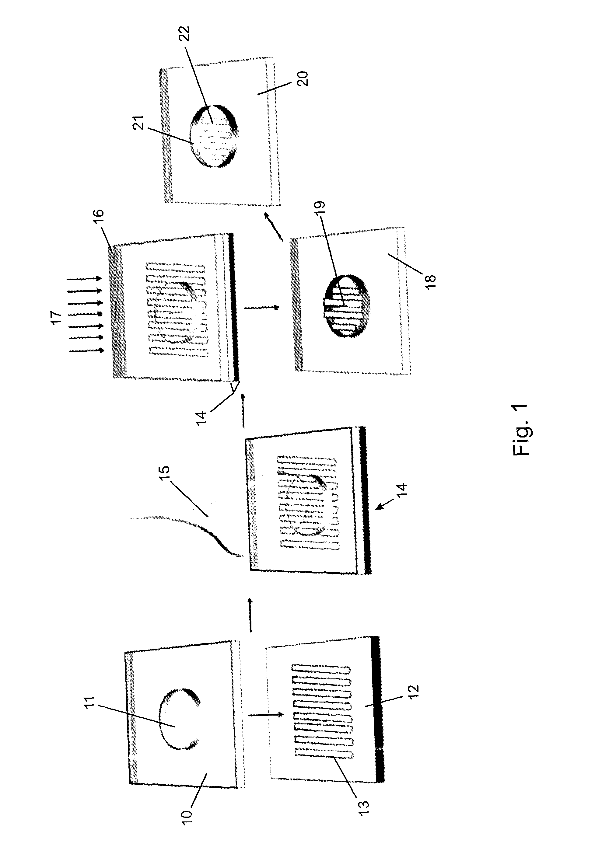
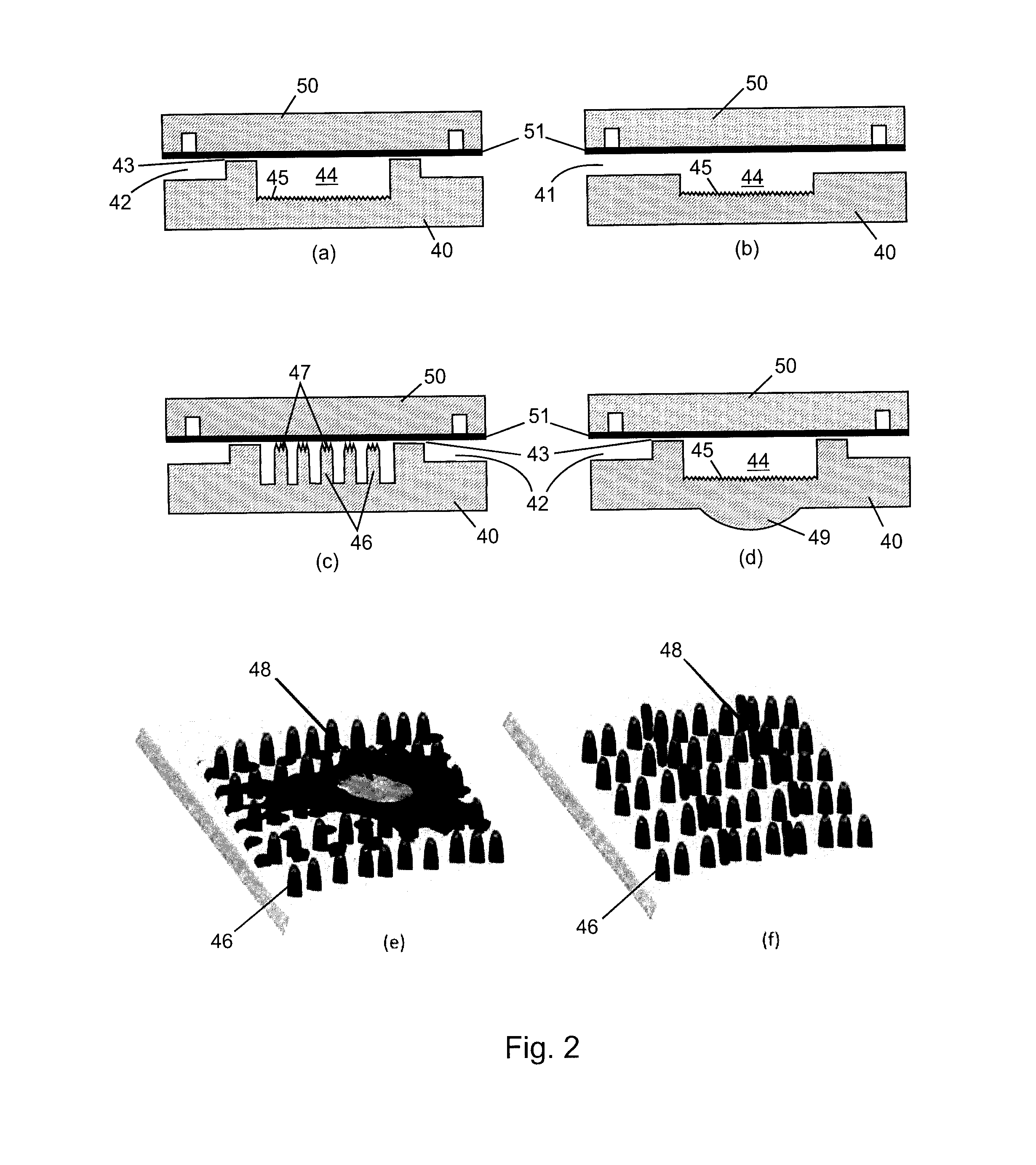
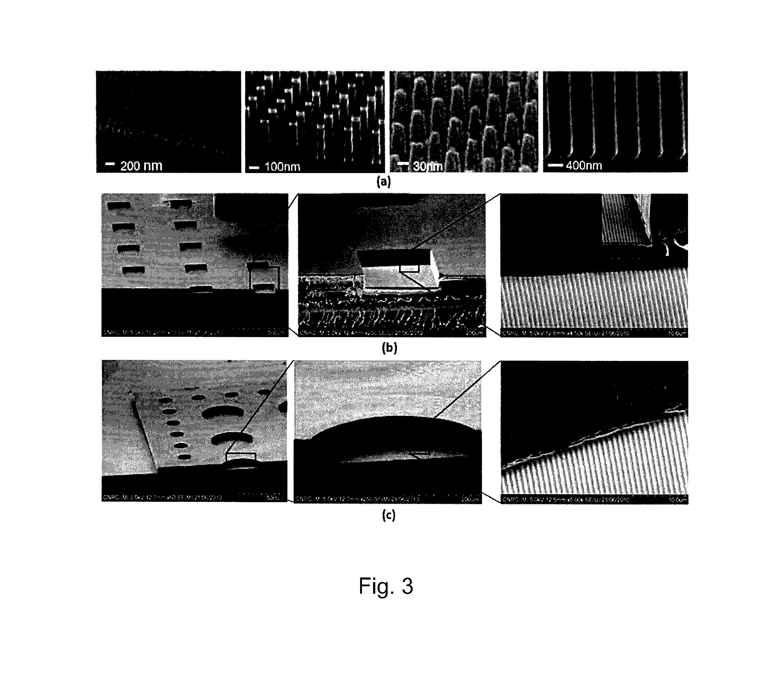
[0044]A monolithic integrated nanoplasmonic microfluidic cell culture system of the present invention may be produced generally as shown in FIG. 1, which illustrates the process showing a single cell chamber and none of the channels, conduits, valves or other microfluidic features for clarity. A soft thermoplastic elastomer film is hot-embossed to form micro-scale and meso-scale features (microchannels, conduits, chambers, etc.) in TPE membrane 10 including through-hole 11. This is performed at an applied pressure ranging from 5 kN to 15 kN, for 5-30 min, at a temperature in a range of from 100° C. to 160° C., depending on the desired features. Hard Zeonor™ substrate 12 is patterned by hot-embossing at an applied pressure of 10 kN to 20 kN for 10-30 min at a temperature ranging from 140° C. to 170° C. depending on the specific Zeonor™ grade to form a regular array of nano-scale grating elem...

example 2

Use of a Monolithic Integrated Nanoplasmonic Microfluidic Cell Culture System in Plasmonic Detection

[0048]In operation a monolithic integrated nanoplasmonic microfluidic cell culture system of the present invention employs pressure-driven flow to transport cells in suspension from a plurality of reservoirs through a plurality of cell-loading channels to a plurality of nanostructured cell culture chambers by closing valves of the perfusion channels and opening valves on the cell-loading channels. A plurality of cell-lines are loaded using a plurality of different reservoirs. Following initial cell attachment on the bottom of the nanostructured chambers, the valves on the cell-loading channels are closed, and fresh media is continuously injected in each of the perfusion channels. Multiple high resistance perfusion conduits ensure equal distribution of the media within the chamber while minimizing the shear-stress exerted on the cells.

[0049]Once the cell culture chambers are loaded wit...


PUM

PropertyMeasurementUnit
Fractionaaaaaaaaaa
Nanoscale particle sizeaaaaaaaaaa

Abstract

A microfluidic system, particularly suited as a cell culture system, is provided having a single monolithic biocompatible substrate with both a surface having an ordered array of nano-scale elements required for plasmonic response monitoring and a network of microchannels for precisely controlling cellular environment. The system has the additional advantages of low-volume consumption, rapid low-cost fabrication of molds with easily interchangeable microfluidic channel layouts, amenability to mass production, and in situ label-free real-time detection of cellular response, viability, behavior and biomolecular binding using plasmonic techniques. A ratio of greater than 0.2 between the cross-sectional dimension and the spacing distance of the nano-scale elements is useful for plasmonic response monitoring. A process for producing such a system involves fabrication of a master mold containing the nano-scale elements etched into a hard substrate, and the micro-scale and meso-scale features, such as channels and chambers, provided in a soft membrane bonded to the hard substrate. A stamp may be created by setting a settable liquid polymer or metal placed in the master mold and then the features of the intended device transferred to a polymeric substrate using the stamp.

Description

CROSS-REFERENCE TO RELATED APPLICATIONS[0001]This application claims the benefit of U.S. Provisional Patent Application U.S. Ser. No. 61 / 452,868 filed Mar. 15, 2011, the entire contents of which is herein incorporated by reference.FIELD OF THE INVENTION[0002]The present invention is related to microfluidic systems and devices having nanoplasmonic features, particularly for use in cell culture applications, and to processes of producing such systems and devices.BACKGROUND OF THE INVENTION[0003]Analysis of molecular binding and cell behavior are important for disease diagnostics, biomedical research, and drug discovery. The vast majority of array-based studies of bioaffinity interactions employ fluorescently labeled biomolecules or enzyme-linked colorimetric assays. However, there is a need for methods that detect bioaffinity interactions without molecular labels, especially for biomolecular and cellular interactions, where labeling is problematic and can interfere with their biologic...

Claims


Application Information

Patent Timeline
IPC IPC(8): G01N21/65
CPCG01N21/658B82Y30/00C12M23/16G01N21/554B01L3/502715B01L2300/0654B01L2300/0822B01L2300/0896B01L2400/086Y10T137/8593B01L3/502707G01N21/65B01L2200/12B01L2300/044B01L2300/0861B01L2300/0887B01L2300/12
Inventor MALIC, LIDIJAMORTON, KEITHVERES, TEODOR
Owner NAT RES COUNCIL OF CANADA
Why Eureka
Try Eureka